Laser-Induced Breakdown Spectroscopy (LIBS): A Comprehensive Review of Principles, Biomedical Applications, and Analytical Advancements
This article provides a comprehensive analysis of Laser-Induced Breakdown Spectroscopy (LIBS), an emerging atomic emission technique renowned for its rapid, multi-elemental analysis capabilities with minimal sample preparation.
Laser-Induced Breakdown Spectroscopy (LIBS): A Comprehensive Review of Principles, Biomedical Applications, and Analytical Advancements
Abstract
This article provides a comprehensive analysis of Laser-Induced Breakdown Spectroscopy (LIBS), an emerging atomic emission technique renowned for its rapid, multi-elemental analysis capabilities with minimal sample preparation. Tailored for researchers, scientists, and drug development professionals, it explores the fundamental principles of LIBS and its operational mechanics. The review systematically covers its diverse methodological applications—from pharmaceutical tablet analysis and quality control to biomedical diagnostics and elemental mapping in tissues. It further addresses key analytical challenges, including signal optimization and matrix effects, while evaluating the technique's performance against established methods like ICP-MS. By synthesizing foundational knowledge with cutting-edge applications and validation studies, this work serves as a critical resource for understanding LIBS's potential as a transformative tool in both industrial and clinical settings.
The Fundamentals of LIBS: From Laser-Plasma Interaction to Spectral Fingerprinting
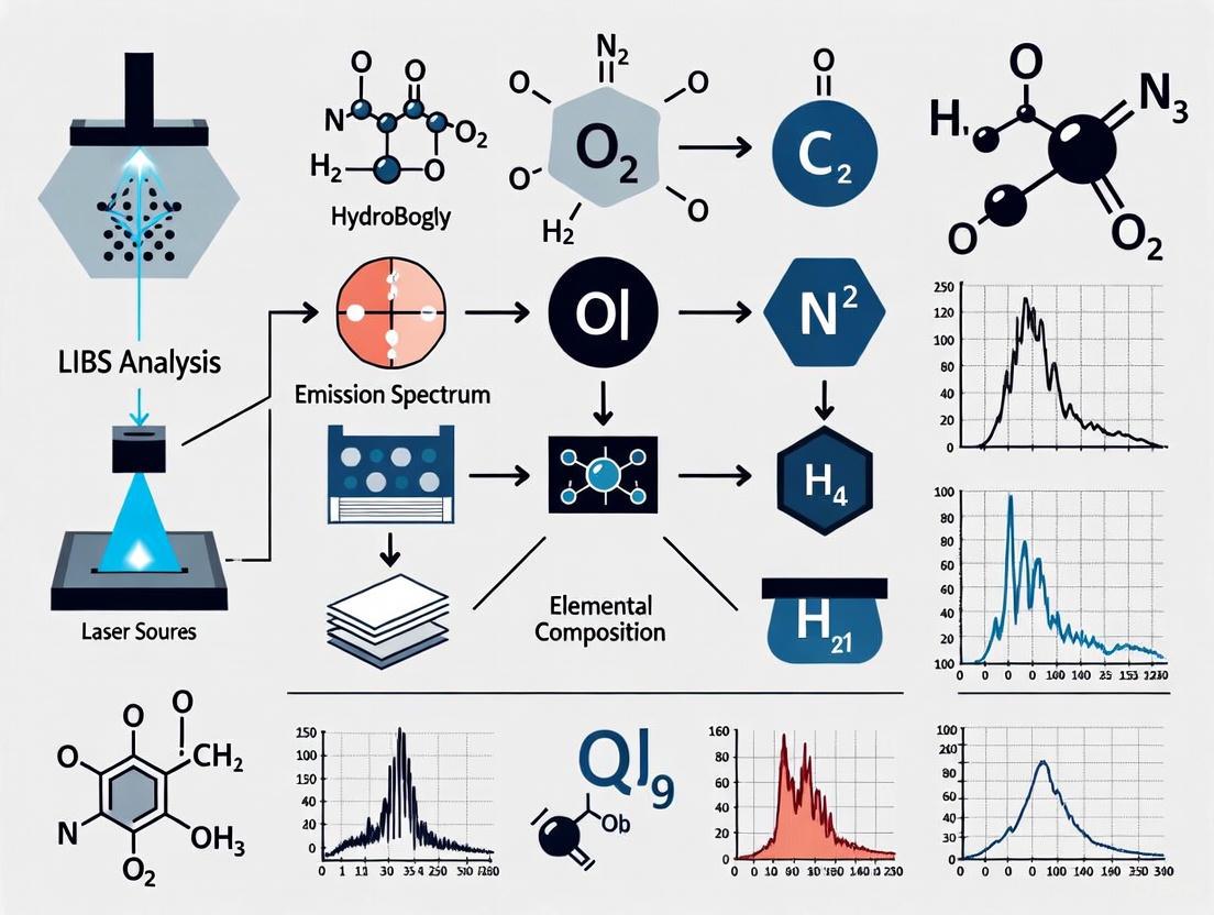
Laser-Induced Breakdown Spectroscopy (LIBS) is a rapid, versatile analytical technique used for the elemental analysis of materials. Its core principle involves using a laser pulse to generate a microplasma on a sample surface. As this plasma cools, it emits atomic and ionic radiation that is characteristic of the elements present, serving as a unique "fingerprint" for their identification and quantification [1]. This technique is valued for its minimal sample preparation, capability for in-situ analysis, and applicability across diverse fields such as biomedical research, environmental monitoring, industrial applications, and geological mining [1].
Core Physical Principles
Laser Ablation and Plasma Formation
The LIBS process is initiated when a high-powered, focused laser pulse is directed at the sample surface. This interaction ablates a minute volume of material (nanograms to picograms), creating a vapor plume. The leading part of the laser pulse further interacts with this vapor, exciting and ionizing its constituents to form a high-temperature plasma plume (often at temperatures of 10,000–20,000 K) [1]. The fundamentals of this laser-matter interaction, various forms of ablated material, and subsequent plasma interaction are governed by complex phenomena including laser parameters (wavelength, pulse duration, energy) and specimen properties [1].
Plasma Expansion and Cooling
Following the laser pulse, the plasma expands rapidly and begins to cool. The lifetime and evolution of this plasma depend significantly on the laser pulse duration. For nanosecond (ns) laser pulses, plasma formation occurs during the pulse, with the trailing part of the pulse reheating the plasma and leading to a longer lifetime (on the order of microseconds, µs). In contrast, for femtosecond (fs) laser pulses, plasma is formed after the laser pulse, evolves faster, and has a shorter lifetime (several hundred nanoseconds) [1]. The absence of laser-plasma interaction in fs-LIBS reduces the heat-affected zone, offering higher ablation efficiency and less dependence on the material matrix [1].
Atomic and Ionic Emission
As the plasma cools, the excited atoms and ions within the plasma decay to lower energy states, emitting photons at specific wavelengths. The emitted light is collected by a spectrometer, which separates it into its constituent wavelengths to produce a spectrum. Each element in the periodic table possesses a unique set of emission lines, determined by its electronic energy level structure. The analysis of these spectral lines, including their presence (for qualitative analysis) and intensity (for quantitative analysis), reveals the elemental composition of the sample [1]. The underlying mechanisms for understanding LIBS analytical outcomes are governed by theoretical models, with local thermodynamic equilibrium (LTE) being the most commonly used for plasma modeling [1].
Experimental Protocols
LIBS Setup and Workflow
A standard LIBS apparatus consists of several key components: a pulsed laser source (commonly Nd:YAG), optics for focusing the laser beam, a sample stage, a system for collecting the plasma light (lens and optical fiber), a spectrometer, and a detector (such as an ICCD or CCD camera) [1]. The general workflow for a LIBS experiment is illustrated below.
Protocol for Custom Color Sample Analysis (Psychophysical Context)
This protocol is adapted for studies requiring highly controlled, closely related color pairs for visual discrimination experiments, such as those investigating low vision [2].
- Objective: To produce and analyze pairs of color patches with minimal, perceptually equidistant color differences using a calibrated inkjet printer and stabilized LED lighting.
Materials & Equipment:
- High-quality inkjet printer (e.g., Canon image PROGRAF PRO-300).
- Matte photo paper.
- Spectrophotometer for color measurement.
- Custom LED lighting system with spectral power distribution (SPD) stabilization.
- Software for color management and data analysis.
Step-by-Step Procedure:
- Color Space Sampling: Generate a set of reference color patches by sampling the CIELAB color space using a non-Euclidean color difference formula (CIEDE2000 or ΔE00). Utilize a pre-calculated, tabulated sampling grid (
CIELABTab00) where each color is equidistant (e.g., ΔE00 = 0.5) from its six neighbors [2]. - Printer Characterization and Calibration: Bypass the operating system's default color management. Use a color management process based on polyharmonic spline and tetrahedral interpolation to create a precise profile linking the printer's native RGB values to the target CIELAB coordinates. This ensures accurate color reproduction [2].
- Sample Printing: Print the generated color patches. For a discrimination experiment, select pairs of colors that are colorimetrically close and equidistant according to the ΔE00 metric [2].
- Lighting Stabilization: Implement a closed-loop feedback system to stabilize the Spectral Power Distributions (SPDs) of the LED lighting unit. This is critical to eliminate fluctuations caused by heat buildup or aging components, which could otherwise introduce unwanted variables in color perception [2].
- Data Collection and Analysis: Present the printed color pairs to study participants under the stabilized light. Use a paired comparison method to gather data on color discrimination thresholds. Analyze the results to determine the just-noticeable differences (JND) under the optimized lighting conditions [2].
- Color Space Sampling: Generate a set of reference color patches by sampling the CIELAB color space using a non-Euclidean color difference formula (CIEDE2000 or ΔE00). Utilize a pre-calculated, tabulated sampling grid (
Protocol for Cement Content Analysis in Concrete
This protocol demonstrates a specific materials science application of LIBS for non-destructive, in-situ analysis [3].
- Objective: To quantitatively estimate the cement content in concrete samples using spatially resolved LIBS.
Materials & Equipment:
- Pulsed laser source (e.g., Nd:YAG).
- Spectrometer with high resolution.
- Motorized sample stage for raster scanning.
- Concrete samples (prepared models and real-world samples).
Step-by-Step Procedure:
- Sample Preparation and Modeling: Begin by creating mesoscale concrete models with known cement content. These models help identify key experimental parameters such as optimal spatial resolution, measurement area, and boundary effects [3].
- LIBS Raster Scanning: Focus the laser pulse on the concrete sample surface. Raster-scan the laser across a defined area to build a spatially resolved chemical map of the surface. The plasma emission at each point provides the local elemental composition, allowing differentiation between cement paste, aggregates, and voids [3].
- Spectral Clustering and Data Processing: Process the collected spectra using multivariate analysis. Combine Principal Component Analysis (PCA) with density-based spectral clustering to achieve clear separation between the different phases of the concrete (cement paste vs. aggregate) [3].
- Quantification: Correlate the classified spectral data with the known composition of the models to estimate the cement content in unknown samples. Under optimized conditions, this method has demonstrated an average relative error of approximately 8%, an improvement over traditional destructive methods [3].
The Scientist's Toolkit: Essential Research Reagents and Materials
Table 1: Key reagents, materials, and equipment for LIBS experiments.
| Item | Function/Description | Application Example |
|---|---|---|
| Pulsed Laser | Generates high-energy pulses for sample ablation and plasma initiation. Common types include Nd:YAG (Nanosecond) and Femtosecond lasers. | Core component in all LIBS setups [1]. |
| Spectrometer | Disperses the collected plasma light into its constituent wavelengths to form a spectrum. | Elemental identification and quantification [1]. |
| Calibrated Printer & Matte Paper | Produces custom, colorimetrically accurate sample patches for psychophysical experiments. | Generating color pairs for vision studies [2]. |
| Stabilized LED Light Source | Provides consistent, flicker-free illumination with controlled Spectral Power Distribution (SPD). | Essential for visual experiments where lighting variability must be minimized [2]. |
| Reference Materials | Samples with known elemental composition (e.g., standard reference materials for CC-LIBS). | Used for calibration and validation of quantitative results [1]. |
| Mesoscale Concrete Models | Laboratory-made concrete samples with precisely defined composition. | Method development and parameter optimization for cement analysis [3]. |
Data Analysis and Quantification Methods
Data Processing Workflow
The journey from raw plasma emission to quantitative results involves several critical steps, increasingly supported by artificial intelligence (AI) and machine learning (ML) models to handle spectral complexity and improve classification accuracy [1]. The following diagram outlines this workflow.
Quantitative Formalism in LIBS
Two primary formalisms are used for elemental quantification, each with distinct advantages and requirements.
Table 2: Comparison of quantitative methods in LIBS.
| Method | Principle | Requirements | Advantages | Limitations |
|---|---|---|---|---|
| Calibration-Curve LIBS (CC-LIBS) | Plots a calibration curve of emission line intensity versus concentration using standard reference materials. | Matrix-matched standard reference materials. | High accuracy when standards are well-matched. | Requires a set of reliable standards; prone to matrix effects [1]. |
| Calibration-Free LIBS (CF-LIBS) | Determines elemental concentration from the plasma emission spectrum based on theoretical models of plasma physics (LTE assumption), without standard references. | No standard references needed; requires accurate plasma temperature measurement. | Eliminates need for calibration standards; useful for unknown samples. | Relies on the validity of the LTE assumption; computationally intensive [1]. |
Advanced Applications in Biomedical and Materials Science
The application of LIBS continues to expand into diverse and complex fields. In oncology, LIBS has been effectively used to differentiate between malignant and normal tissues and to classify cancer stages and types based on the detection of elemental imbalances and biomarkers [1]. For instance, fs-LIBS has enabled high-resolution elemental imaging of melanoma tumour tissue with a spatial resolution of 15 µm [1]. In the analysis of calcified tissues (e.g., teeth, bones), LIBS serves as a powerful tool for inspecting minerals, mapping metabolic markers, and studying disorders that alter the crystallography of hydroxyapatite [1]. In materials science, as demonstrated in the cement analysis protocol, LIBS provides a rapid, non-destructive alternative to traditional methods, enabling quality control and diagnostics in construction and industrial manufacturing [3].
Laser-Induced Breakdown Spectroscopy (LIBS) is a rapid, versatile form of atomic emission spectroscopy used for elemental analysis. The fundamental principle involves using a highly energetic, focused laser pulse to ablate a micro-volume of material, creating a transient plasma. As this plasma cools, the excited atoms and ions within it emit characteristic wavelengths of light; collecting and analyzing this emitted light with a spectrometer and detector allows for the determination of the sample's elemental composition [4] [1]. LIBS stands out for its minimal-to-no sample preparation requirements, capability for simultaneous multi-element detection, and potential for portability and in-situ analysis, making it applicable across fields from industrial sorting to medical diagnostics [1].
The core components of any LIBS system work in a tightly synchronized sequence. The laser serves as the excitation source, generating the optical energy required for plasma formation. The spectrometer acts as the separation tool, dispersing the collected plasma light into its constituent wavelengths. Finally, the detector functions as the measurement device, converting the dispersed optical signals into quantifiable electrical data for analysis. The performance and selection of these three components directly dictate the system's analytical capabilities, including its spectral resolution, limit of detection, and overall sensitivity.
Core Component Analysis
The analytical performance of a LIBS system is fundamentally governed by the technical specifications and synergistic operation of its three key components. The following sections provide a detailed breakdown of each component, with quantitative data summarized for easy comparison.
Lasers
The laser is the primary excitation source in a LIBS system, and its parameters critically influence plasma formation and the resulting spectral emission.
Key Laser Parameters and Typical Specifications
| Laser Parameter | Common Types / Values | Impact on LIBS Performance |
|---|---|---|
| Pulse Duration | Nanosecond (ns), Femtosecond (fs) [1] | ns-pulses: Longer plasma lifetime (µs), plasma-laser interaction, more thermal effects [1].fs-pulses: Shorter plasma lifetime (hundreds of ns), minimal thermal damage, reduced matrix effects, higher spatial resolution [1]. |
| Wavelength | UV to IR (e.g., 1064 nm Nd:YAG) [5] | Shorter wavelengths often lead to better ablation efficiency and plasma coupling with solid surfaces. |
| Pulse Energy | Millijoule (mJ) to Joule (J) range | Higher energy typically increases plasma volume and signal intensity, but can lead to excessive sample damage. |
| Repetition Rate | Single pulse to kHz [1] | Higher rates enable faster analysis and signal averaging for improved precision. |
Nanosecond (ns) Q-switched Nd:YAG lasers are the most commonly used lasers in LIBS due to their maturity and reliability. As utilized in a typical experimental setup for studying aluminum plasma, a Nd:YAG laser operating at its fundamental wavelength of 1064 nm, with a pulse duration of 7 ns and pulse energy of 94 mJ, can produce a laser fluence of approximately 24 J/cm² when focused onto a target [5]. In such ns-laser ablation, the plasma is formed during the laser pulse, and the trailing part of the pulse interacts with and reheats the plasma, leading to a longer plasma lifetime on the order of microseconds [1].
Femtosecond (fs) lasers represent an advanced development, offering significant advantages. Fs laser ablation depth of 6 µm on a thin tissue section of liver metastases from a colorectal cancer patient has been reported, allowing for fast in-depth multi-elemental profiling at cellular spatial resolution [1]. The ultra-short pulse duration means plasma forms after the laser pulse, virtually eliminating plasma-laser interaction. This results in a plasma lifetime of several hundred nanoseconds, significantly reduced thermal effects on the sample, and less dependence on the material's matrix, which is particularly beneficial for analyzing heterogeneous biological tissues [1].
Spectrometers
The spectrometer resolves the broad-spectrum light emitted by the plasma into its constituent wavelengths, creating the unique elemental fingerprint for analysis.
Spectrometer Configurations and Capabilities
| Spectrometer Type | Spectral Resolution | Typical Application | Advantages / Disadvantages |
|---|---|---|---|
| Czerny-Turner | Moderate to High | Broad elemental analysis, research applications [4] | Good flexibility and resolution; can be larger in size. |
| Echelle Spectrometer | High | Simultaneous broad-range analysis with high resolution | Compact design for wide spectral coverage; requires cross-dispersion. |
| Compact / Miniaturized | Moderate | Portable and handheld LIBS systems [6] | Enabled by advancements in miniaturization for field use. |
The choice of spectrometer is a critical trade-off between spectral resolution, wavelength coverage, and physical size. Broadband high-resolution spectrometers, developed and commercialized in the early 2000s, were a key innovation that allowed LIBS systems to maintain sensitivity to chemical elements even at low concentrations [4]. For handheld devices dominating the market, the challenge and achievement have been to miniaturize spectrometer components without completely sacrificing analytical performance, thus enabling real-time, on-site elemental analysis [6]. The spectral window covered (e.g., from deep UV to near-IR) determines which elemental emission lines can be observed.
Detectors
Detectors capture the dispersed light from the spectrometer and convert photons into an electrical signal that is digitized and processed.
Common Detector Types and Characteristics in LIBS
| Detector Type | Principle | Key Features | Suitability |
|---|---|---|---|
| Intensified CCD (ICCD) | Gated intensifier + CCD | High sensitivity, ultrafast gating (ns), time-resolved analysis [1] | Essential for rejecting initial plasma continuum; useful for plasma diagnostics. |
| Non-Intensified CCD/CMOS | Semiconductor photodiodes | Lower cost, rugged, no gating required | Often used with fs-LIBS where plasma continuum is weak, or in portable systems. |
The Intensified CCD (ICCD) is a cornerstone of traditional ns-LIBS. Its ability to be electronically gated is crucial. By activating the detector with a precise time delay (from tens of nanoseconds to several microseconds) after the laser pulse, the initial intense continuum radiation from the hot plasma can be excluded. This allows the detector to collect only the sharper atomic and ionic line emissions that appear as the plasma cools, dramatically improving the signal-to-noise ratio [1]. The temporal resolution of LIBS plasma, which is on the order of a few nanoseconds for ns-laser pulses, makes this gating capability essential [1]. For fs-LIBS, where the plasma continuum is inherently weaker, non-intensified CCD or CMOS detectors can be sufficient, simplifying the system and reducing cost.
Advanced Methodologies and Protocols
Experimental Workflow for Material Analysis
The following diagram outlines the generalized experimental workflow for a LIBS analysis, from sample preparation to data interpretation.
Protocol: Analysis of a Metallic Alloy Using a Bench-Top LIBS System
1. Sample Preparation:
- Objective: Obtain a flat, clean surface for consistent laser ablation.
- Procedure: If the sample is a bulk metal, polish the surface with progressively finer grit sandpapers (e.g., from 400 to 1200 grit) to create a uniform surface. Clean the polished surface with a solvent like isopropanol and allow it to dry to remove any residues or particles [7].
2. Instrument Setup:
- Laser Alignment: Focus the laser pulse (e.g., from a Nd:YAG laser at 1064 nm) onto the sample surface using a plano-convex lens (e.g., 20 cm focal length). The focus should be slightly below the surface to maximize ablation efficiency and stabilize the plasma.
- Spectrometer & Detector Configuration:
- Set the spectrometer to cover a relevant wavelength range (e.g., 200 - 500 nm for most metals).
- For an ICCD detector, set the delay time (td) and gate width (tw). A typical starting point is a delay of 1 µs and a gate width of 5 µs to avoid the strong plasma continuum. Adjust based on the observed signal-to-noise ratio [5].
3. Data Acquisition:
- Set the laser pulse energy (e.g., 50 mJ). Use a laser fluence that is above the ablation threshold but avoids excessive sample damage or signal saturation.
- Position the sample on a motorized stage to allow for analysis at multiple fresh spots.
- Acquire spectra from multiple laser pulses (e.g., 10-50 pulses per spot) and average them to improve precision.
4. Data Analysis:
- Qualitative Analysis: Identify elemental emission lines in the spectrum by comparing their wavelengths to a database of known atomic lines (e.g., Al I at 396.15 nm, Cu I at 324.75 nm).
- Quantitative Analysis: Construct a calibration curve using certified reference materials with known compositions. Plot the intensity (or integrated area) of a characteristic emission line against the concentration of the corresponding element. Use this curve to determine the concentration of the element in the unknown sample.
Protocol for Nanoparticle-Enhanced LIBS (NELIBS)
NELIBS is a powerful signal enhancement technique where metallic nanoparticles (NPs) deposited on a sample surface significantly increase the emission intensity of the analyte.
1. Nanoparticle Preparation and Deposition:
- Materials: Colloidal suspension of nanoparticles (e.g., 40-50 nm Gold NPs in deionized water) [5].
- Procedure: Deposit a controlled volume (e.g., 5-10 µL) of the NP colloidal solution onto the polished sample surface. Allow the droplet to dry evenly at room temperature, forming a layer of NPs on the analysis area.
2. LIBS Analysis with NPs:
- Use the same laser parameters as for conventional LIBS analysis of the bare sample.
- Focus the laser pulse onto the NP-coated region.
- The localized surface plasmon resonance of the NPs enhances the local electromagnetic field, leading to more efficient ablation and atomization, and a subsequent increase in emission intensity. Studies have reported signal enhancement of a few folds to orders of magnitude for metallic samples in NELIBS compared to standard LIBS [5].
3. Data Comparison:
- Directly compare the spectra acquired from the NP-coated spot with those from the bare sample spot. The enhancement factor for a specific emission line (e.g., Al I) can be calculated as the ratio of the peak intensity with NPs to the peak intensity without NPs.
Essential Research Reagent Solutions
Successful execution of LIBS experiments, especially advanced protocols like NELIBS, requires specific reagents and materials.
Key Research Reagents and Materials for LIBS
| Reagent / Material | Function / Application | Example Specification / Note |
|---|---|---|
| Gold Nanoparticles (Au NPs) | Signal enhancement in NELIBS [5] | Colloidal suspension, 40-50 nm diameter. Enhances emission via plasmonic effects. |
| Certified Reference Materials (CRMs) | Calibration and quantitative analysis | Metal alloys, soil, or polymer standards with certified elemental concentrations. |
| Polishing Supplies | Sample preparation for solid targets | Silicon carbide sandpaper (400-1200 grit), polishing cloths, alumina suspension. |
| Calibration Sources | Wavelength calibration of spectrometer | Deuterium-Argon lamps, low-pressure mercury pen lamps. |
| High-Purity Gases | Controlled atmosphere for plasma | Argon purge to improve signal quality for certain elements like carbon [6]. |
Technological Advancements and Future Outlook
The field of LIBS is being rapidly advanced through technological innovation and the integration of sophisticated data processing techniques. A major trend is the miniaturization of LIBS components into portable and handheld devices, which now represent the largest product segment in the market. These devices empower users to perform real-time, on-site elemental analysis in diverse settings, from scrap yards for metal sorting to mining operations for geological surveying [6].
The integration of Artificial Intelligence (AI) and Machine Learning (ML) is revolutionizing data analysis. AI algorithms can process vast and complex spectral datasets to identify patterns and correlations that are difficult to discern with traditional methods. For instance, new regression methods that integrate domain knowledge have been shown to outperform standard methods in LIBS quantification tasks, enhancing the accuracy and reliability of element detection and quantification [6]. This is particularly valuable for classifying complex samples like biological tissues or different rock types [8].
Furthermore, advancements in ultra-fast laser technology, particularly the use of femtosecond lasers, are pushing the boundaries of analytical precision. Techniques like plasma-grating induced breakdown spectroscopy (GIBS) have been developed to overcome traditional sensitivity limitations [6]. The use of fs-lasers minimizes thermal damage to the sample and reduces the matrix effect, enabling high-resolution elemental imaging in delicate materials such as pathological tissues with spatial resolution on the scale of micrometers [1]. These combined advancements in hardware and data science are solidifying LIBS's role as a powerful and adaptable analytical technique across an ever-widening range of scientific and industrial applications.
Laser-Induced Breakdown Spectroscopy (LIBS) has emerged as a rapid chemical analysis technique that revolutionizes elemental composition assessment across diverse fields, including pharmaceutical research, material science, and geological exploration [9]. This analytical technique utilizes a highly focused, pulsed laser to instantaneously vaporize a microscopic portion of a sample, creating a high-temperature plasma whose emitted light provides a unique elemental fingerprint [10]. The core advantage of LIBS lies in its capability for virtually sample preparation-free analysis, delivering comprehensive elemental profiles within seconds, a significant improvement over traditional methods requiring hours of preparation [9] [11]. Furthermore, LIBS offers exceptional versatility, capable of analyzing solid, liquid, and gaseous samples across the entire periodic table, with particular excellence in detecting light elements like lithium, beryllium, and carbon that challenge other spectroscopic methods [9] [12]. This protocol provides a detailed, step-by-step guide to the fundamental LIBS process, from laser ablation to spectral interpretation, framed within the context of advanced research applications.
Fundamental Principles and Physics of LIBS
The LIBS process is governed by the fundamental principles of atomic emission spectroscopy. When a high-energy laser pulse is focused onto a sample surface, it delivers an energy density typically ranging from 10⁸ to 10¹¹ watts per square centimeter, sufficient to cause optical breakdown [9]. This process generates a transient plasma with initial temperatures that can exceed 15,000 Kelvin and may reach up to 30,000 K in the earliest stages of its lifetime [9] [11]. At these extreme temperatures, a small mass of the sample—typically on the order of 1-10 micrograms—is ablated and transformed into a plasma containing free electrons, excited atoms, and ions [9] [10].
As the plasma expands and cools over a period of 1-10 microseconds, the excited electrons within the atoms and ions begin to transition from higher energy states to lower, more stable ground states [9] [13]. During these electronic transitions, energy is released in the form of photons. Critically, the wavelength of each emitted photon is inversely proportional to the energy difference between the excited and ground states, following the relation ( E = hc/\lambda ), where ( E ) is the energy difference, ( h ) is Planck's constant, ( c ) is the speed of light, and ( \lambda ) is the wavelength of the emitted photon [12]. Since each element possesses a unique electronic structure with characteristic energy level differences, the emitted wavelengths serve as unique identifiers for the elements present in the sample [11]. The detection and analysis of these characteristic emissions form the basis for both qualitative identification and quantitative determination of elemental composition in LIBS.
Step-by-Step LIBS Process
Laser Ablation and Plasma Formation
The LIBS analytical sequence begins with laser ablation, a process where a focused, high-energy pulsed laser beam interacts with the sample surface. The most commonly employed lasers are Q-switched Nd:YAG lasers operating at their fundamental wavelength of 1064 nm or at harmonic wavelengths such as 532 nm, 355 nm, or 266 nm [10] [13] [12]. These lasers typically generate pulses with durations of 4-15 nanoseconds, pulse energies from 1 mJ to several hundred millijoules, and repetition rates ranging from single shot to over 20 Hz [9] [12]. When this short, intense laser pulse is focused onto a sample, the electric field in the focal region accelerates naturally present ions and those formed via multiphoton interactions, leading to rapid heating and an explosive boiling process that ejects material from the sample surface [13]. This ablation process creates a microscopic crater measuring only 50-500 micrometers in diameter, removing merely 1-10 micrograms of material per pulse, thus preserving sample integrity for subsequent analyses [9].
The ablated material, consisting of atoms, molecules, and particulates, then interacts with the trailing portion of the laser pulse, leading to the formation of a high-temperature plasma plume. The leading edge of the laser pulse creates the initial conditions for ablation, while the remainder of the pulse energy further heats the ejected material, forming a highly energetic plasma [13]. This plasma, initially in a state of severe disequilibrium with temperatures potentially exceeding 50,000 K, contains free electrons, excited atoms, and ions [11] [13]. The specific characteristics of the plasma, including its temperature, electron density, and lifetime, are influenced by multiple factors including the laser parameters (wavelength, pulse duration, energy) and the sample matrix itself [13]. For nanosecond-class lasers, the ablation process is predominantly thermal, involving melting and vaporization, whereas femtosecond lasers produce more mechanical ablation with minimal thermal effects, though their application remains primarily in research due to cost and complexity [13].
Plasma Cooling and Spectral Emission
Following the termination of the laser pulse, the plasma begins to expand and cool rapidly. The initial stage of plasma cooling (typically < 1 microsecond) is dominated by continuum radiation (Bremsstrahlung emission), where highly excited free electrons slow down and emit a broadband background spectrum [11] [13]. As the plasma continues to cool and approaches local thermodynamic equilibrium (LTE)—generally occurring 0.5-1.0 microseconds after plasma formation for a 100-400 mJ plasma—the continuum emission diminishes, and discrete atomic and ionic emission lines begin to dominate the spectral output [13]. During this phase, excited electrons within atoms and ions transition to lower energy states, emitting photons at specific wavelengths characteristic of each element present [9] [12].
The emission characteristics vary throughout the plasma lifetime. Different types of radiation can be observed, including continuum, atomic, ionic, and molecular emissions, each revealing different components of the plasma [10]. The atomic and ionic emissions containing the analytically useful information typically occur after the plasma has sufficiently cooled, usually in the 1-10 microsecond window following the laser pulse [9] [10]. The timing of emission collection is crucial; detectors are often gated to activate after this initial continuum radiation has subsided to improve the signal-to-noise ratio of the discrete elemental emissions [13]. At this stage, the average plasma temperature typically ranges between 7,500-10,000 K, ideal for generating strong elemental emission lines while minimizing background continuum radiation [13].
Light Collection and Dispersion
The emitted light from the plasma is collected through specialized optical systems. In most laboratory and portable LIBS systems, photons are collected by a lens or lens system positioned near the plasma and transmitted to a spectrometer via an optical fiber [9] [12]. An alternative approach, known as stand-off LIBS, is employed in applications such as planetary exploration, where the analyzed sample may be several meters from the instrument. In these configurations, photon emission is captured by a telescope and transmitted to the spectrometer by fiber optics [14] [12]. This stand-off capability has been successfully implemented in NASA's Mars rovers, including the Curiosity rover's ChemCam instrument [14] [12].
The collected light is then dispersed by a spectrometer to separate it into its constituent wavelengths. Various spectrometer types are employed in LIBS systems, with echelle spectrographs being particularly common due to their high resolution across broad wavelength ranges [13] [12]. The spectrometer distributes the light across different spatial locations according to wavelength, creating a detailed spectrum that is recorded using a detector array. Common detectors include charge-coupled devices (CCD), intensified CCD (ICCD) cameras, or photomultiplier tubes, which convert the photon signals into electrical signals for digital processing [9] [10] [13]. These detectors often feature precise timing capabilities (gating) to selectively collect light during the optimal emission window after the continuum background has diminished [13].
Spectral Analysis and Data Interpretation
The final stage of the LIBS process involves spectral analysis and data interpretation to extract meaningful chemical information. The raw spectral data undergoes preprocessing procedures including dark background subtraction, wavelength calibration, ineffective pixel masking, spectrometer channel splicing, and background baseline removal [14]. The resulting spectrum displays intensity as a function of wavelength, with characteristic peaks representing specific electronic transitions of elements present in the sample [11].
Qualitative analysis involves identifying elements by matching observed emission lines to known spectral fingerprints of elements from reference databases [9] [12]. For instance, lithium emits a characteristic line at 670.8 nm, cobalt at 345.4 nm, and nickel at 352.4 nm [9]. Quantitative analysis utilizes the relationship between spectral line intensity and elemental concentration, typically established through calibration with certified reference materials [11] [12]. Advanced chemometric methods are increasingly employed, including machine learning and deep learning algorithms such as convolutional neural networks (CNNs), which can process entire spectral profiles to classify materials or quantify compositions, even overcoming challenges like spectral variations due to changing measurement distances [14] [15]. These multivariate analysis techniques often provide superior accuracy and precision compared to univariate methods that rely on single emission lines [12].
Schematic Diagram of the LIBS Analytical Process
Key Experimental Parameters and Protocols
Critical LIBS Operational Parameters
Successful implementation of LIBS requires careful optimization of several critical operational parameters that significantly influence analytical performance. The table below summarizes these key parameters and their typical values or considerations for robust method development.
Table 1: Key Operational Parameters in LIBS Analysis
| Parameter Category | Specific Parameter | Typical Values / Considerations | Impact on Analysis |
|---|---|---|---|
| Laser Properties | Wavelength | 1064 nm (fundamental), 532 nm, 355 nm, 266 nm (harmonics) | Shorter wavelengths often provide better ablation efficiency and smaller spot sizes [10] [13] |
| Pulse Duration | Nanosecond (most common), Picosecond, Femtosecond | ns-pulses: thermal ablation; fs-pulses: non-thermal, minimal heat-affected zone [13] | |
| Pulse Energy | 1 mJ to hundreds of mJ | Higher energy increases plasma temperature and emission intensity, but may increase fractionation [9] [13] | |
| Spot Size | 50-500 μm diameter | Smaller spots enable higher spatial resolution; larger spots provide better sampling volume [9] [12] | |
| Temporal Parameters | Gate Delay | 0.5-1.0 μs (for higher energy lasers) | Time between laser pulse and start of signal collection; optimizes signal-to-background ratio [13] |
| Gate Width | 1-10 μs | Duration of signal collection; affects signal intensity and spectral resolution [14] | |
| Sample Considerations | Surface Condition | Fresh, representative surface | LIBS analyzes only the surface; weathered or contaminated surfaces yield non-representative results [9] |
| Homogeneity | Homogeneous vs. heterogeneous | Heterogeneous samples require multiple analysis points for representative bulk composition [9] [12] | |
| Environmental Factors | Atmosphere | Air, Argon, Helium, or Vacuum | Ambient atmosphere affects plasma formation and emission characteristics [13] |
| Distance | Contact to several meters (stand-off) | Distance variations alter laser spot size, energy distribution, and collection efficiency [14] |
Standard LIBS Analysis Protocol
The following protocol outlines a standardized approach for LIBS analysis, adaptable to various sample types and instrument configurations.
Protocol: LIBS Elemental Analysis of Solid Samples
1. Sample Preparation
- For solid samples, ensure a fresh, representative surface is available for analysis. If necessary, clean the surface with solvent or gently abrade to remove oxidation or contamination layers [9] [12].
- For powdered materials, consider compression into pellets using a standardized compression molding process to enhance homogeneity and reproducibility [14].
- Mount the sample securely to minimize movement during analysis, particularly important for automated mapping or depth profiling studies.
2. Instrument Setup and Calibration
- Power up the LIBS instrument, laser system, and detector, allowing sufficient warm-up time for source stability (typically 15-30 minutes).
- If quantitative analysis is required, select appropriate certified reference materials (CRMs) that closely match the sample matrix to establish calibration curves [9] [12].
- Set the laser parameters based on sample properties and analysis requirements:
- Wavelength selection: UV wavelengths (266 nm, 355 nm) for improved spatial resolution and reduced thermal effects; IR wavelengths (1064 nm) for robust plasma formation [13].
- Pulse energy: Adjust to achieve sufficient signal without excessive sample damage (typical range: 1-100 mJ) [9].
- Spot size: Select based on spatial resolution requirements and heterogeneity (typical range: 50-500 μm) [9].
- Optimize temporal parameters:
3. Spectral Acquisition
- Position the sample at the focal point of the laser using the instrument's viewing system or range-finding capability.
- For heterogeneous materials, acquire multiple spectra from different locations (typically 10-100 spots) to obtain representative sampling [12].
- For each analysis point, acquire multiple laser pulses (typically 3-10 pulses) at the same location if depth profiling is desired, or use single pulses at different locations for bulk composition assessment.
- For each spectrum, record the complete emission wavelength range (typically 200-900 nm) with sufficient spectral resolution to resolve element-specific lines [14].
4. Data Processing and Analysis
- Apply preprocessing algorithms to raw spectra:
- Dark background subtraction to remove detector noise [14].
- Wavelength calibration using known emission lines from standard materials [14].
- Background baseline correction to remove continuum background contributions [14].
- Normalization to a reference line or total intensity to minimize pulse-to-pulse variations [12].
- For qualitative analysis: Identify elements by matching characteristic emission lines to reference spectral libraries [9] [11].
- For quantitative analysis: Apply univariate calibration (using intensity-concentration relationship of specific lines) or multivariate calibration (using full spectral information with methods like Partial Least Squares Regression) [12].
5. Quality Control and Validation
- Analyze quality control samples (CRMs not used in calibration) at regular intervals to verify analytical accuracy.
- Monitor key performance metrics including precision (typically ±2-5% RSD for major elements), detection limits (ppm to sub-ppm for many elements), and analytical accuracy [9].
- For research publications, include comprehensive method documentation detailing all instrument parameters, sample preparation procedures, and data processing methods.
The Scientist's Toolkit: Essential LIBS Research Reagents and Materials
Table 2: Essential Research Materials for LIBS Analysis
| Category | Item | Specification / Purpose | Application Notes |
|---|---|---|---|
| Calibration Standards | Certified Reference Materials (CRMs) | Matrix-matched to samples with certified elemental concentrations | Essential for quantitative analysis; should cover expected concentration ranges of analytes [9] [12] |
| Standard Reference Materials | National Institute of Standards and Technology (NIST) or equivalent | Used for method validation and quality control procedures | |
| Sample Preparation | Pellet Press | Hydraulic or manual press with die sets | For compacting powdered samples into homogeneous pellets; typically applies 5-20 tons pressure [14] |
| Binding Agents | High-purity cellulose, polyvinyl alcohol, or wax powders | For enhancing cohesion of powdered samples during pelletization; should be spectroscopically pure | |
| Instrument Calibration | Wavelength Calibration Standards | Mercury/argon lamps or certified spectral calibration slides | For accurate wavelength assignment across the spectral range [14] |
| Response Calibration Standards | NIST-traceable intensity standards | For correcting instrument response function across wavelength range | |
| Quality Control | Quality Control Samples | Certified materials with known compositions, different from calibration set | For verifying analytical accuracy and precision during analysis sequences |
| Sample Mounting Materials | High-purity graphite holders, glass slides, or custom fixtures | For secure and reproducible sample positioning during analysis |
Advanced Applications and Future Directions
LIBS technology has evolved beyond basic elemental analysis to enable sophisticated applications across diverse scientific disciplines. In planetary exploration, LIBS instruments onboard NASA's Curiosity and Perseverance rovers and China's Zhurong rover have demonstrated the capability for stand-off analysis of geological samples at distances of several meters, providing crucial geochemical data for understanding Martian geology [14] [15] [12]. For pharmaceutical analysis, LIBS offers rapid quality control of raw materials and finished products, with the ability to detect metallic impurities and verify composition without extensive sample preparation [16]. The mining and geology sectors utilize portable LIBS systems for real-time field analysis, enabling immediate decisions during exploration and grade control operations with analysis times of 30-60 seconds per measurement point [9].
The integration of artificial intelligence with LIBS represents the most significant advancement in the field. Deep learning algorithms, particularly convolutional neural networks (CNNs), have demonstrated remarkable capabilities in processing complex LIBS spectra, automatically identifying patterns, and overcoming traditional challenges such as the "distance effect" where spectral profiles change with varying measurement distances [14] [15]. Recent research has shown that CNN models with optimized spectral sample weighting can achieve classification accuracies exceeding 92% for geochemical samples analyzed at multiple distances, significantly improving upon traditional chemometric methods [14]. These AI-enhanced LIBS systems are increasingly incorporated into industrial automation and quality control processes, with cloud connectivity enabling real-time data sharing and predictive maintenance alerts [16]. As LIBS technology continues to mature, its combination with complementary analytical techniques such as Raman spectroscopy and the development of standardized calibration protocols across industries will further expand its applications in research and industrial settings [16].
Historical Development and Technological Evolution of LIBS
Laser-Induced Breakdown Spectroscopy (LIBS) has emerged as a robust analytical technique for elemental analysis, experiencing significant technological evolution since its inception. This article traces the historical development of LIBS and details its technological advancements, focusing on its principles, instrumentation, applications, and experimental protocols. The technique's journey from a laboratory curiosity to a tool deployed on Mars exemplifies its growing importance in scientific research and industrial applications. LIBS is now recognized as a "future superstar" of chemical analysis due to its minimal sample preparation requirements and capability for real-time, multi-element analysis [17].
The core principle of LIBS involves using a high-energy laser pulse to generate a micro-plasma on the sample surface, with subsequent spectral analysis of the emitted light to determine elemental composition. This simple yet powerful concept has enabled LIBS to find applications across diverse fields including microbiology, environmental science, pharmaceutical analysis, and geology. The recent integration of machine learning with LIBS has further enhanced its analytical capabilities, addressing earlier limitations and opening new frontiers for quantitative analysis [17].
Historical Development
The foundation of LIBS was laid with the development of lasers in the 1960s, but the technique gained significant research momentum starting in the 1980s. Annual publications and patents containing "LIBS" or "laser-induced breakdown" in their titles have grown dramatically from near zero through the 1970s to approximately 300-400 per year currently [13]. This growth pattern follows a logistic function, suggesting the technology is reaching maturity while continuing to expand into new application areas.
The early 2000s marked a significant turning point for LIBS, with the technique transitioning from primarily academic research to commercial implementation. Major manufacturers began producing commercially available LIBS systems, indicating the technology's maturation and growing acceptance in analytical laboratories [13]. This period also saw LIBS being incorporated into undergraduate teaching and research programs, promoting awareness and familiarity with the technique among emerging scientists [18].
A landmark achievement in LIBS history was the deployment of a LIBS instrument on the Mars Science Laboratory rover, demonstrating the technique's capability for in-situ analysis in extreme environments [19]. This success highlighted LIBS's advantages for space exploration and other challenging applications where traditional analytical methods are impractical.
Fundamental Principles and Physics
Basic LIBS Process and Plasma Formation
The LIBS technique operates through a sequence of physical processes that convert laser energy into analytical information. When a high-energy pulsed laser is focused onto a sample surface, several interconnected events occur in rapid succession. The initial portion of the laser pulse penetrates the sample, causing ablation through thermal and non-thermal mechanisms that eject a small quantity of material [11] [13]. For nanosecond-class lasers, this process involves substantial heating and melting of the sample due to the laser pulse duration being significantly longer than characteristic lattice vibration times in solids [13].
The ejected material subsequently interacts with the trailing portion of the laser pulse, forming a highly energetic plasma with temperatures that can exceed 30,000K in its early stages [11]. This laser-induced plasma contains free electrons, excited atoms, and ions, creating the conditions for elemental emission. The plasma cools rapidly after the laser energy terminates, and during this cooling process, electrons in excited atoms and ions transition to lower energy states, emitting light at characteristic wavelengths [11]. The collected emission spectrum provides a unique fingerprint of the sample's elemental composition, with each element producing distinctive spectral peaks that enable both qualitative identification and quantitative analysis [11].
Figure 1: Fundamental LIBS Process Flow Diagram
Laser-Matter Interaction Mechanisms
The interaction between laser pulses and sample materials varies significantly based on laser parameters, particularly pulse duration. Nanosecond lasers (typically 4-8 ns pulse duration) produce thermally dominated ablation characterized by explosive boiling that ejects both liquid and solid-phase particles, creating crater-like structures on the sample surface [13]. The resulting plasma formation is influenced by laser wavelength, with infrared wavelengths generally producing more robust plasmas than ultraviolet wavelengths due to enhanced absorption in the forming plasma [13].
In contrast, femtosecond laser ablation operates through fundamentally different mechanisms. The extremely short pulse durations (on the order of 10⁻¹⁵ seconds) prevent significant heat transfer, minimizing sample melting and creating more precise ablation craters with minimal residual heat-affected zones [13]. This non-thermal ablation reduces problems of preferential desorption and non-stoichiometric ablation, though the higher cost and complexity of femtosecond lasers have limited their widespread adoption in commercial LIBS systems [13].
Technological Evolution and Instrumentation
LIBS System Components and Configurations
A typical LIBS system consists of several key components: a pulsed laser, beam delivery optics, sample stage, light collection optics, a wavelength-sensitive detector, and data processing electronics [13]. The laser source is most commonly an Nd:YAG laser operating at its fundamental wavelength of 1064 nm or harmonics (532, 355, or 266 nm), selected based on application requirements and cost considerations [20] [13].
Detection systems have evolved significantly, with modern LIBS instruments employing intensified CCD detectors, electron-multiplying CCDs, or photomultiplier tubes with integrating electronics [13]. These detectors often incorporate time-gating capabilities, allowing collection of emission signals after the initial continuum radiation has decayed, when the plasma approaches local thermodynamic equilibrium and characteristic atomic emissions dominate [13]. This temporal resolution is crucial for optimizing signal-to-noise ratios and improving detection limits.
Advancements in LIBS Performance
Recent technological advancements have addressed several traditional limitations of LIBS, particularly regarding quantification capabilities and measurement precision. The development of double-pulse LIBS techniques, where two sequential laser pulses interact with the sample, has demonstrated significant improvements in detection limits by enhancing ablation efficiency and plasma conditions [13]. Additionally, the combination of LIBS with other analytical techniques such as Raman spectroscopy or laser-induced fluorescence has expanded its analytical capabilities for specific applications [18].
The emergence of handheld LIBS instruments represents another significant advancement, enabling field-based analysis for applications including geochemical fingerprinting, forensic science, and industrial quality control [18]. These portable systems maintain analytical performance while offering the convenience of in-situ measurement, with potential impacts comparable to those achieved by handheld XRF instruments [18].
Table 1: Evolution of LIBS Technology and Performance Characteristics
| Time Period | Laser Technology | Detection Systems | Key Applications | Limits of Detection |
|---|---|---|---|---|
| 1980s-1990s | Basic Nd:YAG lasers, primarily 1064 nm | Non-gated detectors, photomultiplier tubes | Laboratory-based elemental analysis | High ppm range for most elements |
| 2000-2010 | Harmonic generation (532, 355, 266 nm), improved stability | Intensified CCD cameras, initial portable systems | Environmental monitoring, industrial sorting | Mid to low ppm range |
| 2011-Present | Compact diode-pumped lasers, handheld systems | High-resolution spectrometers, advanced gating | Mars exploration (Curiosity rover), field analysis | Low ppm to ppb for some elements |
| Future Trends | Femtosecond lasers, hybrid systems | Hyperspectral imaging, AI-enhanced analysis | Medical diagnostics, pharmaceutical quality control | Improved precision and reliability |
Applications in Research and Industry
Biological and Medical Applications
LIBS has emerged as a valuable tool for detecting and identifying microorganisms, including bacteria, molds, yeasts, and spores [20]. The technique's ability to provide rapid, elemental-based identification of pathogens has significant implications for medical diagnostics, food safety, and environmental monitoring. LIBS has successfully detected foodborne pathogens such as Salmonella enterica serovar Typhimurium and Escherichia coli, enabling quicker response to contamination events compared to traditional microbiological methods [20].
In medical science, LIBS shows promise for rapid diagnosis of infectious diseases like tuberculosis, potentially reducing the time between sample collection and treatment initiation [20]. The technique's minimal sample preparation requirements and ability to analyze various sample types (blood, sputum, urine) make it suitable for clinical settings where speed and simplicity are essential.
Pharmaceutical and Material Science Applications
The pharmaceutical industry has adopted LIBS for various applications, including active ingredient distribution analysis, impurity detection, and quality control of raw materials and finished products [21]. When combined with artificial intelligence, particularly deep learning algorithms, LIBS can automatically identify complex patterns in spectral data, enhancing its capabilities for drug development and analysis [21]. The technique's sensitivity to light elements including carbon, hydrogen, nitrogen, and oxygen makes it particularly valuable for organic compound analysis in pharmaceutical contexts.
LIBS has also proven valuable for geochemical fingerprinting and analysis of conflict minerals, where it can verify the geographic origin of materials based on their unique elemental signatures [18]. This application leverages the Earth's crustal heterogeneity, with mineral compositions reflecting their specific geographic origins, enabling discrimination between samples from different mining locations [18].
Table 2: LIBS Applications Across Different Fields
| Field | Specific Applications | Key Advantages | Representative Samples |
|---|---|---|---|
| Microbiology | Pathogen detection, bacterial discrimination, microbial identification | Rapid analysis, minimal sample preparation, no culture required | Bacteria, molds, yeasts, spores |
| Pharmaceuticals | Drug composition analysis, impurity detection, quality control | Sensitivity to light elements, minimal sample preparation | Tablets, powders, raw materials |
| Environmental Science | Soil analysis, water quality monitoring, air particulate matter | Field deployable, real-time monitoring, multi-element capability | Soils, sediments, water, aerosols |
| Geology | Geochemical fingerprinting, conflict mineral identification, ore grading | Handheld operation, light element sensitivity, rapid analysis | Rocks, minerals, ores, soils |
| Forensic Science | Glass analysis, paint characterization, ink and paper analysis | Minimal destruction, spatial resolution, broad element coverage | Glass fragments, paint chips, documents |
Experimental Protocols and Methodologies
Standard LIBS Analysis Protocol
Protocol Title: Standard Operating Procedure for LIBS Elemental Analysis
Principle: A high-energy pulsed laser is focused onto the sample surface to create a transient plasma. The collected light from this plasma is spectrally resolved to identify elemental composition based on characteristic emission lines [20] [11].
Materials and Equipment:
- Pulsed laser system (typically Nd:YAG, 1064 nm or harmonics)
- Spectrometer with broadband detection capability
- Timing electronics for laser and detector synchronization
- Sample presentation stage
- Optical components for laser focusing and light collection
- Computer with data acquisition and analysis software
Procedure:
- Sample Preparation:
- For solid samples: Present with flat, clean surface. Minimal preparation typically required.
- For powder samples: May be pressed into pellets or analyzed loose.
- For liquid samples: Typically analyzed by drying droplets on substrates or using liquid jets.
Instrument Setup:
- Align laser focusing optics to achieve required power density (typically 1-10 GW/cm²).
- Adjust light collection optics to maximize signal from plasma region.
- Set spectrometer parameters (wavelength range, resolution) appropriate for target elements.
- Configure timing parameters (laser pulse duration, detector delay time, gate width).
Data Acquisition:
- Position sample at laser focus point.
- Initiate laser firing sequence (single shots or bursts).
- Collect emission spectra from multiple locations for representative analysis.
- Record background spectra for subtraction if required.
Data Analysis:
- Identify elemental emission lines through spectral database matching.
- Apply calibration models for quantitative analysis.
- Utilize chemometric methods for complex sample discrimination.
Quality Control:
- Analyze certified reference materials with similar matrix to validate accuracy.
- Monitor signal stability through repeated analysis of control samples.
- Verify detector performance using standard light sources.
Microbial Sample Analysis Protocol
Protocol Title: LIBS Analysis for Bacterial Discrimination and Identification
Special Considerations: Microbial samples typically require deposition on specialized substrates and may need pretreatment for optimal analysis [20].
Sample Preparation Variations:
- Filter Deposition: Pass liquid suspensions through membrane filters, analyze filters directly.
- Agar Substrates: Transfer colonies directly from agar plates to suitable substrates.
- Direct Analysis: Analyze bacterial colonies or biofilms growing on surfaces without transfer.
Experimental Parameters:
- Laser wavelength: 266 nm often preferred for reduced background from organic matrix
- Laser energy: Typically 10-50 mJ/pulse
- Detector delay: 1-2 μs to reduce continuum background
- Number of spectra: 30-50 per sample for statistical significance
Data Analysis Approach:
- Utilize multivariate statistical methods (PCA, LDA, PLS-DA) for discrimination
- Employ machine learning algorithms for classification
- Develop spectral libraries for known organisms for future identification
Current Challenges and Future Perspectives
Analytical Challenges and Standardization
Despite significant advancements, LIBS still faces challenges that limit its widespread adoption for routine quantitative analysis. The technique has been traditionally considered only semi-quantitative due to issues with reproducibility, precision, and matrix effects [19] [17]. These limitations stem from the complex nature of laser-matter interactions and plasma formation processes, which are influenced by numerous experimental parameters [19].
A critical challenge for the LIBS community is the lack of standardization across different instruments and laboratories. Interlaboratory comparisons have demonstrated significant variations in results, even when analyzing identical samples, highlighting the influence of both experimental parameters and data processing methods [19]. This variability is reflected in reported limits of detection that can span several orders of magnitude for the same element [19]. Addressing these challenges requires developing standardized protocols, reference materials, and data processing approaches to improve reproducibility and comparability between different LIBS systems.
Machine Learning Integration
The integration of machine learning with LIBS represents one of the most promising avenues for addressing the technique's quantitative challenges [17]. Machine learning methods can automatically extract meaningful information from LIBS spectra, reducing the need for subjective interpretation and manual feature selection [17]. These approaches have demonstrated potential for mitigating matrix effects, self-absorption, signal uncertainty, and spectral line interference – all traditional limitations of LIBS analysis [17].
Initial applications employed linear methods such as multiple linear regression, principal component regression, and partial least squares, which provide good interpretability but may struggle with complex, nonlinear data [17]. More recent approaches have incorporated nonlinear methods including support vector regression, kernel extreme learning machines, and multilayer perceptrons to better capture data nonlinearity and improve quantitative accuracy [17]. The most advanced implementations utilize deep neural networks to extract high-level abstract features from LIBS data, potentially achieving predictive performance beyond traditional methods [17].
Figure 2: Machine Learning Integration in LIBS Analysis
Emerging Applications and Future Directions
Future developments in LIBS technology will likely focus on improving analytical performance while expanding application areas. The continued miniaturization of LIBS systems will enable new field applications in environmental monitoring, planetary exploration, and point-of-care medical diagnostics [18] [19]. Combining LIBS with complementary analytical techniques through data fusion strategies represents another promising direction, potentially providing more comprehensive material characterization than any single technique alone [17].
The development of laser ablation molecular isotopic spectrometry (LAMIS) extends LIBS capabilities to isotopic analysis by leveraging larger isotopic shifts in molecular spectra compared to atomic emissions [18]. This approach could enable LIBS-based measurement of isotope ratios in geomaterials without requiring ultrahigh-resolution spectrometers, with potential applications in geochronology and nuclear materials analysis [18].
Table 3: Key Research Reagent Solutions for LIBS Applications
| Item | Function | Application Notes |
|---|---|---|
| Certified Reference Materials | Calibration and validation | Matrix-matched standards essential for quantitative analysis; available as powders, pellets, or solid disks |
| Sample Preparation Tools | Pellet presses, filters, substrates | Preparation of powders into pellets improves reproducibility; membrane filters used for liquid sample concentration |
| Laser Accessories | Harmonic generators, beam expanders, focusing lenses | Wavelength selection affects plasma characteristics; focusing conditions influence ablation efficiency |
| Spectrometer Calibration Sources | Wavelength calibration, intensity correction | Mercury-argon lamps common for wavelength calibration; standard light sources for intensity correction |
| Specialized Sampling Chambers | Controlled atmosphere analysis | Gas-tight chambers enable analysis under inert gases or reduced pressure to enhance signal for specific elements |
| Data Processing Software | Spectral analysis, chemometrics, machine learning | Commercial and custom software packages for data preprocessing, multivariate analysis, and machine learning implementation |
Laser-Induced Breakdown Spectroscopy (LIBS) is an analytical technique that uses a high-powered laser pulse to create a microplasma on the sample surface. The light emitted from this plasma is then analyzed to determine the elemental composition of the sample [22] [23]. This technique has gained significant attention for its rapid analysis capabilities and minimal sample preparation requirements. LIBS is also known by the term Laser Optical Emission Spectrometry (Laser-OES), which positions it within the broader family of optical emission spectroscopy techniques that include Spark-OES, Arc-OES, and ICP-OES [24].
Traditional elemental analysis methods include Inductively Coupled Plasma Atomic Emission Spectroscopy (ICP-AES, also commonly referred to as ICP-OES), Atomic Absorption Spectroscopy (AAS), and X-ray Fluorescence (XRF). Each of these techniques has established itself in various analytical domains but comes with specific operational requirements and limitations. ICP-AES excels in sensitivity and multi-element detection but requires extensive sample preparation [22]. AAS is known for its precision in trace element analysis but typically handles only one element at a time. XRF provides non-destructive analysis but struggles with light elements and has limited sensitivity for trace-level detection [25] [26].
This application note provides a detailed comparison of these techniques, focusing on the operational advantages of LIBS, particularly in contexts where speed, portability, and minimal sample preparation are critical.
Technical Comparison of Analytical Methods
The following table summarizes the key technical characteristics of LIBS compared to ICP-AES, AAS, and XRF.
Table 1: Technical comparison of LIBS, ICP-AES, AAS, and XRF
| Parameter | LIBS | ICP-AES | AAS | XRF |
|---|---|---|---|---|
| Sample Preparation | Minimal or none [22] [23] | Extensive (typically digestion and dilution) [22] | Extensive (digestion needed for solids) | Minimal (non-destructive) [25] |
| Analysis Speed | Very fast (seconds) [27] | Moderate to fast (minutes including preparation) | Slow (single element at a time) | Fast (seconds to minutes) [25] |
| Detection Limits | ppm range [22] [26] | ppb to ppt range [22] | ppb range | ppm range [25] |
| Elemental Coverage | Wide range (including light elements Li, Be, B) [25] | Very wide range | Limited by light source | Limited for light elements (Z<11) [25] |
| Sample Throughput | High (rapid in-situ analysis) | High (after preparation) | Low | High [25] |
| Portability | Excellent (handheld systems available) [22] [27] | Poor (lab-bound) | Poor (lab-bound) | Good (handheld systems available) [25] |
| Sample State Compatibility | Solids, liquids, gases [22] | Primarily liquids | Primarily liquids | Primarily solids [25] |
| Sample Damage | Minimal (micro-ablation) [27] | Destructive | Destructive | Non-destructive [25] |
| Operational Costs | Low (no consumables) [27] | High (argon gas, tubes) | Moderate (lamp sources) | Low [25] |
Key Advantages of LIBS Over Traditional Techniques
Compared to ICP-AES: LIBS eliminates the need for sample digestion and the use of expensive argon gas, significantly reducing both preparation time and operational costs [22] [27]. This allows for direct analysis of solids in their native state, which is particularly advantageous for field applications and industrial process control.
Compared to AAS: Unlike AAS, which is limited to single-element analysis, LIBS provides simultaneous multi-element detection capabilities [23]. This dramatically improves analytical throughput when comprehensive elemental characterization is required.
Compared to XRF: LIBS demonstrates superior performance for light element detection (lithium, beryllium, boron, carbon) that are challenging for conventional XRF systems [25] [27]. This capability is particularly valuable in industries such as aerospace and battery manufacturing where these elements are critical.
Experimental Protocols
Generic LIBS Analytical Protocol
Scope: This protocol describes the standard procedure for elemental analysis of solid samples using a handheld or benchtop LIBS system.
Equipment and Reagents:
- LIBS analyzer (handheld or benchtop)
- Sample presentation stage (if using benchtop system)
- Compressed air source for lens cleaning
- Standard reference materials for calibration (when quantitative analysis is required)
Procedure:
- Sample Preparation: For solid samples, ensure the analysis surface is accessible. No cutting, polishing, or digestion is typically required. Remove gross contamination if present. For loose powders, consider pressing into pellets for improved reproducibility [26].
- Instrument Calibration: Verify instrument calibration using manufacturer-supplied standards. For quantitative analysis, establish a calibration curve using certified reference materials that match the sample matrix [26].
- Analysis: Position the analyzer probe perpendicular to and in direct contact with the sample surface. For handheld units, apply consistent pressure to maintain contact. Fire the laser for the predetermined number of pulses (typically 10-50 pulses per spot) [27].
- Data Collection: The system will automatically collect spectral emissions from the generated plasma. Multiple shots may be averaged to improve signal-to-noise ratio.
- Data Interpretation: Use built-in software algorithms to identify elements based on characteristic emission lines and calculate concentrations based on calibration curves.
- Quality Control: Analyze a known standard after every 10-20 samples to verify calibration stability. Clean the instrument window regularly with compressed air to prevent sample carryover.
Applications: This general protocol is applicable to various sample types including metals, soils, polymers, and biological materials with minimal modifications.
Specialized Protocol: LIBS Analysis of Gold in Ore Samples
Background: Traditional analysis of gold in ores requires fire assaying followed by AAS or ICP-AES analysis, which is time-consuming and laboratory-bound. LIBS offers rapid screening capability with minimal sample preparation [26].
Specific Equipment:
- LIBS system with enhanced sensitivity for gold (267.59 nm emission line)
- Pellet press for powder samples
- Synthetic standard samples with known gold concentrations (0.5-100 ppm) in relevant matrices
Procedure:
- Sample Preparation: Grind representative ore samples to fine powder (<100 μm). Mix thoroughly to ensure homogeneity. Press approximately 5g of powder into a pellet at 10-15 tons pressure.
- Matrix-Matched Calibration: Establish separate calibration curves for iron-rich (>15% Fe) and silicon-rich (<5% Fe) matrices, as the gold signal intensity varies with matrix composition [26].
- Analysis Parameters: Use 30-50 laser pulses per spot at 1064 nm wavelength. Analyze multiple spots (10-20) per pellet to account for potential heterogeneity of gold distribution.
- Signal Normalization: Normalize the gold line intensity (Au I 267.59 nm) to the integrated spectrum intensity or the spectral background close to the gold line to improve regression fit [26].
- Data Interpretation: Use the appropriate calibration curve (Fe-rich or Si-rich) based on the iron content determined from the continuum emission.
Performance Metrics: With this protocol, limits of detection of 0.8 ppm for Si-rich samples and 1.5 ppm for Fe-rich samples can be achieved, nearly meeting the needs of the mining industry for gold determination (~1 ppm) [26].
Specialized Protocol: LIBS for Bitumen Content in Oil Sands
Background: Traditional Dean-Stark extraction for bitumen content determination in oil sands takes several hours to complete. LIBS provides rapid alternative with minimal sample preparation [26].
Procedure:
- Sample Preparation: Collect representative oil sands samples. No additional preparation is required, though crushing oversized material improves reproducibility.
- Qualitative Screening: Perform initial LIBS analysis to identify spectral features correlated with bitumen content, particularly carbon and hydrogen lines.
- Multivariate Calibration: Use Partial Least Squares (PLS) regression or other multivariate algorithms to correlate spectral features with bitumen content determined by reference methods.
- Validation: Validate the model using independent sample sets not included in the calibration.
Performance: This approach has demonstrated prediction averaged absolute error of <1% for bitumen content, representing a viable alternative to traditional methods [26].
Visual Workflows and Technical Diagrams
LIBS Analytical Process Workflow
The following diagram illustrates the fundamental process of Laser-Induced Breakdown Spectroscopy analysis:
Technique Selection Decision Framework
This decision tree guides the selection of the most appropriate analytical technique based on application requirements:
Advanced Applications and Recent Developments
Machine Learning Enhancement of LIBS
Traditional LIBS quantification has been challenged by matrix effects and signal variability. Recent advances combine LIBS with machine learning to address these limitations [17]. The integration of algorithms such as Partial Least Squares Regression (PLSR), Support Vector Regression (SVR), and neural networks has significantly improved the accuracy and reliability of LIBS quantitative analysis [28] [17].
Table 2: Machine learning approaches for enhancing LIBS performance
| ML Method | Application | Benefit | Reference Example |
|---|---|---|---|
| PLSR | Quantitative analysis of heavy metals in aerosols | Improved calibration model accuracy | [28] |
| Light Gradient Boosting Machine (LGBM) | Spectral data screening | Superior performance compared to standard deviation method | [28] |
| Back Propagation Neural Network | Soil analysis and stream sediment analysis | Improved repeatability and accuracy | [17] |
| Transfer Learning | Analysis under extreme conditions | Adaptation to new tasks with limited data | [17] |
| Recursive Feature Elimination (RFE) | Feature optimization in multivariate models | Enhanced model performance by selecting optimal variables | [28] |
Novel LIBS Configurations
Advanced LIBS configurations continue to emerge, addressing specific limitations of traditional LIBS:
Plasma-Grating-Induced Breakdown Spectroscopy (GIBS): This novel technique uses a plasma grating induced by nonlinear interaction of multiple femtosecond filaments to overcome the laser intensity clamping effect. GIBS enhances signal intensity by more than three times and extends plasma lifetime compared to conventional LIBS [23].
Femtosecond LIBS (fs-LIBS): Using femtosecond laser pulses instead of nanosecond pulses reduces the plasma shielding effect, leading to improved reproducibility and signal-to-noise ratio [23].
Spectral Screening-Assisted LIBS: Combining LIBS with effective spectral selection algorithms improves quantitative analysis of heavy metal elements in challenging matrices like liquid aerosols [28].
The Scientist's Toolkit: Essential Research Reagent Solutions
Table 3: Essential components for LIBS research and application
| Item | Function | Application Notes |
|---|---|---|
| Portable LIBS Analyzer | Field-based elemental analysis | Handheld units available for in-situ measurements; no radiation concerns unlike XRF [27] |
| Benchtop LIBS System | Laboratory-based high-precision analysis | Typically offers higher spectral resolution and better precision than handheld units |
| Matrix-Matched Reference Materials | Calibration and validation | Critical for quantitative analysis; should match sample composition [26] |
| Pellet Press | Sample preparation for powders | Creates uniform surfaces for improved analytical reproducibility [26] |
| Machine Learning Software | Data analysis and modeling | Essential for advanced quantification; PLSR, neural networks, etc. [28] [17] |
| Custom Sampling Chambers | Analysis of specialized samples | Enable analysis of aerosols, liquids, and other challenging sample types [28] |
LIBS represents a significant advancement in elemental analysis technology, offering unique advantages over traditional techniques including minimal sample preparation, rapid analysis capabilities, portability, and the ability to detect light elements. While techniques like ICP-AES maintain superiority for ultra-trace detection and XRF remains preferred for strictly non-destructive testing, LIBS has established its niche in applications ranging from mining and metallurgy to environmental monitoring and industrial process control.
The integration of machine learning algorithms with LIBS has addressed many of the historical limitations associated with quantitative analysis, further expanding its application potential. As LIBS technology continues to evolve with developments such as femtosecond LIBS and plasma-grating-induced breakdown spectroscopy, its role in analytical laboratories and field applications is expected to grow significantly.
For researchers considering implementation of LIBS, the technique offers particular advantages in scenarios requiring rapid screening, analysis of light elements, field-based measurements, and situations where sample preparation must be minimized. The protocols and comparisons provided in this application note serve as a foundation for method development and technique selection in various analytical contexts.
LIBS in Action: Methodologies and Pioneering Applications in Biomedicine and Pharmaceuticals
Laser-Induced Breakdown Spectroscopy (LIBS) is an atomic emission spectroscopy technique emerging as a powerful tool for the rapid, on-site analysis required in modern pharmaceutical quality control (QC). Its capacity for minimal sample preparation and ability to provide molecular and elemental information in a single measurement makes it particularly valuable for verifying Active Pharmaceutical Ingredient (API) composition and detecting inorganic contaminants [29]. The technique operates by focusing a pulsed laser onto a sample, generating a microplasma. As this plasma cools, the emitted light is collected and separated into a spectrum, which serves as a unique elemental fingerprint of the sample [1]. This characteristic is crucial for the strict regulatory environment of pharmaceutical manufacturing, where patient safety depends on medications being free from contamination and possessing consistent, optimal efficacy [30].
The integration of LIBS aligns with the industry's shift toward Process Analytical Technology (PAT), a system for real-time monitoring and control of Critical Quality Attributes (CQAs) during production [30]. Unlike traditional QC methods, which often rely on time-consuming offline testing, LIBS can be implemented for fast on-line process control monitoring, significantly reducing production cycle times and the risk of batch failure [29]. This application note details standardized protocols and application-specific methodologies for deploying LIBS in the pharmaceutical QC laboratory, with a focus on API verification and contaminant screening.
LIBS Fundamentals and Pharmaceutical Relevance
Principles of Laser-Induced Breakdown Spectroscopy
The LIBS analytical process can be summarized in four key steps, as illustrated in the workflow below:
Laser Ablation and Plasma Formation: A high-powered, pulsed laser (e.g., Nd:YAG at 1064 nm or 532 nm) is focused onto the sample surface, ablating a nanogram to microgram amount of material and creating a transient plasma with temperatures reaching 10,000–20,000 K [1] [29]. This plasma consists of excited atoms, ions, and free electrons.
Plasma Emission and Spectral Analysis: As the plasma expands and cools, the excited species emit radiation at characteristic wavelengths. This light is collected and dispersed by a spectrometer, generating a spectrum where elemental composition is determined from emission line identities and intensities [1] [29]. The presence of specific inorganic elements common in excipients or contaminants, such as Iron (Fe), Magnesium (Mg), or Titanium Dioxide (TiO₂), can be readily identified from their unique emission lines [29].
Addressing Technical Challenges in Pharmaceutical Analysis
The analysis of pharmaceutical samples via LIBS presents specific challenges that must be managed for reliable QC.
- Matrix Effects: The organic molecular structure of APIs and excipients can influence plasma properties, affecting emission intensity and complicating quantification [1]. This is a known challenge in analyzing complex biological and organic matrices.
- Signal Reproducibility: Factors like laser energy fluctuation, sample surface inhomogeneity, and plasma–sample interaction can lead to signal variance [31] [1].
- Self-Absorption: This phenomenon occurs when emitted light is re-absorbed by cooler atoms in the plasma periphery, causing non-linear calibration curves and reduced sensitivity for major elements [32] [33].
Mitigation strategies include robust sample preparation, precise control of experimental parameters, and the application of advanced data processing techniques.
Experimental Protocols
Protocol 1: Sample Preparation for Solid Dosage Forms
Principle: Consistent and representative sampling is critical for reliable LIBS analysis. This protocol ensures a homogeneous, flat surface to minimize signal variation and improve reproducibility.
Materials:
- Pharmaceutical tablets or powder
- Hydraulic press (e.g., capable of 35 MPa pressure)
- Stainless-steel pellet die
- Agate mortar and pestle
- Desiccator (optional)
Procedure:
- Grinding: If using intact tablets, grind them into a fine, homogeneous powder using an agate mortar and pestle [31].
- Pelletization: Place approximately 400–1000 mg of the powdered sample into a pellet die. Press at 35 MPa for 1–2 minutes to form a solid pellet [31] [33].
- Storage: Store pellets in a desiccator if not used immediately to prevent moisture absorption.
- Coating Handling: For coated tablets, the coating should be removed using a standardized protocol prior to grinding to avoid signal interference, as coatings often contain inorganic elements like Titanium [29].
Protocol 2: LIBS Instrumental Analysis
Principle: This protocol outlines the standard instrumental parameters for acquiring LIBS spectra from pharmaceutical pellets, balancing signal intensity and reproducibility.
Materials:
- Q-switched Nd:YAG Laser (e.g., 1064 nm or 532 nm)
- Spectrometer with ICCD detector
- Motorized X-Y translation stage
- Fiber optic for light collection
- Focusing lens
Procedure:
- Laser Setup: Place the pellet on a motorized translation stage to present a fresh surface for each laser shot. Set the laser parameters. Typical settings include:
- Optical Alignment: Focus the laser beam onto the sample surface using a plano-convex lens. Position the collection fiber optic at a 45°–90° angle to the laser path to collect plasma emission [33] [29].
- Detection Parameters: Set the spectrometer gate delay and width to optimize signal-to-noise ratio. A typical gate delay of 0.5–3 µs and a gate width of 1–20 µs are effective for isolating the atomic emission signal from the continuous background radiation [32] [29].
- Spectral Acquisition: Acquire spectra by averaging multiple laser shots (e.g., 10–20 shots) per location and scanning multiple locations (e.g., 3–5) per pellet to account for sample heterogeneity [31] [29].
Protocol 3: Data Processing and Chemometric Analysis
Principle: Converting raw spectral data into meaningful chemical information requires preprocessing and multivariate analysis to handle the complexity of pharmaceutical samples.
Materials:
- Computer with chemometric software (e.g., MATLAB, PLS_Toolbox, or custom scripts)
- LIBS spectral data
Procedure:
- Data Preprocessing: Normalize spectra to the total emission intensity or a background region to minimize pulse-to-pulse laser energy variation. Apply background correction algorithms to subtract the spectral continuum [34].
- Model Development:
- Unsupervised Learning (PCA): Use Principal Component Analysis (PCA) for exploratory data analysis to identify natural clustering of samples based on their spectral profiles without prior knowledge of their class [29].
- Supervised Learning (SIMCA/PLS): For classification (e.g., authentic vs. counterfeit), use Soft Independent Modeling of Class Analogy (SIMCA). For quantification (e.g., contaminant concentration), use Partial Least Squares (PLS) regression [31] [29].
- Model Validation: Validate models using a separate set of samples not included in the model training (test set). Report standard metrics such as coefficient of determination (R²) and Root Mean Square Error (RMSE).
Key Applications in Pharmaceutical QC
API Verification and Tablet Discrimination
LIBS, combined with chemometrics, can successfully discriminate between different pharmaceutical tablets based on their inorganic excipient profile and API, a crucial capability for counterfeit detection and product identification.
Table 1: Classification of Pharmaceutical Tablets using LIBS and Chemometrics
| Sample Name | Primary Component | Key Identified Elements | Chemometric Method | Classification Result | Reference |
|---|---|---|---|---|---|
| Brufen | Ibuprofen (C₁₃H₁₈O₂) | C, H, N, O, Fe | PCA & SIMCA | Successfully discriminated from other classes | [29] |
| Glucosamine | Glucosamine (C₆H₁₃NO₅) | C, H, N, O, Mn | PCA & SIMCA | Successfully discriminated from other classes | [29] |
| Paracetamol | Paracetamol (C₈H₉NO₂) | C, H, N, O, Ca | PCA & SIMCA | Successfully discriminated from other classes | [29] |
The application of SIMCA for this task has demonstrated excellent prospective classification accuracy, proving LIBS's potential for fast on-line process control [29].
Detection and Quantification of Contaminants
LIBS is highly effective at detecting and quantifying inorganic contaminants, such as heavy metals, in complex organic matrices. The following table summarizes performance data from food and environmental studies, which are directly relevant to pharmaceutical impurity analysis.
Table 2: LIBS Performance in Quantifying Elements in Complex Matrices
| Analyte | Sample Matrix | Concentration Range | Calibration Technique | Limit of Detection (LOD) | Reference |
|---|---|---|---|---|---|
| Cadmium (Cd) | Cocoa Powder | 70 - 5000 ppm | Univariate Calibration | 0.08 - 0.4 μg/g | [34] |
| Sodium (Na) | Bakery Products | 0.025 - 3.5 % | Partial Least Squares (PLS) | Not Specified | [31] |
| Lithium (Li) | Natural Brines | 10 - 1000 ppm | τ-LIBS (univariate with self-absorption correction) | Not Specified | [33] |
The Impact of Sample State on Analytical Performance
The physical state of the sample during analysis significantly impacts the quality of LIBS data. Controlling this variable is a powerful method for enhancing analytical performance.
Table 3: Effect of Sample Temperature on LIBS Signal Quality for Fat Analysis
| Parameter | Liquid State (37 °C) | Frozen State (-2 °C) | Improvement Factor |
|---|---|---|---|
| Emission Signal Intensity | Base | 4x Increase | 4x |
| Signal-to-Noise Ratio (SNR) | Base | 10x Increase | 10x |
| Repeatability (RSD of Se I line) | 40% | 18% | ~2.2x |
| Repeatability (RSD of K I line) | 37% | 16% | ~2.3x |
| Self-Absorption | Significant | Decreased Significantly | - |
While this data is from a fat matrix, the principle is universally applicable: cryogenic cooling of samples via a thermoelectric system can drastically improve signal intensity, SNR, and reproducibility for a wide range of materials [32].
The Scientist's Toolkit
Essential Research Reagent Solutions
Table 4: Key Materials and Reagents for LIBS-based Pharmaceutical QC
| Item | Function / Application | Example / Specification |
|---|---|---|
| Calibration Standards | Quantitative model development | Pellets with known concentrations of APIs or contaminants (e.g., Cd, Na, Li). |
| Pellet Die Set | Sample preparation for solid dosage forms | Stainless-steel die, capable of withstanding >35 MPa pressure. |
| Calcium Oxide (CaO) | Liquid-to-solid matrix conversion | Used as a binder for preparing solid pellets from liquid samples [33]. |
| Chemometric Software | Multivariate data analysis | Software packages capable of PCA, SIMCA, and PLS regression. |
This application note demonstrates that LIBS is a robust and versatile analytical technique well-suited to the demands of modern pharmaceutical quality control. The provided protocols for sample preparation, instrumental analysis, and chemometric data processing provide a framework for implementing LIBS for two critical QC tasks: verifying API composition and detecting contaminants. The ability to provide rapid, on-line analysis with minimal sample preparation positions LIBS as an ideal PAT tool, supporting the industry's goals of continuous manufacturing, real-time release, and ultimate assurance of drug safety and efficacy. Future advancements in instrument portability, standardized chemometric models, and regulatory acceptance will further solidify its role in the pharmaceutical QC laboratory.
Elemental Mapping for Tablet Homogeneity and Process Analytical Technology (PAT)
Laser-Induced Breakdown Spectroscopy (LIBS) has emerged as a powerful analytical technique for elemental analysis in pharmaceutical research and development. LIBS operates by using a short-pulse laser focused onto a sample surface to create a micro-plasma; the emitted light from this plasma is collected and analyzed to determine the elemental composition of the sample [11]. This technique offers rapid, preparation-free measurement and capabilities for spatial and depth profiling, making it particularly suitable for analyzing pharmaceutical solid dosage forms [35] [11].
Process Analytical Technology (PAT) is a framework endorsed by regulatory agencies for designing, analyzing, and controlling manufacturing through timely measurement of Critical Process Parameters (CPP) that affect Critical Quality Attributes (CQA) [36]. PAT enables real-time quality control and facilitates Quality by Design (QbD) principles by building quality into products rather than testing it into them after production [37]. The integration of LIBS within PAT frameworks provides manufacturers with a powerful tool for real-time elemental analysis during pharmaceutical production, leading to improved process understanding and control.
The combination of LIBS and PAT represents a significant advancement in pharmaceutical manufacturing, allowing for enhanced quality assurance, reduced production cycle times, and increased automation [37] [36]. This application note details the implementation of LIBS for elemental mapping of tablet homogeneity within a PAT framework.
Theoretical Foundations of LIBS Analysis
Principles of Laser-Induced Breakdown Spectroscopy
The LIBS technique is based on several fundamental physical processes that occur when a high-energy laser pulse interacts with a material surface. The process begins when a short-pulse laser beam (typically nanoseconds) is focused onto a small area of the sample, producing power densities exceeding 1 GW/cm² [11]. This focused energy causes localized ablation (removal of a small quantity of material) from the sample surface through both thermal and non-thermal mechanisms.
The ablated material subsequently interacts with the trailing portion of the laser pulse, forming a high-temperature plasma (>15,000K) that contains free electrons, excited atoms, and ions [11]. As the laser pulse terminates, the plasma begins to cool, and electrons from excited atomic states return to lower energy states, emitting light with discrete spectral peaks characteristic of the elements present. The collected light is then dispersed by a spectrometer and detected to generate a LIBS spectrum, where each element produces unique spectral lines that serve as fingerprint identification [14].
LIBS Capabilities for Pharmaceutical Analysis
LIBS offers several distinctive advantages for pharmaceutical analysis, particularly for tablet homogeneity assessment. It enables broad elemental coverage, including light elements such as hydrogen, carbon, nitrogen, oxygen, sodium, and magnesium that are challenging to analyze with other techniques [35] [11]. The technique requires minimal sample preparation, as solid samples can be analyzed directly without extensive processing [35].
LIBS provides versatile sampling protocols including surface rastering for 2D elemental mapping and depth profiling for coating thickness assessment [38] [35]. It also features extremely fast measurement times, typically seconds per analysis point, enabling high-throughput analysis suitable for real-time process monitoring [11]. Furthermore, LIBS has excellent spatial resolution capable of micron-scale mapping, allowing detailed characterization of ingredient distribution within tablets [39] [35].
LIBS for Powder Blend Homogeneity Analysis
Experimental Protocol for Blend Uniformity Assessment
Objective: To determine the optimal blending time and ensure uniform distribution of Active Pharmaceutical Ingredients (APIs) and excipients in powder blends.
Materials and Equipment:
- Laboratory-scale rotary blender
- Powder blends containing API (e.g., Chlorpheniramine maleate) and excipients (e.g., magnesium stearate, croscarmellose sodium) [39]
- LIBS instrument equipped with Nd:YAG laser (typically 266 nm or 1064 nm) [35]
- Chemometric software for data analysis
Procedure:
- Prepare powder blends according to established formulations [39]
- Operate the blender at constant speed (e.g., 38 rpm) and collect samples at predetermined intervals (e.g., every 20 rotations) [39]
- From each sampling point, collect five sub-samples from different locations in the blender
- Analyze each sub-sample using LIBS with the following typical instrument parameters:
- Laser wavelength: 266 nm or 1064 nm
- Pulse energy: 9 mJ (adjustable based on material)
- Pulse repetition rate: 1-3 Hz
- Gate delay: 0 μs to 1 μs
- Gate width: 1000 μs [14]
- For each sampling time, monitor characteristic elemental spectral lines of the API and excipients
- Calculate the Percent Relative Standard Deviation (%RSD) of the LIBS spectral signals across the five sub-samples for each blending time [39]
- Plot %RSD versus blending time to identify the point where the blend achieves homogeneity
Data Interpretation: Blend homogeneity is typically achieved when the %RSD stabilizes at a low value, generally below 5% [39]. The blending time where this stabilization occurs represents the minimum required blending time for optimal process efficiency and consistent product quality.
Representative Data and Acceptance Criteria
Table 1: Blend Uniformity Assessment Using LIBS
| Blending Time (Rotations) | %RSD of LIBS Signal | Interpretation |
|---|---|---|
| 20 | 15.8% | Poor uniformity |
| 40 | 9.2% | Non-uniform |
| 60 | 6.5% | Approaching uniformity |
| 80 | 4.3% | Uniform |
| 100 | 3.8% | Uniform |
| 120 | 4.1% | Uniform |
Data adapted from Spectroscopy Online application note [39]
The data demonstrates that between 60 and 80 rotations, the %RSD drops below 5%, indicating the blend has reached acceptable homogeneity. Continuing blending beyond this point provides diminishing returns and reduces production efficiency.
LIBS for Tablet Coating Uniformity Assessment
Coating Thickness and Uniformity Protocol
Objective: To quantify tablet coating thickness and assess inter-tablet and intra-tablet coating uniformity.
Materials and Equipment:
- Film-coated tablets
- LIBS instrument with high-resolution XYZ stage
- Reference tablets with known coating thickness (verified by SEM)
Procedure:
- Mount individual tablets on the LIBS sample stage
- Program the instrument to perform multiple LIBS measurements at predetermined locations across the tablet surface, including:
- Upper convex surface
- Lower convex surface
- Central band region
- Set LIBS parameters for coating penetration:
- Laser wavelength: 266 nm or 1064 nm
- Pulse repetition rate: 1-10 Hz
- Number of shots per location: until coating penetration is achieved
- Record the number of laser shots required to penetrate the coating at each location, indicated by the appearance of core material elemental signatures
- Convert laser shot numbers to coating thickness using established correlation factors (e.g., 2.58 μm per laser shot) [38]
- For comprehensive uniformity assessment, analyze multiple tablets from the same batch (inter-tablet) and multiple locations on each tablet (intra-tablet)
Data Analysis:
- Calculate average coating thickness for each tablet and for the entire batch
- Determine coating thickness variability using statistical measures (RSD, range)
- Generate 2D and 3D chemical images to visualize coating distribution [38]
Coating Thickness Correlation Data
Table 2: LIBS Coating Thickness Assessment Correlation with SEM
| Tablet Sample | LIBS Penetration (Laser Shots) | LIBS Thickness (μm) | SEM Thickness (μm) | Deviation (%) |
|---|---|---|---|---|
| A | 15 | 38.7 | 39.2 | 1.3% |
| B | 18 | 46.4 | 45.8 | 1.3% |
| C | 22 | 56.8 | 57.5 | 1.2% |
| D | 25 | 64.5 | 65.1 | 0.9% |
| E | 29 | 74.8 | 75.3 | 0.7% |
Data correlation based on published methodology [38]
The strong correlation between LIBS penetration shots and actual coating thickness verified by SEM demonstrates the quantitative capability of LIBS for coating assessment. The consistent relationship of approximately 2.58 μm per laser shot provides a reliable conversion factor for thickness determination [38].
LIBS for Ingredient Distribution Mapping in Tablets
Spatial Mapping Protocol
Objective: To create two-dimensional elemental maps showing the spatial distribution of API and excipients within tablet formulations.
Materials and Equipment:
- Finished tablets (both uniformly and non-uniformly blended)
- LIBS instrument with automated X-Y translation stage
- High-sensitivity spectrometer covering UV-Vis-NIR ranges (200-900 nm)
Procedure:
- Secure the tablet on the translation stage to prevent movement during analysis
- Define the mapping area and resolution (e.g., 500×500 μm area with 50 μm step size)
- Set LIBS acquisition parameters:
- Laser energy: Optimized for minimal ablation while maintaining sufficient signal
- Spot size: Typically 10-100 μm, depending on resolution requirements
- Points per line: Determined by step size and analysis area
- Spectra per point: 1-3 for averaging
- Program the instrument to automatically collect spectra at each predefined location
- Select characteristic elemental emission lines for:
- API: Element-specific lines (e.g., chlorine at 247 nm for Chlorpheniramine maleate) [39]
- Excipients: Element-specific lines (e.g., magnesium for magnesium stearate)
- Collect full spectral data at each position and store for processing
Data Processing and Visualization:
- Preprocess spectra (dark background subtraction, wavelength calibration, background removal) [14]
- Extract peak intensities for selected elemental lines at each spatial location
- Normalize intensity values across the mapping area
- Generate 2D false-color maps showing spatial distribution of each component
- Calculate homogeneity indices based on intensity variation across the tablet surface
Representative Distribution Data
Table 3: Elemental Distribution Analysis in Non-uniformly Blended Tablets
| Tablet Region | Chlorine Intensity (API) | Magnesium Intensity (Excipient) | Homogeneity Index |
|---|---|---|---|
| 1 | 12560 | 8560 | 0.45 |
| 2 | 3850 | 12450 | 0.23 |
| 3 | 15680 | 7450 | 0.52 |
| 4 | 2950 | 13280 | 0.18 |
| 5 | 14200 | 8120 | 0.49 |
Data representative of LIBS analysis from published studies [39]
The data demonstrates significant variation in API distribution (as indicated by chlorine intensity) across different tablet regions, confirming non-uniform blending. The Homogeneity Index calculated from LIBS data provides a quantitative measure of distribution uniformity, with values closer to 1.0 indicating better homogeneity.
PAT Implementation and Integration
LIBS Integration within PAT Framework
The successful implementation of LIBS within a PAT framework requires systematic integration with process control systems. PAT is defined as "a mechanism to design, analyze, and control pharmaceutical manufacturing processes through the measurement of critical process parameters (CPP) which affect critical quality attributes (CQA)" [36]. LIBS serves as an ideal PAT tool for real-time monitoring of CPPs related to elemental composition.
Implementation Strategy:
- Define Critical Quality Attributes: Identify specific tablet homogeneity parameters that affect product quality (e.g., API distribution, coating thickness uniformity)
- Establish CPP-CQA Relationships: Determine how process parameters (blending time, coating application rate) affect the CQAs measured by LIBS
- Develop Multivariate Calibration Models: Create chemometric models that correlate LIBS spectral data with product quality attributes [37]
- Implement Control Strategies: Use real-time LIBS data to adjust process parameters and maintain quality within specified limits
PAT Integration Workflow
PAT-LIBS Integration Workflow
The workflow illustrates the continuous feedback loop enabled by LIBS integration within PAT. Real-time LIBS data informs process adjustments, which in turn optimize the Critical Process Parameters to maintain consistent product quality.
Essential Research Reagent Solutions and Materials
Table 4: Essential Research Materials for LIBS Analysis of Pharmaceutical Tablets
| Material/Reagent | Function in LIBS Analysis | Application Examples |
|---|---|---|
| Certified Reference Materials | Calibration and method validation | Quantitative analysis of element concentrations [14] |
| Hydrocarbon-Rich Solids | Model systems for method development | Core and shale samples for hydrocarbon analysis [40] |
| Pharmaceutical Excipients | Matrix-matched calibration | Magnesium stearate, croscarmellose sodium [39] |
| Elemental Standards | Spectral line identification | Pure elements for characteristic emission lines [35] |
| Pelletizing Press | Sample preparation for powders | Creating uniform pellets from powder blends [40] |
| Nd:YAG Laser Systems | LIBS excitation source | 266 nm or 1064 nm lasers for plasma generation [14] |
| Multivariate Analysis Software | Chemometric data processing | Classification and quantification of complex spectra [37] |
Analytical Considerations and Method Validation
Method Validation Parameters
When implementing LIBS for tablet homogeneity analysis within a PAT framework, several validation parameters must be established to ensure data reliability and regulatory compliance.
Key Validation Parameters:
- Specificity: Ability to detect specific elements in the presence of excipients and other tablet components
- Accuracy: Agreement between LIBS results and reference methods (e.g., SEM for coating thickness)
- Precision: Repeatability (intra-day) and intermediate precision (inter-day) of homogeneity measurements
- Linearity: Ability to obtain results proportional to analyte concentration across the working range
- Range: Interval between upper and lower concentration/amount of analyte where method has suitable precision and accuracy
- Limit of Detection (LOD): Lowest element concentration that can be detected but not necessarily quantified
- Limit of Quantification (LOQ): Lowest element concentration that can be quantified with acceptable precision and accuracy
- Robustness: Capacity to remain unaffected by small, deliberate variations in method parameters
Quality Control Measures
Implement appropriate quality control measures including system suitability tests, periodic calibration verification, and control charts to monitor LIBS performance over time. Establish procedures for handling out-of-specification results according to regulatory guidelines.
LIBS technology provides a powerful analytical solution for assessing tablet homogeneity and implementing effective Process Analytical Technology strategies in pharmaceutical manufacturing. The techniques outlined in this application note enable comprehensive characterization of powder blend uniformity, coating thickness and distribution, and spatial mapping of ingredients within solid dosage forms.
The integration of LIBS within PAT frameworks facilitates real-time quality assurance, reduces production cycle times, and enables continuous manufacturing processes [37]. The minimal sample preparation requirements, rapid analysis capabilities, and broad elemental coverage of LIBS make it an ideal technique for pharmaceutical development and manufacturing environments.
As regulatory emphasis on Quality by Design continues to grow, the implementation of LIBS for elemental mapping and homogeneity assessment will play an increasingly important role in ensuring pharmaceutical product quality while optimizing manufacturing efficiency.
Laser-Induced Breakdown Spectroscopy (LIBS) has emerged as a powerful analytical technique for elemental analysis in biomedical research. As a rapid, minimally destructive technique that requires little to no sample preparation, LIBS offers significant advantages for analyzing biological tissues and biomarkers. This application note details the use of LIBS for elemental analysis of hard biological tissues—teeth, bone, hair, and nails—within the broader context of LIBS research for disease diagnosis and health monitoring. These tissues serve as excellent biomarkers because they accumulate trace elements over time, providing a historical record of nutritional status, metabolic changes, and exposure to toxic elements [41] [42]. The elemental composition of these tissues can reflect systemic changes associated with various pathological conditions, making LIBS a valuable tool for biomedical diagnostics [43].
LIBS Fundamentals and Advantages for Biomedical Analysis
LIBS operates on the principle of atomic emission spectroscopy. A high-energy laser pulse is focused onto a sample surface, ablating a microscopic amount of material and creating a transient plasma. As the plasma cools, excited atoms and ions emit characteristic wavelengths of light. This emitted light is collected and spectrally resolved to identify elemental composition based on unique atomic emission lines [43] [44].
The technique offers several compelling advantages for biomedical analysis:
- Minimal Sample Preparation: Unlike traditional techniques like ICP-MS or AAS, LIBS typically requires no complex digestion or preparation procedures [41] [42].
- Rapid, Multi-element Analysis: LIBS simultaneously detects multiple elements from a single laser pulse, enabling high-throughput analysis [41].
- Spatial Resolution: LIBS can perform micro-analysis and elemental mapping with resolution down to the micrometer scale, allowing visualization of element distribution in tissues [45].
- Minimally Destructive: The laser ablates only nanograms to micrograms of material, preserving sample integrity for subsequent analyses [41].
Table 1: Comparison of LIBS with Other Elemental Analysis Techniques for Biomedical Samples
| Technique | Sample Preparation | Detection Limits | Multi-element Capability | Spatial Resolution |
|---|---|---|---|---|
| LIBS | Minimal or none | ppm to ppb | Yes | ~µm scale |
| ICP-MS | Extensive digestion | ppb to ppt | Yes | Limited |
| AAS | Extensive digestion | ppb | No (single element) | Limited |
| XRF | Minimal | ppm | Yes | ~mm scale |
LIBS Analysis of Teeth and Bone
Calcified tissues like teeth and bone incorporate elements during their formation and remodeling processes, providing a long-term record of element exposure and metabolism.
Elemental Signatures and Diagnostic Relevance
Teeth and bone primarily consist of calcium and phosphorus, but trace elements can provide crucial diagnostic information. Strontium and lead can accumulate in bone, reflecting long-term exposure. Zinc and magnesium levels correlate with bone formation and metabolic activity [41]. Lithium has been investigated for its role in bone metabolism. Heavy metals like lead and cadmium accumulate in calcified tissues, providing a historical record of exposure [41].
LIBS has been successfully applied to detect caries in dental tissue by monitoring changes in calcium intensity ratios and identifying increased zinc levels in carious regions [43]. In bone research, LIBS helps understand element-based boundaries in pathological conditions like brain tumors and provides insights into particle clearance and element content in tissue [41].
Experimental Protocol for Teeth and Bone Analysis
Sample Preparation:
- Extract and clean teeth/bone samples with deionized water to remove surface contaminants.
- For sectioning, embed samples in epoxy resin or freeze at -20°C.
- Prepare cross-sections (100-500 µm thick) using a diamond saw or microtome.
- Mount sections on glass slides or custom holders.
- For surface analysis, polish with progressively finer abrasives (up to 0.3 µm alumina).
- Store in a desiccator until analysis to prevent moisture absorption.
LIBS Instrumental Parameters:
- Laser Source: Nd:YAG laser (1064 nm, 5-100 mJ, 5-10 ns pulse width)
- Repetition Rate: 1-10 Hz
- Spot Size: 10-100 µm
- Spectrometer Range: 200-900 nm
- Detection Delay: 0.5-2 µs (to minimize continuum background)
- Atmosphere: Air or argon (for enhanced sensitivity)
Data Acquisition:
- Perform preliminary survey scans to identify regions of interest.
- Acquire spectra from multiple points (typically 10-50) per sample.
- Use raster mapping for elemental distribution analysis.
- Accumulate 3-5 spectra per spot to improve signal-to-noise ratio.
- Include calibration standards (e.g., NIST bone ash) for quantitative analysis.
Figure 1: Experimental workflow for LIBS analysis of teeth and bone samples
Key Research Findings
Table 2: Key Elemental Changes in Dental and Bone Pathology Identified by LIBS
| Tissue | Pathological Condition | Elemental Changes | Diagnostic Significance |
|---|---|---|---|
| Teeth | Dental Caries | Decreased Ca/P ratio, Increased Zn | Early detection of demineralization |
| Bone | Brain Tumors | Altered Cu, Fe, Zn levels | Defining tumor boundaries |
| Bone | Osteoporosis | Decreased Ca, Sr, Mg | Bone density assessment |
| Bone | Heavy Metal Exposure | Elevated Pb, Cd, Hg | Historical exposure record |
LIBS Analysis of Hair and Nails
Hair and nails are particularly valuable biomarkers for LIBS analysis due to their keratinized structure, which incorporates elements during growth, creating a temporal record of element exposure and metabolism [42].
Elemental Signatures and Diagnostic Relevance
Hair and nail analysis provides information about essential nutrients (Zn, Cu, Se) and toxic element exposure (Pb, Cd, As, Hg) over weeks to months, depending on the sample length [42]. The slow growth rate of nails (0.9-1.5 mm/month) and their resistance to external contamination make them particularly reliable biomarkers [42].
LIBS applications for hair and nails include:
- Nutritional Status Assessment: Monitoring essential element levels to identify deficiencies or excesses.
- Heavy Metal Poisoning: Detecting elevated levels of toxic elements like lead, mercury, and arsenic.
- Disease Diagnosis: Identifying characteristic element patterns associated with metabolic disorders, cancers, and other pathologies [42].
- Forensic Toxicology: Establishing historical exposure patterns in legal and occupational medicine.
Experimental Protocol for Hair and Nail Analysis
Sample Preparation:
- Collect hair samples (preferably from the nape of the neck) and nail clippings using ceramic or stainless steel tools.
- Wash samples sequentially with acetone, deionized water, and acetone again (1% Triton X-100 can be used for hair) to remove external contaminants.
- Dry in an oven at 60-80°C for 2-4 hours.
- For LIBS analysis, either:
- Press clipped samples into pellets using a hydraulic press (5-10 tons for 1-2 minutes)
- Mount aligned strands on double-sided adhesive tape on glass slides
- Store in a desiccator until analysis to prevent moisture absorption.
LIBS Instrumental Parameters:
- Laser Source: Nd:YAG laser (1064 nm, 10-50 mJ, 5-10 ns pulse width)
- Repetition Rate: 1-20 Hz
- Spot Size: 50-200 µm
- Spectrometer Range: 200-900 nm
- Detection Delay: 0.1-1 µs
- Atmosphere: Air or argon
Data Acquisition and Analysis:
- Acquire spectra from multiple points along the hair/nail length to assess temporal variations.
- Use 5-10 accumulations per spot to improve signal-to-noise ratio.
- Apply chemometric methods (PCA, PLS-DA) for pattern recognition and classification.
- For quantitative analysis, use CF-LIBS or prepare matrix-matched standards.
Figure 2: Experimental workflow for LIBS analysis of hair and nail samples
Key Research Findings
Table 3: Elemental Biomarkers in Hair and Nails for Disease Diagnosis
| Condition | Elemental Changes | Sample Type | Diagnostic Utility |
|---|---|---|---|
| Breast Cancer | Altered Zn, Cu, Fe, Pb | Hair | Early detection and monitoring |
| Diabetes | Elevated Cr, decreased Zn, V | Nails | Metabolic dysfunction marker |
| Alcoholism | Elevated Cd, Pb, As | Hair/Nails | Substance abuse monitoring |
| Neurodegenerative Diseases | Altered Cu, Fe, Se, Mn | Hair | Oxidative stress assessment |
| Smoking Status | Elevated Cd, Pb | Nails | Exposure verification |
Essential Research Reagents and Materials
Table 4: Essential Research Reagents and Materials for LIBS Analysis of Biological Tissues
| Item | Specification | Application | Notes |
|---|---|---|---|
| Nd:YAG Laser | 1064 nm, 5-100 mJ, 5-10 ns | Plasma generation | Fundamental wavelength most common |
| Spectrometer | 200-900 nm range, 0.1 nm resolution | Spectral detection | Echelle spectrometers provide wide range |
| Sample Embedding Media | Epoxy resin | Tissue support for sectioning | Low elemental background preferred |
| Sample Mounting | Glass slides, double-sided tape | Sample presentation | Chemically clean surfaces required |
| Calibration Standards | NIST bone ash, human hair | Quantitative analysis | Matrix-matched standards ideal |
| Cleaning Solvents | Acetone, deionized water, Triton X-100 | Sample preparation | Remove external contaminants |
| Hydraulic Press | 5-15 ton capacity | Pellet preparation | For powdered or clipped samples |
Data Processing and Chemometric Analysis
LIBS generates complex, high-dimensional data requiring sophisticated processing. Key steps include:
Spectral Preprocessing:
- Dark background subtraction
- Wavelength calibration
- Intensity normalization (e.g., to C I 247.8 nm line or total spectrum area)
- Background removal (e.g., modified polynomial fitting)
- Peak identification using standard databases (NIST)
Chemometric Techniques:
- Principal Component Analysis (PCA): For dimensionality reduction and outlier detection [46]
- Partial Least Squares-Discriminant Analysis (PLS-DA): For classification of pathological states [41]
- Random Forest (RF): For feature selection and classification [41] [47]
- Calibration-Free LIBS (CF-LIBS): For quantitative analysis without standards [46] [42]
- Deep Learning Approaches: Convolutional Neural Networks (CNN) for advanced pattern recognition [14]
Advanced feature selection methods like Minimum Redundancy Maximum Relevance (mRMR) have shown effectiveness in improving model performance by reducing spectral data dimensionality while preserving diagnostically relevant information [47].
LIBS technology represents a transformative approach for elemental analysis of teeth, bone, hair, and nails in biomedical diagnostics. Its minimal sample preparation requirements, rapid analysis capabilities, and capacity for spatial resolution make it particularly valuable for both research and clinical applications. As LIBS instrumentation advances and data processing techniques become more sophisticated, the technique is poised to play an increasingly important role in understanding the relationship between elemental distribution and human health. Future developments will likely focus on standardized protocols for biomedical applications, improved quantification methods, and the integration of LIBS with complementary techniques like Raman spectroscopy for comprehensive molecular and elemental characterization.
Cancer Tissue Identification and Elemental Biomarker Discovery
Laser-Induced Breakdown Spectroscopy (LIBS) has emerged as a powerful analytical technique for rapid elemental analysis in biomedical research. Within the broader context of LIBS research, its application for cancer tissue identification and elemental biomarker discovery represents a significant advancement toward improving oncological diagnostics and surgical outcomes. LIBS functions by using a laser pulse to ablate a micro-volume of material, creating a plasma whose emitted light provides a unique elemental fingerprint of the sample [1] [48]. This technique offers several advantages for clinical applications, including speed, minimal sample preparation requirements, multi-element detection capability, and the potential for real-time analysis [1] [49]. The fundamental premise underlying LIBS for cancer detection is that malignant transformations alter the elemental composition of tissues, creating distinct spectral signatures that can differentiate pathological from healthy cells [50] [51]. This application note details standardized protocols and analytical frameworks for implementing LIBS in cancer research, focusing specifically on tissue identification and biomarker discovery.
LIBS Fundamentals and Cancer Diagnostic Principles
The LIBS process involves four fundamental steps: (1) laser ablation of the target material, (2) plasma formation and expansion, (3) plasma emission during cooling, and (4) spectral analysis of the emitted light [1] [48]. The resulting spectrum contains characteristic emission lines that correspond to the elemental composition of the ablated tissue. In oncological applications, these elemental profiles serve as diagnostic biomarkers, as cancerous tissues frequently exhibit altered concentrations of elements such as calcium (Ca), potassium (K), sodium (Na), magnesium (Mg), zinc (Zn), and iron (Fe) compared to healthy tissues [50] [52].
The analytical capabilities of LIBS can be implemented through two primary formalisms: Calibration Curve LIBS (CC-LIBS), which requires standard reference materials for quantification, and Calibration-Free LIBS (CF-LIBS), which determines elemental concentration without standard references [1] [48]. The choice between nanosecond (ns) and femtosecond (fs) laser pulses significantly impacts analytical performance. Fs-LIBS offers advantages for biomedical applications, including reduced thermal damage, higher spatial resolution (on the order of microns), and less matrix dependence due to the absence of plasma-laser interaction [51] [48]. Research demonstrates that fs-LIBS can achieve ablation depths of approximately 6 µm on thin tissue sections, enabling multi-elemental profiling at cellular spatial resolution [48].
Table 1: Key Elemental Biomarkers in Cancer Tissue Identification
| Element | Biological Role | Alteration in Cancer | Characteristic Emission Lines (nm) |
|---|---|---|---|
| Calcium (Ca) | Cell signaling, structure | Often increased | 422.67, 393.37, 396.85 |
| Potassium (K) | Osmotic balance, electrical properties | Typically decreased | 766.49, 769.90 |
| Sodium (Na) | Osmotic balance, membrane potential | Often increased | 588.99, 589.59 |
| Magnesium (Mg) | Enzyme cofactor, DNA stability | Varies by cancer type | 279.55, 280.27, 285.21 |
| Zinc (Zn) | Antioxidant defense, transcription | Frequently decreased | 334.50, 481.05, 636.23 |
| Iron (Fe) | Oxygen transport, metabolism | Often dysregulated | 358.12, 371.99, 374.95 |
| Lithium (Li) | Exogenous labeling agent | Signal indicates labeled regions | 670.78, 670.79 |
Experimental Protocols
Protocol 1: LIBS Analysis of Thin Tissue Sections
This protocol describes the procedure for identifying cancerous regions in thin pathological samples using fs-LIBS, adapted from studies on liver and breast tissue [51].
Sample Preparation
- Tissue Processing: Obtain human tissue samples (e.g., liver metastases of colorectal cancer, breast primary tumors) through ethical approval protocols. Fix tissues in formalin and embed in paraffin blocks [51].
- Sectioning: Prepare serial sections of 10 µm thickness using a microtome. For each sample set, include additional sections for histological reference [51].
- Deparaffinization: Remove paraffin by subsequent dissolution with xylene, alcohol, and water following standard pathological protocols [51].
- Substrate Mounting: Place deparaffinized tissue sections on high-purity quartz glass substrates to minimize spectral interference. Standard microscopy slides are unsuitable due to significant elemental impurities that interfere with LIBS analysis [51].
- Reference Staining: Apply standard Hematoxylin and Eosin (H&E) staining to the outermost slices of each sample stack to enable correlation between LIBS results and histological identification of healthy and cancerous regions [51].
Instrumental Parameters
- Laser System: Use an amplified Ti:Sapphire laser system generating pulses of 30 fs duration at 785 nm central wavelength with 1 kHz repetition rate [51].
- Pulse Selection: Employ a Pockels cell as an electro-optic pulse selector to reduce repetition rate to single-shot regime [51].
- Beam Delivery: Focus laser pulses using a 10X Mitutoyo Plan Apo objective with numerical aperture (NA) of 0.28 and 34 mm working distance, producing a measured beam radius of 3.5 µm at sample surface [51].
- Pulse Energy: Maintain energy per pulse at (7\pm 0.5 \mu \mathrm{J}), corresponding to a peak intensity of approximately (5\times 10^{14}\mathrm{ W}/{\mathrm{cm}}^{2}) [51].
- Spectral Acquisition: Use a spectrometer with a grating of 400 lines/mm and 500 nm blaze, providing spectral resolution of approximately 1 nm. Employ an intensified CCD camera synchronized with the laser system [51].
- Acquisition Timing: Implement a 23 ns delay after the laser pulse and a gate time of 500 ns to suppress supercontinuum emission and broadband background [51].
- Spatial Sampling: For each selected location, ablate a 10 × 10 matrix in single-shot regime with spot-to-spot distance of 25 µm [51].
Data Collection
- Spectral Recording: Collect approximately 200-300 spectra per sample type (healthy and cancerous), ensuring equal representation across tissue types [51].
- Substrate Exclusion: Remove spectra with significant silicon (Si) signals from the analysis, as these indicate ablation of the quartz substrate rather than tissue [51].
- Quality Control: Verify signal-to-noise ratio and spectral quality before proceeding with data analysis. Exclude spectra with poor signal characteristics.
Protocol 2: Serum Analysis for Cancer Detection
This protocol outlines the procedure for detecting multiple cancer types from serum samples using LIBS with ensemble machine learning, suitable for liquid biopsy applications [49].
Sample Preparation
- Serum Collection: Collect blood samples from patients (liver cancer, lung cancer, esophageal cancer, and healthy controls) following ethical approval and informed consent protocols [49].
- Processing: Centrifuge blood samples at 3000 rpm for 10 minutes to separate serum. Transfer supernatant serum to clean containers [49].
- Storage: Store serum samples at -80°C until analysis. Avoid repeated freeze-thaw cycles to preserve elemental integrity [49].
- Presentation: For LIBS analysis, deposit 10 µL serum droplets on clean substrate surfaces and allow to dry at room temperature, forming uniform thin films [49].
Instrumental Parameters
- Laser System: Utilize a Q-switched Nd:YAG solid-state pulsed laser with wavelength of 532 nm, pulse width of 8 ns, and repetition frequency of 10 Hz [49].
- Focusing Optics: Employ a focusing mirror with 150 mm focal length to direct the laser beam onto the sample surface [49].
- Spectral Range: Collect emissions from 180 to 900 nm to capture major elemental lines including metallic elements (K, Ca, Na, Mg) and non-metallic elements (C, H, O, N) [49].
- Ambient Conditions: Conduct all experiments under standard atmospheric conditions without specialized environmental controls [49].
Data Analysis
- Spectral Preprocessing: Normalize spectra to correct for intensity fluctuations. Apply background subtraction to remove continuum emission [49].
- Feature Extraction: Identify and integrate peak areas for major elemental emission lines. Use manual line selection or automated feature extraction algorithms [49].
- Machine Learning: Implement the Bagging-Voting Fusion (BVF) model, which integrates five base classifiers: Support Vector Machine (SVM), Artificial Neural Network (ANN), k-Nearest Neighbors (KNN), Quadratic Discriminant Analysis (QDA), and Random Forest (RF) [49].
- Model Validation: Employ k-fold cross-validation (typically k=10) to assess model performance and prevent overfitting. Use independent test sets for final evaluation [49].
Table 2: Performance Comparison of Machine Learning Models for Cancer Detection via LIBS
| Model | Average Accuracy (%) | Average Recall (%) | Key Advantages | Implementation Considerations |
|---|---|---|---|---|
| Bagging-Voting Fusion (BVF) | 95.8 | 95.8 | Highest performance, robust to overfitting | Computational complexity, longer training time |
| Support Vector Machine (SVM) | 89.6 | 89.6 | Effective in high-dimensional spaces | Sensitive to kernel selection and parameters |
| Artificial Neural Network (ANN) | 90-95 (tissue) | 90-95 (tissue) | High non-linear mapping capability | Requires large datasets, risk of overfitting |
| Random Forest (RF) | 91.7 (blood) | 91.7 (blood) | Handles mixed data types, robust to outliers | Less interpretable than single trees |
| k-Nearest Neighbors (KNN) | 85.4 | 85.4 | Simple implementation, no training phase | Computationally intensive for large datasets |
Advanced Applications
Lithium-Mediated Cancer Cell Detection and Ablation
A novel approach in LIBS-based cancer detection involves the use of lithium chloride (LiCl) as a tumor labeling agent to enhance signal specificity. This method enables simultaneous detection and ablation of cancerous tissues, potentially facilitating development of compact, dual-purpose platforms for tumor resection surgery [50].
Protocol for Lithium-Mediated Detection:
- Cell Culture: Incubate PC-3 prostate cancer cells and L929 mouse fibroblast cells with LiCl at varying concentrations to assess cytotoxicity and optimal labeling conditions [50].
- LIBS Analysis: Perform LIBS analysis on labeled cells and tissues, specifically monitoring the lithium emission line at 670.78 nm [50].
- Ablation Validation: Use the LIBS system to ablate lithium-labeled regions, confirming precise targeting of cancerous areas while minimizing damage to healthy tissue [50].
- Quantitative Assessment: Compare lithium signals between cancerous and healthy tissues to establish detection thresholds and optimize labeling protocols [50].
Calcified Tissue Analysis
LIBS applications extend to calcified tissues (teeth, bones), which serve as elemental archives due to hydroxyapatite's affinity for toxic metals and metabolic markers. Pathological conditions alter elemental composition in calcified tissues, providing diagnostic opportunities [1] [48].
Protocol for Calcified Tissue Analysis:
- Sample Preparation: Section calcified tissues to 100-500 µm thickness using precision saws. Polish surfaces to ensure uniform ablation [48].
- Laser Parameters: Use fs-laser pulses (wavelength 515 nm, repetition rate 250 kHz) to achieve maximum ablation rates of 0.66 mm³/s in calcified matrices while minimizing thermal damage [48].
- Elemental Mapping: Implement spatial scanning to create two-dimensional elemental distribution maps, identifying regions of demineralization or pathological calcification [48].
- Data Interpretation: Correlate elemental ratios (e.g., Ca/P, Sr/Ca) with clinical findings to establish diagnostic thresholds for conditions such as osteoporosis or dental caries [48].
The Scientist's Toolkit
Table 3: Essential Research Reagent Solutions for LIBS Cancer Studies
| Reagent/Material | Function | Application Notes | Key References |
|---|---|---|---|
| High-Purity Quartz Substrates | Sample mounting with minimal spectral interference | Critical for thin tissue sections; eliminates contaminant signals | [51] |
| Lithium Chloride (LiCl) | Exogenous labeling agent for cancer cells | Enhances LIBS specificity; enables detection and ablation | [50] |
| Formalin-Fixed Paraffin-Embedded (FFPE) Tissue | Standard pathological preservation | Enables correlation with histological analysis | [51] |
| H&E Staining Reagents | Reference tissue identification | Provides gold-standard correlation for LIBS data | [51] |
| Ash-Free Filter Paper | Sample carrier for liquid specimens | Minimal elemental background for serum/fluid analysis | [52] |
| Standard Reference Materials | Calibration and quantification | Essential for CC-LIBS approach; matrix-matched preferred | [53] |
| KCl/NaCl Solutions | Electrolyte calibration standards | Establishes quantitative curves for biological elements | [52] |
Data Analysis and Computational Methods
Effective analysis of LIBS spectral data requires sophisticated computational approaches to extract meaningful diagnostic information from complex spectral datasets.
Machine Learning Implementation
The integration of machine learning algorithms has significantly enhanced the discriminatory power of LIBS for cancer detection. The Bagging-Voting Fusion (BVF) model represents a state-of-the-art approach, combining multiple classifiers to improve accuracy and robustness [49].
BVF Model Implementation:
- Base Classifiers: Simultaneously train five heterogeneous classifiers (SVM, ANN, KNN, QDA, RF) on the same LIBS spectral dataset [49].
- Bagging Phase: Generate multiple training subsets through bootstrap sampling to increase diversity and reduce variance [49].
- Voting Mechanism: Apply majority voting or weighted averaging to combine predictions from all base classifiers, with the consensus determining the final classification [49].
- Performance Optimization: Fine-tune hyperparameters for each base classifier through grid search or Bayesian optimization, maximizing overall ensemble performance [49].
Spectral Data Processing
Raw LIBS spectra require careful preprocessing before analysis to ensure reliable results:
- Peak Area Calculation: Employ Lorentzian peak fitting to calculate integrated peak areas rather than relying solely on peak intensities, providing more robust quantification [52].
- Background Subtraction: Remove continuum background emission using polynomial fitting or wavelet transform techniques [52].
- Normalization: Apply internal standardization using ubiquitous elements (e.g., carbon, oxygen) or total spectrum area to correct for shot-to-shot fluctuations [52].
- Element Identification: Verify elemental assignments using reference spectra from pure materials or the NIST atomic spectra database [52].
LIBS technology has demonstrated significant potential for cancer tissue identification and elemental biomarker discovery, offering rapid, multi-element analysis capabilities that complement existing diagnostic modalities. The protocols outlined in this application note provide standardized methodologies for implementing LIBS in oncological research, from basic tissue analysis to advanced machine learning applications. As the field progresses, ongoing technical developments in laser technology, spectral analysis, and computational methods will further enhance the clinical utility of LIBS. Future directions include miniaturization of LIBS systems for intraoperative use, integration with other spectroscopic techniques, and validation through large-scale clinical trials to establish standardized diagnostic thresholds for various cancer types.
Laser-Induced Breakdown Spectroscopy (LIBS) is an analytical technique that uses a high-powered laser pulse to create a microplasma on a sample surface, whose emitted light is analyzed to determine the elemental composition. While LIBS has been extensively applied to solid materials, its application to liquid samples like blood, serum, and biological fluids presents unique challenges and opportunities for researchers and drug development professionals. The direct analysis of liquids introduces complications including splashing, reduced plasma temperature, and shorter plasma lifetime, which can diminish spectral intensity and analytical precision [54] [55]. Despite these challenges, ongoing methodological advancements are enhancing LIBS capabilities for liquid analysis, particularly through sample preparation techniques that convert liquids into stable solid forms for improved reliability [56].
This application note details current protocols and analytical approaches for LIBS analysis of biological fluids, focusing on practical methodologies to overcome the inherent difficulties of liquid matrices. We provide structured experimental workflows, data processing techniques, and implementation guidelines to enable reliable elemental analysis in pharmaceutical and biomedical research contexts.
Fundamental Challenges in Liquid LIBS Analysis
Analyzing biological fluids with LIBS presents several distinct technical challenges that must be addressed for successful quantification:
The fundamental issue in liquid LIBS involves the physical properties of the sample matrix. When a laser pulse interacts with a liquid surface, it typically results in splashing and surface turbulence, which alters the subsequent laser-sample interaction and reduces analytical precision [55]. Additionally, the cooling effect of the liquid medium leads to lower plasma temperatures and significantly shorter plasma lifetimes compared to solid samples. This directly translates to reduced spectral line intensity and poorer signal-to-noise ratios [55].
The matrix effects are particularly pronounced in complex biological fluids like blood and serum. These samples contain organic compounds and dissolved salts that create a complex chemical environment, potentially interfering with the accurate quantification of target elements [57]. The variable viscosity and surface tension of biological fluids further complicate the production of consistent plasmas. Self-absorption effects, where emitted radiation is re-absorbed by surrounding atoms in the plasma, can also cause nonlinear calibration curves and must be carefully addressed through appropriate calibration strategies [55].
Analysis Approaches and Methodologies
Sample Preparation Techniques
Converting liquid samples into solid forms through encapsulation or substrate deposition represents the most effective strategy for overcoming the fundamental challenges of liquid LIBS.
Liquid-to-Solid Conversion via Encapsulation: Recent methodologies have focused on encapsulating liquid samples in solid matrices to create homogeneous pellets suitable for LIBS analysis. This approach effectively stabilizes the sample, prevents splashing, and improves plasma characteristics [56]. One effective protocol involves mixing the biological fluid with a binding agent such as powdered cellulose or starch in specific ratios, followed by compression under hydraulic pressure (typically 5-10 tons) to form stable pellets. This method has shown particular promise for serum analysis, where maintaining elemental distribution is critical for accurate quantification.
Substrate Deposition and Drying: An alternative approach involves depositing a measured volume of liquid sample onto a specialized substrate with low elemental background, followed by controlled drying. The choice of substrate is critical - common options include agar-based plates, filter paper, or metallic substrates with specific surface treatments to enhance sample distribution homogeneity [55]. For biological fluids with high protein content (e.g., blood), adding a gentle fixation step using methanol or ethanol can prevent crack formation during drying. This method preserves the elemental composition while creating a stable surface for laser ablation.
Liquid Jets and Flow-Cell Systems: For continuous analysis applications, flowing liquid jet systems provide a fresh surface for each laser pulse, minimizing the splashing issue and enabling real-time monitoring of elemental concentrations [55]. These systems require precise control of flow rates and nozzle geometry to maintain a stable liquid stream. While more complex to implement, this approach offers advantages for high-throughput screening applications in pharmaceutical development.
Experimental Setup Optimization
Optimal LIBS parameters for liquid analysis differ significantly from standard solid analysis conditions and require careful optimization:
Laser Parameter Selection: For biological fluids, shorter laser wavelengths (e.g., 266 nm) often provide better coupling with organic matrices compared to the fundamental 1064 nm line. Laser pulse energy should be optimized to balance sufficient plasma formation against excessive splashing - typically in the range of 20-50 mJ for most biological fluids [55]. Double-pulse LIBS configurations can significantly enhance sensitivity for trace elements in aqueous matrices by creating a more robust plasma.
Spectral Acquisition Parameters: The transient nature of liquid-produced plasmas necessitates shorter gate delays (often 0.1-1 μs) and gate widths compared to solid analysis. This temporal resolution helps capture the spectral emission before plasma quenching occurs in the liquid environment [14]. Increasing the number of accumulations (typically 50-100 shots per spectrum) is essential to compensate for the lower single-shot reproducibility, though careful site selection is necessary to avoid depth profiling issues in heterogeneous samples.
Data Processing and Calibration Strategies
Advanced data processing is essential for extracting meaningful quantitative information from liquid LIBS spectra:
Spectral Preprocessing: Effective preprocessing for biological fluid analysis typically includes background subtraction to remove continuum radiation, wavelength calibration to ensure accurate peak assignment, and intensity normalization to correct for pulse-to-pulse fluctuations [58] [59]. For complex biological matrices, normalization against the entire spectral area or a reference element (such as carbon from organic components) has proven more effective than single-line normalization [55].
Multivariate Calibration: Univariate calibration methods often prove inadequate for complex biological matrices due to significant matrix effects. Partial Least Squares (PLS) regression has emerged as the most robust quantitative approach, effectively handling the high-dimensional and collinear nature of LIBS spectral data [55]. More recently, machine learning methodologies including Support Vector Machine Regression (SVMR) and Back Propagation Neural Networks (BPNN) have demonstrated superior performance for quantifying trace elements in complex organic matrices [55]. These methods can effectively model the nonlinear relationships between spectral features and analyte concentrations in biological samples.
Table 1: Comparison of Quantitative Calibration Methods for Liquid LIBS
| Method | Principle | Advantages | Limitations | Suitable Elements |
|---|---|---|---|---|
| Univariate Calibration | Uses intensity of a single spectral line | Simple implementation, easy interpretation | Susceptible to matrix effects, limited accuracy | Major elements (Na, K, Ca) |
| Partial Least Squares (PLS) | Latent variable regression | Handles collinearity, reduces noise | Requires large calibration set | Multiple elements simultaneously |
| Support Vector Machine (SVM) | Non-linear kernel-based regression | Effective for complex matrices | Parameter tuning critical | Trace elements in complex matrices |
| Artificial Neural Networks (ANN) | Multi-layer nonlinear modeling | Handles strong non-linearity | Requires large datasets, risk of overfitting | All elements, especially with complex interactions |
Experimental Protocols
Protocol 1: Serum Analysis via Pellet Preparation
This protocol describes a standardized method for preparing human serum samples for LIBS analysis through pelletization, optimizing both handling stability and analytical performance.
Materials and Equipment:
- Fresh or frozen human serum samples (100-500 μL per analysis)
- Powdered cellulose or microcrystalline cellulose (binding agent)
- Hydraulic pellet press (capable of 5-10 tons pressure)
- 13 mm diameter pellet die
- Micro-pipettes and sterile tips
- Laboratory vortex mixer
- Lyophilizer (optional for moisture control)
Procedure:
- Sample Preparation: If using frozen serum, thaw completely at room temperature and mix thoroughly using a vortex mixer for 30 seconds to ensure homogeneity.
- Mixing with Binding Agent: Combine 200 μL of serum with 300 mg of powdered cellulose in a small mixing vial. The optimal serum-to-cellulose ratio is typically 1:1.5 by weight. Mix thoroughly for 60 seconds until a homogeneous paste forms.
- Pellet Formation: Transfer the mixture to a 13 mm diameter pellet die. Apply 7 tons of pressure using a hydraulic press and maintain for 3 minutes to form a stable pellet.
- Storage: Store prepared pellets in a desiccator until analysis to prevent moisture absorption. Analyze within 24 hours of preparation for optimal results.
- LIBS Analysis: Acquire spectra using a minimum of 50 laser shots per pellet, with 3-5 replicate pellets per sample. Use laser parameters optimized for organic matrices: 266 nm wavelength, 35 mJ pulse energy, 5 ns pulse width.
Quality Control:
- Include certified reference materials (e.g., NIST SRM 1598 Inorganic Elements in Serum) prepared using identical methodology to validate each batch.
- Monitor carbon emission lines (247.8 nm) as an internal standard for normalization, as carbon content remains relatively constant across biological samples.
Protocol 2: Direct Liquid Analysis for High-Throughput Screening
This protocol enables direct analysis of biological fluids in a high-throughput screening context, suitable for pharmaceutical applications requiring rapid elemental profiling.
Materials and Equipment:
- Custom or commercial liquid analysis chamber
- Peristaltic pump with pulse dampener
- Quartz or sapphire window flow cell
- Automated sample handling system
- LIBS system with UV laser capability
Procedure:
- System Setup: Configure the flow cell with a peristaltic pump to deliver sample at a constant rate of 1.0 mL/min. Incorporate a pulse dampener to minimize flow fluctuations.
- Laser Alignment: Precisely align the laser focus 0.5-1.0 mm below the liquid surface within the flow cell to optimize plasma formation while minimizing splashing on the window.
- Parameter Optimization: Set laser repetition rate to 10 Hz with gate delay of 200 ns and gate width of 1 μs to capture the transient liquid plasma emission.
- Automated Analysis: Program the automated sampler to introduce samples with a 30-second wash step between samples. Acquire 100 spectra per sample with 10 laser shots per spectrum.
- Data Processing: Implement real-time preprocessing including background subtraction and spectral normalization to the oxygen line at 777 nm, which serves as an internal standard for aqueous matrices.
Quality Control:
- Monitor system stability using a quality control standard every 10 samples.
- Perform regular cleaning of the flow cell with 10% nitric acid between sample batches to prevent carryover and biofilm formation.
Data Analysis and Interpretation
Quantitative Analysis Workflow
The complex nature of biological fluid spectra requires a systematic approach to data processing and interpretation to ensure accurate quantification.
Table 2: Key Spectral Lines for Biological Fluid Analysis
| Element | Primary Wavelength (nm) | Secondary Wavelength (nm) | Typical Concentration Range in Serum | Potential Interferences |
|---|---|---|---|---|
| Sodium (Na) | 589.0 | 589.6 | 135-145 mM | Low - strong, easily identifiable |
| Potassium (K) | 766.5 | 769.9 | 3.5-5.0 mM | Self-absorption at high concentrations |
| Calcium (Ca) | 422.7 | 393.4 | 2.1-2.6 mM | Matrix effects from proteins |
| Magnesium (Mg) | 285.2 | 279.6 | 0.7-1.0 mM | Spectral overlap in complex matrices |
| Iron (Fe) | 358.1 | 371.9 | 10-30 μM | Multiple lines, complex spectra |
| Zinc (Zn) | 334.5 | 481.1 | 12-18 μM | Low intensity, requires sensitive detection |
| Copper (Cu) | 324.7 | 327.4 | 11-22 μM | Spectral interference from calcium |
| Lithium (Li) | 670.8 | 610.4 | Therapeutic: 0.5-1.2 mM | Minimal interference |
The data processing workflow begins with spectral preprocessing to correct for instrumental effects and noise, followed by feature selection to identify relevant emission lines, and culminates in multivariate regression to establish robust calibration models. For biological applications, special attention must be paid to the limit of detection (LOD) and limit of quantification (LOQ) calculations, which should be determined according to ICH guidelines using the standard error of the regression and the slope of the calibration curve.
Method Validation Parameters
For pharmaceutical and clinical applications, LIBS methods must undergo rigorous validation:
- Precision: Determine both intra-day (repeatability) and inter-day (intermediate precision) variability, with acceptance criteria of ≤15% RSD for precision and ≤20% RSD at LOQ.
- Accuracy: Assess through spike recovery experiments using certified reference materials, with recovery targets of 85-115% for most elements.
- Linearity: Establish over the expected physiological or pathological concentration range, with correlation coefficients (R²) ≥0.990.
- Robustness: Evaluate by deliberately varying method parameters (laser energy, delay time) and assessing the impact on quantitative results.
The Scientist's Toolkit
Table 3: Essential Research Reagent Solutions for Biological LIBS Analysis
| Reagent/Material | Function | Application Notes | Supplier Examples |
|---|---|---|---|
| Powdered Cellulose | Binding agent for pellet preparation | Provides consistent matrix; low elemental background | Sigma-Aldrich, Millipore |
| Certified Reference Serum | Method validation and quality control | Essential for accuracy assessment | NIST, UTAK Laboratories |
| High-Purity Water | Sample dilution and system cleaning | Required to minimize contamination | Millipore, Thermo Fisher |
| Nitric Acid (TraceMetal Grade) | System cleaning and sample digestion | Prevents elemental contamination | Fisher Scientific, Sigma-Aldrich |
| Specialized Substrates | Sample support for direct analysis | Low background for trace element work | Premade substrates from various LIBS manufacturers |
Workflow Visualization
LIBS Analysis Workflow for Biological Fluids
The following diagram illustrates the complete analytical procedure for LIBS analysis of biological fluids, from sample preparation to data interpretation:
Data Processing Pipeline
The data processing workflow for LIBS analysis of biological fluids involves multiple steps to ensure accurate quantification:
LIBS technology presents a promising approach for elemental analysis of biological fluids including blood and serum, with particular advantages for multi-element screening and rapid analysis. The successful application of LIBS to these complex matrices requires careful attention to sample preparation, specifically through liquid-to-solid conversion techniques that mitigate the inherent challenges of liquid analysis. Current research demonstrates that encapsulation methods combined with multivariate calibration or machine learning approaches can provide the accuracy and precision required for pharmaceutical and clinical research applications.
As LIBS technology continues to evolve, with advancements in laser sources, detection systems, and data processing algorithms, its application to biological fluid analysis is expected to expand significantly. The development of standardized protocols, such as those presented in this application note, provides a foundation for implementing LIBS in drug development workflows where elemental profiling can offer valuable insights into metabolic processes, drug mechanisms, and biochemical pathways.
Overcoming LIBS Challenges: Strategies for Signal Enhancement and Quantitative Accuracy
Addressing the Matrix Effect and Sample Heterogeneity
Laser-Induced Breakdown Spectroscopy (LIBS) is a rapid, versatile analytical technique capable of real-time, multi-element analysis with minimal sample preparation. However, its quantitative accuracy is fundamentally challenged by two interconnected issues: the matrix effect and sample heterogeneity. The matrix effect refers to the dependence of an element's emission intensity not only on its concentration but also on the physical and chemical properties of the sample matrix. Sample heterogeneity, encompassing both chemical (uneven element distribution) and physical (variations in surface roughness, density, particle size) non-uniformity, introduces significant spectral variance that confounds calibration models [60] [61]. Within the broader context of LIBS research, developing robust protocols to mitigate these effects is paramount for transitioning the technique from a qualitative tool to a reliable quantitative methodology, especially for complex materials like alloys, pharmaceuticals, and geological samples. This document provides detailed application notes and experimental protocols to address these challenges.
Understanding the Core Challenges
Matrix Effects in LIBS
Matrix effects in LIBS arise from the complex, nonlinear interplay between the laser pulse and the sample material, which influences the ablation process and subsequent plasma formation and dynamics [60]. These effects can be categorized as follows:
- Physical Matrix Effects: Caused by variations in sample properties such as thermal conductivity, heat capacity, absorption coefficient, and density. These properties affect the laser-sample coupling efficiency, the mass of material ablated, and the energy transferred to the plasma [60] [62].
- Chemical Matrix Effects: Related to the chemical composition and bonding of the sample. The presence of certain elements can influence the excitation conditions of the plasma (e.g., electron temperature and density) through mechanisms such as changing the ionization potential of the plasma or the formation of stable compounds, thereby altering the emission intensity of the analyte [60].
The Impact of Sample Heterogeneity
Heterogeneity introduces a primary source of uncertainty for point-analysis techniques like LIBS. A single laser shot may not be representative of the bulk sample composition [63] [61].
- Chemical Heterogeneity: The uneven spatial distribution of elements means the measured spectrum from a small spot is a composite signal that may not reflect the average bulk concentration. This is a common challenge in analyzing geological materials, industrial powders, and composite pharmaceuticals [61].
- Physical Heterogeneity: Variations in surface morphology (roughness, hardness), particle size, and packing density can lead to changes in the laser energy density reaching the sample, affecting ablation yield and plasma properties, and consequently, spectral line intensities [63] [64]. This is particularly problematic for in-situ analysis of irregular surfaces [64].
Summarized Data on Correction Techniques
The following table summarizes the key performance metrics and characteristics of several advanced techniques for addressing matrix effects and heterogeneity, as identified in recent literature.
Table 1: Summary of Techniques for Addressing LIBS Matrix Effects and Heterogeneity
| Technique | Core Principle | Reported Performance Metrics | Key Advantages | Key Limitations |
|---|---|---|---|---|
| 3D Ablation Morphology Calibration [60] | Quantifies ablation crater volume via 3D reconstruction and integrates it into a multivariate calibration model. | R² = 0.987, RMSE = 0.1 for Co in WC-Co alloy [60]. | Directly accounts for laser-sample coupling efficiency; high precision. | Requires integrated imaging system; complex setup. |
| Multi-distance CNN with Weight Optimization [14] | Uses a deep Convolutional Neural Network with a distance-based sample weighting strategy to classify spectra from varying distances. | Testing accuracy of 92.06%; Precision, Recall, and F1-score improved by ~6-8 percentage points [14]. | Robust to varying experimental parameters; no need for explicit distance correction. | Requires large, well-labeled datasets; "black box" model. |
| Significance-Testing-Based Data Screening [65] | Applies statistical significance tests (variance and mean homogeneity) to filter out aberrant spectra from multiple measurements. | Prediction RMSE of 0.049% for Pb in soil; handles data with up to 40-50% RSD [65]. | Significantly improves repeatability; suitable for high-variance scenarios. | Time-consuming due to multiple measurements; complex statistical workflow. |
| Multi-line Internal Calibration [64] | Uses the ratio of the sum of multiple analyte lines to the sum of multiple internal standard lines for calibration. | Improved calibration curve correlation coefficient [64]. | Mitigates effects of irregular surfaces and laser fluctuations. | Requires identification of multiple, non-interfering lines. |
| LIBS Mapping for Heterogeneous Samples [63] | Collects spectra from hundreds of points across a sample surface to achieve representative averaging. | Requires extensive (~hundreds) sampling points for reliable quantification [63]. | Provides a true representation of bulk composition for heterogeneous materials. | Generates large datasets; longer analysis times. |
Detailed Experimental Protocols
Protocol 1: 3D Ablation Morphology for Matrix Effect Calibration
This protocol is designed for high-precision quantitative analysis of materials where physical matrix effects are significant, such as metal alloys and pressed pellets [60].
1. Sample Preparation:
- Prepare standard samples with a known gradient of the analyte element (e.g., 4% to 32% Co in WC powder) [60].
- For powder samples, mix the standard solution with the powder, dry, grind evenly, and press into pellets under a controlled pressure range (e.g., 40-110 MPa) to ensure consistent density and surface integrity [60].
2. LIBS and Morphological Data Acquisition:
- LIBS System: Utilize a Q-switched Nd:YAG laser (e.g., 1064 nm). Set appropriate gate delay and width for plasma emission collection [60] [14].
- Integrated Visual Platform: Employ a system that integrates an industrial CCD camera with a microscope. Calibrate the camera using a customized microscale calibration target [60].
- Data Collection: Fire a series of laser shots on the sample surface. For each ablation crater, collect the LIBS spectrum and subsequently obtain a sequence of images at different focal planes for 3D reconstruction [60].
3. 3D Reconstruction and Ablation Volume Calculation:
- Based on the pinhole imaging model, perform pixel matching on the multi-focal images to generate a disparity map and reconstruct the high-precision 3D morphology of the ablation crater [60].
- Precisely calculate the ablation volume from the reconstructed 3D morphology [60].
4. Model Building and Quantitative Analysis:
- Establish a multivariate regression model. Use the calculated ablation volume, key spectral line intensities (e.g., Co I 340.512 nm), and other plasma parameters (e.g., temperature) as input variables to predict the analyte concentration [60].
- Validate the model using independent validation samples not used in the calibration set.
The workflow for this protocol is outlined below.
Protocol 2: Significance-Testing-Based Data Screening for Improved Repeatability
This protocol is ideal for field or portable LIBS applications where sample presentation cannot be controlled, and spectral variance is high [65].
1. Intensive Spectral Data Collection:
- For each calibration sample, perform M measurement sessions (e.g., M=10). In each session, collect N spectra (e.g., N=50) from different, randomly selected positions on the sample surface [65].
- In real-time, after each spectrum is acquired, apply a quick quality check. For example, if the intensity of key characteristic lines falls below a set threshold, discard the spectrum to ensure only high-quality, clear spectra are retained [65].
2. Preprocessing and Statistical Descriptor Calculation:
- Preprocess all retained spectra (smoothing, background subtraction, normalization) [65].
- For each of the M sessions for a sample, calculate the mean, variance, and relative standard deviation (RSD) of the characteristic line intensities of the analyte [65].
3. Significance Testing and Data Filtering:
- Filter 1 - RSD Threshold: Discard any session where the RSD of any key spectral line exceeds a predetermined threshold (e.g., 40-50%) [65].
- Filter 2 - Variance Homogeneity Test (Bartlett's Test): On the remaining sessions, perform a variance homogeneity test. Sequentially remove the session with the largest variance until the ratio of the maximum to minimum variance in the remaining dataset falls below a critical threshold [65].
- Filter 3 - Mean Homogeneity Test (t-test/ANOVA): Check for significant differences between the mean intensities of the remaining sessions. Remove outlier sessions until no significant difference exists between the means of the remaining groups [65].
4. Final Model Building:
- Average all spectra from the filtered sessions (M' sessions) to produce a final, robust spectrum for each calibration sample [65].
- Use these final averaged spectra to build the quantitative calibration model (e.g., using Partial Least Squares regression) [65].
The workflow for this robust statistical screening protocol is as follows.
The Scientist's Toolkit: Essential Research Reagents & Materials
Table 2: Key Reagents and Materials for LIBS Studies on Matrix Effects
| Item Name | Function & Application | Example Specifications / Notes |
|---|---|---|
| Certified Reference Materials (CRMs) | Essential for building and validating quantitative calibration models. Used to account for matrix matching. | Chinese national reference materials (GBW series) [14]; NIST Standard Reference Materials. |
| High-Purity Powder Precursors | For preparing matrix-matched calibration samples with graded analyte concentrations. | e.g., Tungsten Carbide (WC) powder, 99.99% purity, 200 nm average particle size [60]. |
| Hydraulic Pellet Press | To process powdered samples into solid, flat pellets, improving surface homogeneity and presentation. | Pressures of 40-110 MPa applied with a die set to create pellets of consistent density (e.g., 40 mm diameter) [60]. |
| Microscale Calibration Target | For calibrating the imaging system used in 3D ablation morphology analysis, ensuring dimensional accuracy. | A customized target with precise features for calibrating intrinsic and extrinsic camera parameters [60]. |
| Nanoparticle Suspensions | Used in signal enhancement methods (nanoparticle-enhanced LIBS) to boost sensitivity and reduce matrix effects. | e.g., Au or Ag nanoparticle colloids deposited on the sample surface [62]. |
| Ultrapure Water & Solvents | For sample preparation, cleaning, and formulating standard solutions to prevent contamination. | Used in steps like mixing standard solutions with powder samples [60]. |
Addressing the matrix effect and sample heterogeneity is not a one-size-fits-all endeavor. The protocols detailed herein—ranging from advanced morphological correlation and intelligent data screening to robust statistical filtering—provide a toolkit for researchers to enhance LIBS quantitative accuracy. The choice of method depends on the specific sample type, analytical requirements, and available instrumentation. As LIBS continues to evolve towards standardization [66], integrating these complementary strategies, often in conjunction with machine learning and multi-sensor data fusion, will be crucial for unlocking the full potential of LIBS as a reliable quantitative analytical technique across diverse fields.
Laser-Induced Breakdown Spectroscopy (LIBS) has solidified its position as a versatile technique for elemental analysis across diverse fields, from extraterrestrial exploration with Mars rovers to deep-sea resource investigation [67]. Despite its advantages of rapid, in-situ, and multi-element detection capability with minimal sample preparation, its widespread commercial adoption has been hampered by limitations in signal repeatability and measurement accuracy [67]. The core of this challenge lies in the dynamic nature of the laser-induced plasma, whose signal source varies dramatically with short-time spatiotemporal evolution, leading to signal uncertainty [67]. Consequently, signal optimization is not merely an improvement but a fundamental requirement for advancing LIBS quantitative analysis and applications. This application note details three principal experimental methods for optimizing the original LIBS signal: Double-Pulse LIBS, Spatial Confinement, and Nanoparticle Enhancement. We provide a structured comparison of their performance, detailed experimental protocols, and essential toolkits for researchers aiming to implement these techniques, particularly within the context of improving analytical precision for drug development and material science research.
Signal optimization in LIBS is broadly achieved through experimental methods that enhance plasma generation and emission characteristics. This section introduces the three techniques covered in this document, summarizing their fundamental principles and comparative performance.
Double-Pulse LIBS (DP-LIBS) employs two sequential laser pulses to ablate the sample and re-excite the generated plasma. This separation of ablation and excitation processes typically leads to a larger plasma volume, increased temperature, and higher analyte emission intensity compared to conventional single-pulse LIBS [67] [68]. Spatial Confinement is a cost-effective method that uses a physical cavity (e.g., hemispherical, cylindrical) placed around the ablation site. The cavity wall reflects the shock wave generated by the expanding plasma back onto the plasma plume. This compression increases the collision rate among particles, reheating the plasma and enhancing emission intensity [67] [69]. Nanoparticle Enhanced LIBS (NELIBS) involves depositing metallic nanoparticles (e.g., Ag, Au) on the sample surface before analysis. When the laser pulse irradiates the nanoparticles, it induces a collective oscillation of conduction electrons, known as surface plasmon resonance. This profoundly enhances the local electromagnetic field, leading to more efficient ablation, faster breakdown, and a significant intensification of the emission signal [70].
Table 1: Comparative Performance of LIBS Signal Optimization Techniques
| Technique | Reported Enhancement Factor | Key Analytical Improvement | Optimal Conditions |
|---|---|---|---|
| Double-Pulse LIBS | Signal-to-background ratio enhancement for Al and Cu targets [69] | Overcomes rapid plasma quenching in liquids [68] | Inter-pulse delay: 1-4.5 μs; Collinear or orthogonal geometry [67] |
| Spatial Confinement | 2.05 to 6.7x for Cr lines in steel, depending on pressure [69] | Improved detection sensitivity [69] | Hemispherical cavity (5mm diameter); Delay time 1-4.5 μs (pressure-dependent) [69] |
| Nanoparticle Enhancement (NELIBS) | 1-2 orders of magnitude on conductive samples [70] | Lower LOD for minor/trace elements [71] | Resonant laser wavelength; Homogeneous nanoparticle deposition [70] |
Table 2: Impact on Fundamental Plasma Parameters
| Technique | Effect on Plasma Temperature | Effect on Electron Density |
|---|---|---|
| Double-Pulse LIBS | Increases temperature via reheating [67] | Increases electron density [67] |
| Spatial Confinement | Reheats and maintains higher temperature [69] | Increases collision rate and electron density [69] |
| Nanoparticle Enhancement | More efficient excitation [70] | Increased seed electrons from nanoparticle ablation [70] |
Detailed Experimental Protocols
Double-Pulse LIBS (DP-LIBS) Protocol
1. Principle: DP-LIBS uses two temporally separated laser pulses. The first pulse ablates the material and generates a primary plasma, while the second pulse (either from the same or a different laser) re-heats and further excites the expanding plasma. This leads to a larger plasma volume and increased emission intensity, which is particularly beneficial for analyzing aqueous samples or overcoming rapid plasma quenching [68].
2. Materials and Equipment:
- Two Q-switched Nd:YAG lasers (e.g., operating at 1064 nm or 532 nm).
- Digital delay generator (e.g., Stanford Research System DG535) for precise inter-pulse timing.
- Spectrometer with ICCD detector for time-resolved spectral acquisition.
- Dichroic mirrors and lenses for beam combining and focusing.
- Computer for system control and data acquisition.
3. Step-by-Step Procedure: 1. Laser Setup: Configure the two lasers in a collinear or orthogonal beam geometry. The collinear setup is common for solid samples, while orthogonal is often used for liquids. 2. Ablation and Excitation: Focus the first laser pulse onto the sample surface to ablate material and create the initial plasma. 3. Inter-pulse Delay: Trigger the second laser pulse after a precisely controlled inter-pulse delay (∆t). This delay is critical and typically ranges from 1 microsecond to 4.5 microseconds, optimized for the specific sample and laser energy [67] [69]. 4. Plasma Re-excitation: The second pulse interacts with the expanding plasma from the first pulse, causing reheating and a significant boost in emission intensity. 5. Spectral Acquisition: Set an appropriate gate delay and width on the ICCD to collect the enhanced plasma emission after the second pulse. Accumulate emissions over multiple laser shots (e.g., 20 shots) to average out pulse-to-pulse fluctuations [69].
Spatial Confinement Protocol
1. Principle: This technique places a physical cavity (e.g., hemispherical, cylindrical) around the laser ablation point on the sample. As the plasma expands, it generates a shock wave that reflects off the cavity walls. The reflected shock wave compresses the plasma plume, increasing particle collision rates, reheating the plasma, and enhancing emission intensity [69].
2. Materials and Equipment:
- Confinement cavity (e.g., 5 mm diameter hemispherical cavity made of aluminum or other refractory material) [69].
- Single Q-switched Nd:YAG laser.
- Spectrometer with ICCD detector.
- Vacuum chamber and pump (if investigating pressure effects).
- Sample translation stage.
3. Step-by-Step Procedure: 1. Cavity Positioning: Align the confinement cavity so that the laser beam passes through a guide hole (e.g., 2 mm) at its top and is focused onto the sample surface at the center of the hemisphere. 2. Laser Ablation: Fire a single laser pulse (e.g., 80 mJ at 532 nm) to ablate the sample and generate plasma. 3. Shock Wave Reflection: The expanding plasma and its shock wave travel to the cavity wall and are reflected back toward the plasma core. 4. Plasma Compression: The reflected shock wave compresses the plasma, leading to reheating and enhanced emission. Note that the optimal acquisition delay for the maximum enhancement factor is pressure-dependent, shifting from 1 μs at 0.1 kPa to 4.5 μs at 100 kPa [69]. 5. Spectral Acquisition: Use the ICCD to collect spectra at the optimized delay time. Accumulate signals from 20 laser shots to improve the signal-to-noise ratio [69].
Nanoparticle Enhanced LIBS (NELIBS) Protocol
1. Principle: Metallic nanoparticles (NPs) deposited on the sample surface act as a dense array of nano-antennas. When irradiated by a laser pulse resonant with their surface plasmon frequency, they locally enhance the electromagnetic field by several orders of magnitude. This leads to more efficient ablation, a higher initial electron density, and a more intense plasma emission [70] [71].
2. Materials and Equipment:
- Colloidal solution of metallic nanoparticles (e.g., Ag or Au NPs, 20-80 nm diameter).
- Micropipette for precise droplet deposition.
- Single Q-switched Nd:YAG laser.
- Spectrometer with ICCD detector.
- A clean, dry environment for sample preparation.
3. Step-by-Step Procedure: 1. Sample Preparation: - Clean the sample surface to remove contaminants. - Deposit one or more droplets of the NP colloidal solution onto the surface (e.g., 2 µL of Ag NPs). - Allow the solvent to evaporate completely at room temperature, leaving a layer of NPs on the surface. Avoid the "coffee ring" effect by ensuring the laser spot is focused within the central, homogeneous region of the deposited circle [70]. 2. Laser Ablation on NP-coated surface: Fire the laser pulse onto the NP-treated area. The NPs will dramatically enhance the local electric field, leading to a more efficient and intense plasma breakdown. 3. Plasma Generation and Enhancement: The enhanced field facilitates easier electron ejection and more effective sample vaporization, resulting in a plasma with stronger emission. 4. Spectral Acquisition: Acquire the NELIBS spectrum using standard LIBS parameters. The enhancement allows for improved limits of detection, potentially enabling single-shot analysis of valuable samples like archaeological artifacts [71].
The Scientist's Toolkit: Research Reagent Solutions
This section lists key reagents, materials, and equipment essential for implementing the LIBS optimization techniques described above.
Table 3: Essential Research Reagents and Materials
| Item Name | Specification/Example | Primary Function in LIBS Optimization |
|---|---|---|
| Nd:YAG Laser | Q-switched, e.g., 1064/532 nm, 8 ns, >50 mJ | Primary energy source for sample ablation and plasma generation. |
| ICCD Detector | Gated, e.g., Andor DH320T | Time-resolved detection of plasma emission, crucial for rejecting early continuum radiation. |
| Digital Delay Generator | e.g., Stanford Research DG535 | Precisely controls timing between laser pulses and detector gate. |
| Metallic Nanoparticles | Ag or Au colloidal solution, 20-80 nm diameter | Enhances local electromagnetic field via surface plasmon resonance (NELIBS). |
| Confinement Cavity | Hemispherical, Aluminum, 5 mm diameter | Reflects shock wave to compress and reheat the plasma (Spatial Confinement). |
| Standard Reference Materials | e.g., Certified steel alloys (YSBS series) | Essential for calibration and validation of quantitative models [72]. |
The strategic application of Double-Pulse LIBS, Spatial Confinement, and Nanoparticle Enhancement provides powerful pathways to overcome the inherent challenges of signal uncertainty and low sensitivity in conventional LIBS. DP-LIBS excels in challenging environments like liquids, Spatial Confinement offers a simple and cost-effective intensity boost, and NELIBS provides dramatic signal enhancement for a wide range of solid samples. The choice of technique depends on the specific analytical requirements, sample type, and available resources. By integrating these optimized experimental protocols with robust data analysis methods, researchers can significantly advance the capabilities of LIBS for precise quantitative analysis in demanding applications, including pharmaceutical development and industrial quality control.
Improving Sensitivity and Limit of Detection for Trace Element Analysis
Laser-Induced Breakdown Spectroscopy (LIBS) has emerged as a versatile technique for elemental analysis due to its rapid, minimally destructive nature and minimal sample preparation requirements [1] [67]. Despite its advantages, wider adoption, particularly for trace element analysis, has been hindered by limitations in measurement repeatability, accuracy, and sensitivity compared to established techniques like ICP-MS [67] [73]. The core challenge lies in the intrinsic uncertainty of laser-produced plasma, which is influenced by complex laser-matter and laser-plasma interactions, along with matrix and self-absorption effects [67]. This application note details recent methodological advances that directly enhance LIBS analytical performance, providing researchers with practical protocols to significantly improve sensitivity and achieve lower limits of detection (LOD) for trace elements.
Current Optimization Methods for LIBS
Researchers have developed numerous experimental strategies to optimize the original LIBS signal. These methods can be systematically categorized into four primary scenarios based on their operational mode Table 1: energy injection, spatial confinement, experimental environment control, and technology fusion [67].
Table 1: Classification of LIBS Signal Optimization Methods
| Optimization Scenario | Description | Example Techniques |
|---|---|---|
| Energy Injection | Introducing additional energy to reheat or sustain the plasma. | Double-pulse LIBS, Microwave Assistance, Spark Discharge [67]. |
| Spatial Confinement | Restricting plasma expansion to enhance emission intensity and lifetime. | Spatial Confinement, Magnetic Confinement [67]. |
| Experimental Environment | Controlling the ambient conditions around the plasma. | Vacuum/Inert Atmosphere, Pressure Control [67]. |
| Technology Fusion | Combining LIBS with other enhancement strategies. | Nanoparticle-Enhanced LIBS, Sample Surface Structuring [67] [74]. |
Advanced Protocols for Enhanced Sensitivity and LOD
Protocol: Annular Beam Configuration for Solid Sample Analysis
The use of an annular (ring-shaped) laser beam, as opposed to a conventional Gaussian beam, has been demonstrated to significantly improve plasma spatial stability and analytical performance [75].
- Principle: Converting a circular Gaussian beam into an annular profile creates a larger and more stable plasma region with a flat spatial distribution. This configuration enhances the laser-sample interaction, leading to more stable and intense emission [75].
- Experimental Setup:
- Laser Source: A Q-switched Nd:YAG laser is standard.
- Beam Shaping: An axicon (a conical lens) in combination with a spherical lens is used to transform the Gaussian beam into a collimated annular beam.
- Sample Irradiation: The annular beam is focused onto the solid sample (e.g., alloy steel) to generate plasma.
- Signal Collection: Plasma emission is collected with a spectrometer and ICCD camera.
- Key Findings: Research has shown that the annular beam produces a 2–3 times enhancement in spectral stability. Furthermore, it increases detection sensitivity by 2.1 times and reduces the Limit of Detection (LOD) by 38.5% compared to the Gaussian beam [75].
Protocol: Hydrophobic-Hydrophilic Substrate for Liquid Analysis
Analyzing trace elements in liquids directly is challenging due to splashing and reduced plasma lifetime. This protocol uses a structured substrate to pre-concentrate analytes [74].
- Principle: A femtosecond laser is used to engineer a hybrid superhydrophobic and hydrophilic pattern on a metal substrate (e.g., aluminum). When a liquid droplet is placed on this surface, it is confined to the hydrophilic area and evaporates uniformly, suppressing the "coffee-ring" effect and concentrating the analytes into a small, dense spot for analysis [74].
- Experimental Workflow:
- Key Findings: This method has achieved LODs for toxic elements like Chromium (Cr), Lead (Pb), and Arsenic (As) at the parts-per-billion (ppb) level (below 3 μg/L). When combined with the Partial Least Squares Regression (PLSR) model for quantification, it provides high accuracy with determination coefficients (R²) exceeding 0.98 [74].
Protocol: Signal Enhancement via Spatial Confinement
Spatial confinement is a versatile method to enhance LIBS signals without complex sample preparation.
- Principle: Placing a physical cavity (e.g., hemispherical walls) or a magnetic field around the plasma plume confines its expansion. This confinement increases the collision rate between plasma species, leading to higher plasma temperatures and longer emission lifetimes, which boosts signal intensity [67].
- Experimental Setup:
- Cavity Design: A small, hemispherical cavity made of a material with high thermal stability (e.g., ceramics) is positioned just above the sample ablation point.
- Laser Ablation: The laser is focused through an opening in the cavity to generate plasma.
- Confinement: The expanding plasma is reflected and confined by the cavity walls, enhancing the signal.
- Key Findings: Studies have reported significant emission intensity enhancement factors, particularly for ionic lines, and improved signal-to-noise ratios, leading to lower LODs [67].
Performance Comparison and Data Presentation
The effectiveness of these advanced protocols is evident in the quantitative improvement of key analytical figures of merit. Table 2 summarizes the performance enhancements reported in the cited research.
Table 2: Quantitative Performance Enhancement from Advanced LIBS Protocols
| Method/Parameter | Reported Enhancement | Key Outcome |
|---|---|---|
| Annular Laser Beam [75] | 2–3x spectral stability; 2.1x sensitivity | LOD reduced by 38.5% |
| Hydrophilic-Hydrophobic Substrate [74] | LOD for Cr, Pb, As < 3 μg/L (ppb) | High quantitative accuracy (R² > 0.98) with PLSR |
| Calibration-Free LIBS for Food [73] | LOD for >50 elements: 0.01–100 μg/g | Evaluated LOD for non-detected elements |
The Scientist's Toolkit: Essential Research Reagents and Materials
Successful implementation of sensitivity enhancement protocols requires specific materials and analytical tools.
Table 3: Essential Research Reagents and Materials
| Item | Function/Description |
|---|---|
| Q-switched Nd:YAG Laser | Standard laser source for generating plasma (e.g., 532 nm, 8 ns pulse duration) [76]. |
| Axicon Lens | A conical optical element used to transform a Gaussian laser beam into an annular beam [75]. |
| Structured Aluminum Substrate | A substrate engineered with femtosecond laser to create hydrophobic-hydrophilic patterns for liquid pre-concentration [74]. |
| Spectrometer with ICCD | A spectrometer (e.g., 1200 l/mm grating) coupled with an Intensified CCD camera for time-resolved spectral collection [76]. |
| Hemispherical Confinement Cavity | A physical cavity placed around the plasma to confine its expansion and enhance emission intensity [67]. |
| Partial Least Squares Regression (PLSR) Model | A chemometric model used for quantitative analysis, improving accuracy and reliability from complex spectral data [74]. |
The protocols outlined herein—utilizing annular beams, engineered substrates for pre-concentration, and spatial confinement—demonstrate significant and practical pathways to overcome traditional LIBS limitations. By adopting these methods, researchers can push the sensitivity of LIBS into the ppb range for liquids and achieve marked improvements in stability and LOD for solids. This progress firmly positions LIBS as a more competitive and reliable technique for trace element analysis in demanding fields such as biomedical research, environmental monitoring, and pharmaceutical development.
Managing Plasma Instability and Improving Measurement Reproducibility
Laser-Induced Breakdown Spectroscopy (LIBS) is a rapid optical spectroscopy technique renowned for its minimal sample preparation and capacity for real-time, multi-element analysis [77] [31]. Despite its considerable promise, the technique faces a significant barrier to commercial and routine analytical adoption: poor measurement reproducibility and accuracy [67] [78]. This instability primarily stems from the inherent volatility of the laser-induced plasma, which is sensitive to fluctuations in experimental parameters and the sample's physical-chemical matrix [31] [78].
Critical sources of instability include laser energy fluctuation, instrumental drift, changes in the experimental environment, and the matrix effect, where the signal from an analyte element is influenced by the overall composition of the sample [77] [78]. These factors lead to unsatisfactory long-term reproducibility, causing calibration models to become unreliable over time and necessitating frequent re-calibration, which negates the technique's advantage of speed [77]. This application note details practical protocols and data analysis strategies to manage plasma instability and enhance the reproducibility of LIBS measurements.
Experimental Protocols for Signal Stabilization
Protocol 1: Multi-Period Data Fusion Calibration
This protocol uses data collected over multiple time periods to build a robust calibration model that is less sensitive to time-varying factors [77].
- Aim: To improve the long-term reproducibility of quantitative LIBS analysis.
- Materials:
- LIBS system with a pulsed laser (e.g., Nd:YAG)
- Set of certified standard samples (e.g., 14 alloy steel standards)
- Computer with multivariate analysis software (e.g., capable of Genetic Algorithm and Back-Propagation Artificial Neural Network, GA-BP-ANN)
- Procedure:
- Sample Preparation: Prepare or procure a set of standard samples with certified concentrations of the analyte(s) of interest.
- Data Collection:
- Collect LIBS spectra from the standard samples once daily over an extended period (e.g., 20 days).
- For each sample and day, acquire multiple spectra from different locations to account for heterogeneity.
- Data Set Construction:
- Calibration Set: Use data from the first
Ndays (e.g., days 1-10) and a subset of the samples (e.g., 12 standards) to train the model. - Test Set: Use data from the subsequent
Mdays (e.g., days 11-20) and all samples to validate the model's long-term performance.
- Calibration Set: Use data from the first
- Model Establishment:
- Perform Principal Component Analysis (PCA) on the spectral data from the calibration set to extract features and reduce dimensionality [77].
- Establish a GA-BP-ANN quantitative model. The genetic algorithm optimizes the initial weights and thresholds of the neural network, improving prediction accuracy [77].
- Model Validation:
- Use the test set to evaluate the model's performance.
- Calculate the Average Relative Error (ARE) and Average Standard Deviation (ASD) of the predictions to quantify accuracy and reproducibility [77].
Protocol 2: Comparison of Calibration Techniques for Food Analysis
This protocol compares different calibration methods for quantifying elements in a complex matrix, such as sodium in bakery products [31].
- Aim: To identify the most effective calibration technique for minimizing matrix effects in food samples.
- Materials:
- LIBS system with spectrometer gated for Na detection at 588.6 nm.
- Bakery product samples (e.g., bread).
- Standard samples with known NaCl concentrations (e.g., 0.025%–3.5%).
- Pellet press.
- Software for Partial Least Squares (PLS), Artificial Neural Network (ANN), and standard calibration curve analysis.
- Procedure:
- Sample Preparation:
- Prepare standard bread samples according to a defined baking method with varying salt concentrations [31].
- Dry and powder the bread samples, then press into pellets.
- Prepare commercial bakery product samples using the same method.
- LIBS Measurement:
- Acquire LIBS spectra for all standard and commercial samples.
- Perform triplicate measurements, scanning five different locations with four excitations per location.
- Reference Analysis:
- Determine the "true" sodium concentration in all samples using a reference method like Atomic Absorption Spectroscopy (AAS) [31].
- Data Analysis & Comparison:
- Performance Evaluation:
- Calculate the coefficient of determination (R²), relative error of prediction (REP), and relative standard deviation (RSD) for each method [31].
- Compare the predictive accuracy and precision for both the standard and commercial samples.
- Sample Preparation:
Protocol 3: Stable Variable Selection for Steel Analysis
This protocol employs a variable selection method to identify the most stable and informative spectral lines, enhancing model robustness against different sample set partitions [72].
- Aim: To select stable spectral variables for reliable quantitative analysis of steel samples under different data set partitions.
- Materials:
- LIBS system.
- Certified steel standard samples (e.g., 10 different grades).
- Computer with Matlab or similar software for running the VSC-mIPW-PLS algorithm.
- Procedure:
- Sample Preparation & Data Collection:
- Acquire LIBS spectra from multiple locations on each steel standard.
- Randomly divide the entire sample set into multiple different partitions for training and testing (e.g., 9 different partition scenarios).
- Spectral Preprocessing:
- Perform normalization and wavelet denoising on the spectral data [72].
- Variable Stability Correction (VSC):
- Calculate the stability factor for each spectral variable (wavelength or intensity). The stability factor
c_jis defined as the absolute value of the mean intensity of the variabled_jdivided by its standard deviations(d_j)[72]. - Multiply each variable by its corresponding stability factor.
- Calculate the stability factor for each spectral variable (wavelength or intensity). The stability factor
- Modified Iterative Predictor Weighting-PLS (mIPW-PLS):
- Perform PLS regression on the stability-corrected data.
- Iteratively calculate the importance
Z_jof each variable and remove variables with importance below a calculated threshold [72]. - Continue the cycle until the number of remaining variables and the root mean square error of cross-validation (RMSECV) are optimized.
- Validation:
- Validate the model with the selected variables across all different sample partitions.
- Compare the Root Mean Square Error of Prediction (RMSEP) with results from other variable selection methods like Successive Projections Algorithm (SPA) and Uninformative Variable Elimination (UVE) [72].
- Sample Preparation & Data Collection:
Signaling Pathways and Workflows
LIBS Quantitative Analysis Workflow
The following diagram illustrates the comprehensive workflow for a robust LIBS quantitative analysis, integrating signal optimization, data processing, and modeling steps.
Multi-Period Data Fusion Process
This diagram details the process of fusing data from multiple time periods to create a calibration model with improved long-term stability.
Data Presentation
Table 1: Performance Comparison of Calibration Techniques for NaCl in Bakery Products
This table compares the performance of Standard Calibration Curve (SCC), Partial Least Squares (PLS), and Artificial Neural Network (ANN) for quantifying sodium in bread samples, demonstrating the superiority of multivariate methods, particularly PLS [31].
| Calibration Method | Sample Type | Coefficient of Determination (R²) | Relative Error of Prediction (REP %) | Relative Standard Deviation (RSD %) |
|---|---|---|---|---|
| Standard Calibration Curve (SCC) | Standard Bread | 0.961 | Not Reported | Not Reported |
| Artificial Neural Network (ANN) | Standard Bread | Not Reported | Not Reported | Not Reported |
| Partial Least Squares (PLS) | Standard Bread | 0.999 | Not Reported | Not Reported |
| Standard Calibration Curve (SCC) | Commercial Products | 0.788 | Not Reported | Not Reported |
| Partial Least Squares (PLS) | Commercial Products | 0.943 | Not Reported | Not Reported |
Note: The PLS model shows a significant improvement in R² for both standard and commercial samples, indicating its enhanced ability to handle matrix effects compared to the univariate SCC method [31].
Table 2: Quantitative Analysis Results of Steel Samples Using VSC-mIPW-PLS
This table presents the prediction accuracy for chromium, nickel, and manganese in steel samples using the stable variable selection method (VSC-mIPW-PLS) across nine different sample set partitions, demonstrating its robust performance [72].
| Element | Number of Partitions | Maximum Root Mean Square Error of Prediction (RMSEP) |
|---|---|---|
| Chromium (Cr) | 9 | 5.1817 |
| Nickel (Ni) | 9 | 1.9759 |
| Manganese (Mn) | 9 | 2.5848 |
Note: The low and consistent RMSEP values across multiple partition scenarios confirm the method's stability and adaptability for quantitative analysis [72].
Table 3: Experimental Methods for LIBS Signal Optimization
This table summarizes various experimental techniques used to enhance LIBS signal quality and stability, categorized by their optimization scenario [67].
| Optimization Scenario | Method | Key Principle | Reported Outcome |
|---|---|---|---|
| Energy Injection | Multi-pulse LIBS | Uses consecutive laser pulses to reheat or pre-ablate the sample. | Enhances signal intensity and stability. |
| Energy Injection | Discharge-Assisted LIBS | Applies an external electrical discharge to the laser plasma. | Increases emission intensity and signal-to-noise ratio. |
| Spatial Confinement | Spatial Confinement | Places physical barriers near the plasma to confine its expansion. | Increases plasma temperature and emission intensity. |
| Experimental Environment | Pressure & Gas Control | Adjusts the ambient pressure or composition of the surrounding gas. | Can significantly enhance signal lifetime and intensity. |
| Experimental Environment | Nanoparticle Enhancement | Deposits nanoparticles on the sample surface to enhance laser absorption. | Can greatly improve signal intensity (e.g., NELIBS). |
| Technology Fusion | Femtosecond-Nanosecond DP-LIBS | Combines ultra-fast and conventional lasers. | Improves signal for specific elements like trace carbon. |
The Scientist's Toolkit: Key Research Reagent Solutions
Table 4: Essential Materials and Methods for LIBS Reproducibility Research
This table lists key reagents, samples, and computational methods crucial for developing and testing protocols aimed at improving LIBS reproducibility.
| Item / Method | Function in Research | Specific Example / Note |
|---|---|---|
| Certified Reference Materials (CRMs) | Essential for accurate calibration and validation of quantitative models. | Alloy steel standards (e.g., YSBS37354-18-S1) [72]; Standard bread samples with known NaCl content [31]. |
| Chemometric Software | For implementing advanced multivariate calibration and variable selection algorithms. | PLS, ANN, GA-BP-ANN, VSC-mIPW-PLS algorithms implemented in platforms like Matlab [77] [31] [72]. |
| Genetic Algorithm (GA) | An optimization algorithm used to select optimal parameters for other models, such as ANN. | Used to optimize the initial weights and thresholds of a BP-ANN, leading to a more accurate MPDF model [77]. |
| Principal Component Analysis (PCA) | A dimensionality reduction technique used to extract key features from complex spectral data. | Extracts characteristic quantities from LIBS spectral data before model establishment [77] [3]. |
| Variable Stability Factor (c_j) | A defined metric used to identify and weight stable spectral variables. | c_j = |mean(d_j)| / stdev(d_j); used in the VSC-mIPW-PLS method to improve model robustness [72]. |
| Partial Least Squares (PLS) | A multivariate regression method that projects the predictive variables and the observable variables to a new space. | Effectively handles multicollinearity in spectral data and improves prediction accuracy over univariate methods [31] [72]. |
Advanced Surface Treatments and Experimental Configurations for Signal Enhancement
Laser-Induced Breakdown Spectroscopy (LIBS) is a versatile analytical technique used for elemental analysis across various scientific and industrial fields. However, its broader application is often constrained by inherent challenges such as signal instability, matrix effects, and relatively weak detection sensitivity for trace elements [79]. To overcome these limitations, researchers have developed numerous signal enhancement strategies. This application note details advanced surface treatments and experimental configurations that significantly improve LIBS signal intensity, stability, and overall detection sensitivity, providing validated protocols for researchers in drug development and related fields.
Advanced Surface Treatments for Signal Enhancement
Nanoparticle-Enhanced LIBS (NELIBS)
Nanoparticle-enhanced LIBS involves depositing metallic nanoparticles (e.g., gold) onto the sample surface. The enhancement mechanism is primarily attributed to the plasmonic coupling between the nanoparticles and the incident laser's electromagnetic field, leading to improved atomization and excitation efficiency within the plasma [5].
Experimental Protocol:
- Sample Preparation: Clean the target substrate (e.g., an aluminum sample) to remove any surface contaminants.
- Nanoparticle Deposition: Deposit a colloidal solution of gold nanoparticles (e.g., 40-50 nm diameter) onto the sample surface and allow it to dry. The concentration and volume should be optimized for uniform coverage.
- LIBS Analysis: Ablate the prepared sample using a Q-switched Nd:YAG laser. Typical parameters include a laser fluence of ~24 J/cm² and a wavelength of 1064 nm.
- Data Acquisition: Collect plasma emission spectra at various delay times (e.g., from 100 ns to 2000 ns) to study the temporal evolution of the signal enhancement.
Key Findings: Studies on aluminum samples show that NELIBS can significantly enhance the intensity of both atomic and ionic lines. The signal enhancement is more pronounced in a vacuum (4 × 10⁻² mbar) compared to ambient air conditions, and the enhancement factor evolves with time, often peaking at specific delay times [5].
Laser Surface Microstructuring
Laser surface microstructuring is a pretreatment method where the sample surface is textured with microstructures using a laser before LIBS analysis. This process alters the surface's physical properties, which can enhance laser absorption and plasma formation.
Experimental Protocol:
- Microstructuring: Use a pulsed laser system to create a predefined pattern of microstructures on a metal surface (e.g., copper). This process is often referred to as "laser darkening" due to the increased light absorption of the treated surface.
- LIBS Analysis: Perform LIBS measurements on both the microstructured and pristine areas of the sample.
- Comparison: Compare the spectral line intensities from the treated and untreated surfaces to quantify the enhancement.
This method has been successfully demonstrated on copper, where the microstructured surface led to a notable increase in the LIBS signal [80].
Experimental Configurations for Signal Enhancement
Arc Discharge-Assisted LIBS (AD-LIBS)
Integrating a controlled arc discharge with LIBS provides external energy to the laser-induced plasma, leading to increased plasma temperature and electron density.
Experimental Protocol:
- Setup Configuration: Design an arc discharge circuit with simple, low-cost components and integrate it with a standard LIBS setup. Ensure proper synchronization between the laser pulse and the arc discharge trigger.
- Laser Ablation: Ablate the sample (e.g., silicon) using either nanosecond (ns) or femtosecond (fs) laser pulses at varying energies.
- Discharge Activation: Simultaneously or immediately after laser ablation, activate the arc discharge to re-heat and sustain the plasma.
- Parameter Measurement: Record spectral data to calculate key plasma parameters such as plasma temperature and electron density. Compare these values with those from conventional LIBS.
Key Findings: Research demonstrates that AD-LIBS significantly improves spectral intensity and the signal-to-noise ratio (SNR) in both ns- and fs-LIBS regimes. The enhancement is particularly pronounced at lower laser energies in fs-LIBS. The plasma temperature and electron density are consistently higher when arc discharge is applied [81].
Double-Pulse LIBS (DP-LIBS)
DP-LIBS employs two sequential laser pulses to interact with the sample. The first pulse generates the initial plasma, while the second pulse re-heats and further excites it.
Experimental Protocol:
- Laser Configuration: Utilize two synchronized pulsed lasers. Configurations can be:
- Collinear: Both laser pulses travel along the same path to the sample.
- Orthogonal (Preheating or Reheating): The second pulse is oriented perpendicularly to interact with the plasma or the sample surface.
- Timing Optimization: Systematically vary the inter-pulse delay (the time between the two pulses) to find the optimal value for signal enhancement.
- Spectral Analysis: Acquire emission spectra and compare the intensity with single-pulse LIBS data.
Key Findings: DP-LIBS can enhance spectral intensity by several-fold (2-32 times in some studies) and improve the Limit of Detection (LOD). The collinear long-short DP-LIBS variant, which uses a microsecond-long second pulse, has been shown to produce a more stable and sustained plasma, improving the R² of calibration curves for elements like Mn in steel from 0.810 to 0.988 [79].
Crater Confinement and Signal Stabilization
This method uses the crater formed by repeated laser ablation in the same location to spatially confine subsequent plasmas, thereby improving signal stability.
Experimental Protocol:
- Ablation Pit Formation: Fire a series of consecutive laser pulses at a fixed location on the sample surface (e.g., a high-pressure insulating board).
- Plasma Monitoring: For each pulse, record the emission spectra and calculate plasma characteristic parameters like plasma temperature and electron density.
- Crater Dimension Analysis: After the experiment, use a laser confocal microscope to measure the dimensions (area and depth) of the ablation crater corresponding to different pulse counts.
- Stability Correlation: Correlate the signal stability, expressed as the Relative Standard Deviation (RSD) of spectral line intensity, with the crater dimensions.
Key Findings: Stable plasma conditions and significantly improved signal stability (lower RSD) were found within specific crater dimensions, such as areas of 0.400 mm² to 0.443 mm² and depths of 0.357 mm to 0.412 mm. This approach does not require additional hardware and is effective for on-site application [82].
Table 1: Comparison of Signal Enhancement Techniques
| Enhancement Technique | Reported Signal Enhancement Factor | Key Improvement | Optimal Conditions / Notes |
|---|---|---|---|
| Nanoparticle-Enhanced (NELIBS) | Varies with element & conditions [5] | Improved sensitivity & LOD | More effective in vacuum; uses Au nanoparticles. |
| Arc Discharge-Assisted (AD-LIBS) | Significant improvement in SNR [81] | Higher plasma temperature & electron density | Simple design; most effective at lower fs-laser energies. |
| Double-Pulse (DP-LIBS) | 2x to 32x [79] | Higher plasma temperature & electron density | Requires two synchronized lasers; optimal inter-pulse delay is critical. |
| Long-Short DP-LIBS | 3x to 7x [79] | Improved plasma stability & calibration | Uses a µs-range second pulse for sustained plasma. |
| Crater Confinement | Significant RSD reduction [82] | Improved signal stability | No extra equipment; stable within specific crater dimensions (0.400-0.443 mm²). |
Table 2: Essential Research Reagent Solutions
| Item | Function in LIBS Enhancement | Example Application |
|---|---|---|
| Gold Nanoparticle Colloid | Plasmonic coupling for enhanced laser absorption and plasma generation. | NELIBS for signal enhancement of aluminum and other metals [5]. |
| Inert Gases (Argon, Helium) | Control atmosphere to reduce plasma quenching and increase emission intensity. | Atmosphere control method for analyzing metals like Al and Cu [79]. |
| Calibration Standard Materials | For building quantitative models and correcting for matrix effects. | Used with chemometrics (e.g., PCA, SIMCA) for pharmaceutical tablet classification [29]. |
| Specialized Substrates | Provide consistent surface for analysis of liquids or powders. | Slippery surface-aggregated substrate for trace heavy metal detection in liquids [5]. |
Workflow and Logical Diagrams
The advanced surface treatments and experimental configurations detailed herein provide robust methodologies for significantly enhancing LIBS performance. Techniques such as NELIBS, AD-LIBS, DP-LIBS, and crater confinement address the core challenges of signal intensity and stability through distinct physical mechanisms. The provided protocols, quantitative data, and workflows serve as a practical guide for researchers aiming to integrate these enhancements into their spectroscopic analyses, particularly in demanding fields like pharmaceutical development where precision and detection of minor components are critical.
Validating LIBS Performance: Comparative Studies and Integration with Advanced Data Analysis
Benchmarking LIBS Against ICP-MS and ICP-OES for Biosample Analysis
Elemental analysis of biosamples is a critical component of biomedical research, toxicology, and clinical diagnostics. The quantitative assessment of essential and toxic elements in biological tissues and fluids provides invaluable insights into physiological processes, disease mechanisms, and metabolic disorders. For decades, inductively coupled plasma mass spectrometry (ICP-MS) and inductively coupled plasma optical emission spectrometry (ICP-OES) have served as the reference techniques for elemental analysis in biological matrices due to their exceptional sensitivity and accuracy [83]. However, the emergence of laser-induced breakdown spectroscopy (LIBS) as a competitive alternative has prompted rigorous benchmarking studies to evaluate its analytical capabilities for biosample analysis [41] [84].
This application note provides a comprehensive comparative analysis of LIBS, ICP-MS, and ICP-OES specifically for the elemental characterization of biomedical samples. We present structured performance data, detailed experimental protocols, and practical implementation workflows to guide researchers in selecting the appropriate analytical technique based on their specific research objectives, sample types, and analytical requirements. The data presented herein aims to establish the validity of LIBS as a complementary technique to plasma-based methods for specific biomedical applications, particularly where rapid analysis, minimal sample preparation, or spatial information are prioritized.
Performance Benchmarking and Comparative Analysis
Technical Specifications and Analytical Capabilities
The analytical performance of LIBS, ICP-MS, and ICP-OES varies significantly across key parameters, necessitating careful consideration for specific applications.
Table 1: Comparison of Key Analytical Characteristics
| Parameter | LIBS | ICP-OES | ICP-MS |
|---|---|---|---|
| Detection Limits | ~0.1 - 100 ppm [85] | ~0.1 - 10 ppb [83] | ~0.001 - 0.1 ppb (ppt for some elements) [83] |
| Working Range | µg/g - % | ~0.1 ppb - 100s ppm [86] | sub-ppt - 100s ppm [86] |
| Precision (% RSD) | ~1 - 17% [87] | Typically < 5% [83] | Typically < 5% [83] |
| Sample Throughput | Very High (seconds per point) [88] | High (minutes per sample) | High (minutes per sample) |
| Sample Preparation | Minimal to none [41] [88] | Extensive (digestion required) [83] | Extensive (digestion required) [83] |
| Sample Consumption | Minimal (ng - µg) | mL volumes typically | mL volumes typically |
| Spatial Resolution | Yes (µm-mm) | No (bulk analysis) | No (bulk analysis), unless coupled with LA |
| Elemental Coverage | Metals & Metalloids, C, H, O, N [85] | Metals & Metalloids | Primarily metals & metalloids |
Analytical Validation in Biomedical Applications
Correlative studies have demonstrated the laboratory and clinical validity of LIBS for analyzing a wide range of biosamples. The technique has been successfully applied to profile trace elements and minerals in tissues including teeth, bone, nails, hair, blood, and cancerous tissues [84]. For instance, studies analyzing human teeth showed a satisfactory correspondence between LIBS and ICP-based methods for several toxic elements: As (81-93%), Pb (94-98%), and Cd (50-94%) [84]. Similarly, analysis of hair samples demonstrated strong correlation for Cu (97-105%), Fe (117%), and Zn (88-117%) [84].
Table 2: Analysis of Biomedical Samples: Technique-Specific Applications and Performance
| Biosample Type | Analytes Detected | LIBS Performance | ICP-MS/OES Utility |
|---|---|---|---|
| Teeth | As, Ag, Ca, Cd, Cr, Cu, Fe, Hg, Mg, Ni, P, Pb, Sn, Sr, Ti, Zn [84] | Good correlation with ICP for As, Pb, Cd [84] | Reference method for accurate quantification, especially at trace levels |
| Bone | Al, Ba, Ca, Cd, Cr, K, Mg, Na, Pb, Sr [84] | Suitable for major elements and bone-seeking toxins | Superior for monitoring low-level exposure to toxic metals like Cd and Pb |
| Hair & Nails | Al, As, Ca, Cu, Fe, K, Mg, Na, P, Pb, Si, Sr, Ti, Zn [84] | Effective for longitudinal exposure assessment | Gold standard for validating findings and achieving highest accuracy |
| Cancer Tissues | Ca, Cu, Fe, Mg, K, Na, Zn [41] [84] | Can discriminate between healthy and cancerous tissue [41] | Confirms elemental fingerprints associated with pathological states |
| Blood | Al, Ca, Co, Cd, Cu, Fe, Mg, Mn, Ni, Pb, Si, Sn, Zn [84] | Emerging application with simpler prep | Remains the primary choice for clinical blood metal tests due to sensitivity |
Experimental Protocols
Protocol 1: LIBS Analysis of Solid Biosamples
Principle: A pulsed laser is focused on the sample surface, producing a microplasma. The collected emission light from excited atoms and ions is spectrally resolved to provide qualitative and quantitative elemental information [41] [88].
Materials & Reagents:
- High-purity silicon wafers or glass slides (for sample mounting)
- Cryostat or microtome (for tissue sectioning)
- Pellet press (for powdering solid samples, if needed)
Procedure:
- Sample Preparation:
- Tissue Samples: Freeze fresh or fixed tissue and section to 5-20 µm thickness using a cryostat. Mount sections on high-purity silicon wafers.
- Bone/Teeth/Nails: Clean surfaces with high-purity solvents or water. Analyze directly or crush into a fine, homogeneous powder using a pestle and mortar. Press into pellets if necessary.
- Hair: Wash sequentially with acetone, deionized water, and ethanol to remove external contaminants. Air dry and bundle strands for analysis.
Instrumental Setup:
- Laser: Utilize a Q-switched Nd:YAG laser (typically 1064 nm, 266 nm). Set pulse energy between 1-100 mJ and pulse duration to ~5-10 ns.
- Spectrometer: Employ a Czerny-Turner spectrometer with a CCD detector. Wavelength range should cover 200-900 nm.
- Ablation Chamber: Ensure chamber is purged with an inert gas (Ar or He) at atmospheric pressure to enhance plasma emission and stability.
Data Acquisition:
- Focus the laser beam onto the sample surface using a lens.
- Set the laser repetition rate to 1-20 Hz and use a gate delay of 0.5-2.0 µs to minimize continuum background radiation.
- Collect spectrum for each laser shot. For quantitative analysis, accumulate multiple spectra (50-100 shots) per site and average.
Data Analysis:
- Identify elements based on characteristic emission lines using the NIST atomic spectra database.
- For quantitative analysis, use univariate calibration with internal standardization (e.g., a known element) or multivariate chemometric models (e.g., Partial Least Squares Regression - PLSR) built from certified reference materials.
Protocol 2: ICP-MS Analysis of Digested Biosamples
Principle: The sample is digested to create a liquid solution, which is nebulized into an argon plasma. Elemental ions are generated, separated by mass, and quantified with exceptional sensitivity [83].
Materials & Reagents:
- High-purity nitric acid (HNO₃, 65-69%)
- High-purity hydrogen peroxide (H₂O₂, 30%)
- Certified single-element or multi-element stock standard solutions
- Internal standard mix (e.g., Sc, Ge, Y, In, Tb, Bi)
- Certified Reference Material (CRM), e.g., NIST SRM 1577c (Bovine Liver)
Procedure:
- Sample Digestion:
- Accurately weigh ~0.2-0.5 g of sample into a clean PTFE digestion vessel.
- Add 5-7 mL of concentrated HNO₃.
- Heat on a hot block or use a microwave-assisted digestion system: ramp to 180°C over 15 min and hold for 20 min.
- Allow to cool, then add 1-2 mL of H₂O₂. Heat again if necessary until the solution becomes clear.
- Cool and dilute to a final volume (e.g., 25 mL or 50 mL) with deionized water. A final acid concentration of 2% is typical.
Instrumental Setup:
- ICP-MS: Use a quadrupole or MS/MS instrument. Set RF power to 1550 W. Use nebulizer gas flow of ~0.9-1.1 L/min.
- Data Acquisition: Operate in spectrum or peak hopping mode. Use a reaction/collision cell (e.g., with He or H₂ gas) to mitigate polyatomic interferences.
Quantification:
- Prepare a calibration curve using at least 4 standard solutions (e.g., blank, 1, 10, 100 µg/L) covering the expected concentration range.
- Add the internal standard mix online to all standards and samples to correct for instrumental drift and matrix effects.
- Analyze CRMs with each batch to verify accuracy.
Protocol 3: ICP-OES Analysis of Digested Biosamples
Principle: Similar to ICP-MS, a liquid sample is introduced into an argon plasma, where elements are excited and emit light at characteristic wavelengths, which is measured for quantification [83].
Materials & Reagents: (Largely identical to Protocol 2)
Procedure:
- Sample Preparation: Follow the same digestion procedure as for ICP-MS (Protocol 2, Step 1).
Instrumental Setup:
- ICP-OES: Set RF power to 1150-1400 W. Use nebulizer gas flow of ~0.6-0.8 L/min. Use a cyclonic or Scott-type spray chamber.
- Wavelength Selection: Choose analytical emission lines for each element that are free from spectral overlaps. For example: K 766.490 nm, Mg 285.213 nm, Cu 324.754 nm, Zn 206.200 nm.
Quantification:
- Prepare a calibration curve as described for ICP-MS.
- Internal standardization (e.g., using Y or Sc) is recommended for complex matrices like biological digests.
- Analyze CRMs for quality control.
Implementation Workflow and Data Integration
The selection and application of these techniques can be visualized as a logical workflow. The following diagram guides the researcher from the initial sample consideration to the final choice of technique, highlighting the key decision points.
The Scientist's Toolkit: Essential Research Reagent Solutions
Successful elemental analysis requires not only instrumentation but also high-quality reagents and materials to ensure accuracy and prevent contamination.
Table 3: Essential Research Reagents and Materials
| Item | Function/Application | Technical Notes |
|---|---|---|
| High-Purity Acids (HNO₃, HCl) | Sample digestion for ICP-MS/OES | Essential for low procedural blanks; use trace metal grade. |
| Certified Reference Materials (CRMs) | Quality control, method validation, calibration | Use matrix-matched CRMs (e.g., NIST SRM 1577c Bovine Liver). |
| Multi-Element Stock Standards | Calibration curve preparation | Certified, acid-matched standards are critical for accurate quantification. |
| Internal Standard Mix | Corrects for instrumental drift & matrix effects in ICP-MS/OES | Should contain elements not present in the sample (e.g., Sc, Y, In, Tb, Bi). |
| High-Purity Silicon Wafers | Sample substrate for LIBS and LA-ICP-MS | Provides a clean, low-background mounting surface. |
| Q-switched Nd:YAG Laser | Plasma generation in LIBS | Wavelengths of 1064 nm (fundamental) or 266 nm (quadrupled) are common. |
| Chemometric Software Packages | Data processing for LIBS (PCA, PLSR, RF) | Crucial for extracting qualitative and quantitative information from complex LIBS spectra [41]. |
ICP-MS remains the undisputed reference technique for applications demanding the highest sensitivity and accuracy for trace element quantification in biosamples. ICP-OES serves as a robust and cost-effective tool for analyzing major and minor elements. LIBS has firmly established itself as a powerful and complementary analytical technique, whose primary strengths lie in its rapid analysis, minimal sample preparation requirements, and capability for spatial analysis [41] [84].
The choice between these techniques is not a matter of identifying a single "best" option, but rather of selecting the most fit-for-purpose tool. For high-throughput screening, spatial mapping of elements in tissues, or the analysis of samples where digestion is impractical, LIBS offers distinct advantages. For the most demanding trace element analysis where the highest level of accuracy and sensitivity is paramount, ICP-MS is the recommended choice. The ongoing development of hyphenated techniques, such as tandem LA-ICP-MS/LIBS, further blurs the lines between these methods, promising even more powerful and information-rich analytical capabilities for future biomedical research [89].
Laser-Induced Breakdown Spectroscopy (LIBS) has emerged as a versatile analytical technique for the elemental analysis of diverse materials, from geological samples to biological tissues and food products [1] [88]. The technique operates by focusing a high-energy laser pulse onto a sample surface, creating a microplasma whose emitted light contains characteristic atomic fingerprints of the elements present [1]. While LIBS offers advantages of rapid, minimally destructive, and stand-off analysis capabilities, the resulting spectra are complex, containing thousands of data points with subtle patterns that are difficult to interpret through manual inspection alone [90] [91].
This is where chemometrics plays a transformative role. Chemometrics applies statistical and mathematical methods to extract meaningful chemical information from complex instrumental data [92]. In LIBS analysis, multivariate chemometric methods have become indispensable for overcoming challenges such as matrix effects, spectral noise, and the need for rapid classification [1] [91]. These methods enable researchers to identify subtle spectral patterns that serve as fingerprints for different sample classes, allowing for accurate classification even when visual spectrum inspection reveals minimal differences [90].
The integration of chemometrics with LIBS has opened new frontiers in analytical chemistry, particularly for scenarios requiring rapid classification of complex samples. From Mars rovers analyzing geological samples to food safety laboratories authenticating olive oil, the synergy between LIBS and chemometrics has demonstrated remarkable capabilities for solving challenging classification problems across diverse scientific fields [93] [90].
Fundamental Chemometric Methods for LIBS Classification
Principal Component Analysis (PCA)
Principal Component Analysis (PCA) serves as a fundamental dimensionality reduction technique in LIBS data analysis. PCA transforms the original high-dimensional spectral data into a new set of variables called principal components (PCs), which capture the maximum variance in the data while reducing redundancy [91]. This transformation allows researchers to visualize and interpret complex spectral datasets in a simplified dimensional space.
In practical LIBS applications, PCA has demonstrated remarkable effectiveness for sample classification. In soil analysis, researchers successfully employed PCA on LIBS spectra from 17,173 wavelength channels reduced to just 7 characteristic emission lines (Si, Al, Fe, Mg, Na, Ca, and K). The first two principal components explained 94.49% of the total spectral variance (PC1: 65.69%, PC2: 28.80%), revealing clear clustering of six different soil types in the PCA score plot [91]. Similarly, in geographical origin discrimination of olive oils, PCA enabled the visualization of distinct clusters corresponding to samples from Crete, Lesvos, and Peloponnese, forming the foundation for successful classification [90].
The PCA workflow typically involves multiple stages, from spectral preprocessing to pattern recognition, as illustrated below:
Soft Independent Modeling of Class Analogy (SIMCA)
Soft Independent Modeling of Class Analogy (SIMCA) represents a supervised classification approach that builds upon PCA principles. Unlike PCA's unsupervised nature, SIMCA develops separate PCA models for each predefined class in the training dataset, establishing class boundaries in the reduced principal component space [94]. This method allows for class-specific modeling, making it particularly valuable for applications where different sample classes exhibit distinct chemical characteristics.
In LIBS-based soil classification, SIMCA demonstrated a 90% correct discrimination rate when applied to six different soil types, utilizing the same 7 emission lines that proved effective for PCA [91]. The "soft" classification capability of SIMCA means that samples can be assigned to multiple classes or none at all, providing flexibility for real-world applications where some samples may not fit neatly into predefined categories [94].
SIMCA's strength lies in its ability to handle complex, high-dimensional data while providing transparent interpretability through its PCA foundations. The method has found particular utility in quality control and authenticity verification applications, where established product categories exist and new samples must be evaluated against these reference standards [91] [94].
Machine Learning and Deep Learning Approaches
Modern LIBS classification has increasingly embraced advanced machine learning and deep learning techniques, particularly for challenging classification tasks where traditional methods show limitations. These approaches can automatically learn complex patterns and hierarchical features directly from raw or preprocessed LIBS spectra without requiring manual feature selection [93] [14].
Convolutional Neural Networks (CNNs) have demonstrated exceptional performance in LIBS classification, especially for handling complex scenarios such as varying detection distances. In a study utilizing a MarSCoDe duplicate instrument for planetary exploration, a deep CNN model achieved 92.06% classification accuracy for geochemical samples across eight different detection distances, significantly outperforming conventional methods [14]. The CNN architecture effectively learned to recognize distance-invariant features, eliminating the need for specialized distance correction protocols.
Transfer learning has emerged as a powerful strategy for addressing the limited availability of labeled LIBS data in specific applications. In ChemCam Mars data analysis, researchers pre-trained a CNN model on 59,760 laboratory LIBS spectra, then fine-tuned the model using only 175 in-situ spectra from Mars calibration targets. This approach yielded quantification accuracy comparable to sophisticated methods developed by the ChemCam team, demonstrating how knowledge transfer from related domains can boost performance with limited target data [93].
Other machine learning algorithms successfully applied to LIBS classification include:
- Support Vector Machines (SVM): Effective for high-dimensional data, achieving 100% correct discrimination in soil classification studies [91]
- Linear Discriminant Analysis (LDA): Successfully discriminated olive oil geographical origins with 100% accuracy [90]
- Random Forests: Utilized for both classification and regression tasks in LIBS analysis [88]
The table below summarizes the performance of different chemometric methods in specific LIBS applications:
Table 1: Performance Comparison of Chemometric Methods in LIBS Classification
| Method | Application | Accuracy | Key Features | Reference |
|---|---|---|---|---|
| PCA | Soil type discrimination | 94.49% variance (PC1+PC2) | Unsupervised, visual clustering | [91] |
| SIMCA | Soil type classification | 90% correct discrimination | Class-specific modeling | [91] |
| LDA | Olive oil origin | 100% | Linear separation, maximizes class distance | [90] |
| LS-SVM | Soil variety discrimination | 100% | Handles nonlinear relationships | [91] |
| CNN | Multi-distance geochemical classification | 92.06% | Automatic feature learning | [14] |
| Transfer Learning CNN | ChemCam Mars data quantification | Comparable to expert schemes | Effective with limited labeled data | [93] |
Experimental Protocols and Application Notes
Protocol 1: Multi-Distance LIBS Classification Using CNN
Purpose: To classify geochemical samples from LIBS spectra collected at varying distances using a Convolutional Neural Network, mimicking planetary exploration conditions.
Materials and Equipment:
- MarSCoDe duplicate LIBS instrument or equivalent [14]
- Nd:YAG laser (1064 nm, 9 mJ, 4 ns pulse width, 1-3 Hz) [14]
- 37 certified geochemical reference materials (GBW series) [14]
- Three spectral channel spectrometer (240-340 nm, 340-540 nm, 540-850 nm) [14]
Sample Preparation:
- Select 37 geochemical samples as certified reference materials
- Process powdered materials into tablets using standardized compression molding
- Categorize samples into six classes: Carbonate Mineral, Regular Rock, Clay, Regular Soil, Metal Ore, and High-silica Rock based on K-Means clustering and PCA results [14]
LIBS Acquisition Parameters:
- Laser pulse energy: 9 mJ
- Wavelength: 1064 nm
- Pulse repetition rate: 1-3 Hz
- Gate delay: 0 μs
- Gate width: 1000 μs (1 ms)
- Detection distances: 2.0, 2.3, 2.5, 3.0, 3.5, 4.0, 4.5, and 5.0 m [14]
- 60 spectra per sample per distance, total 17,760 spectra [14]
Spectral Preprocessing:
- Perform dark background subtraction
- Apply wavelength calibration
- Mask ineffective pixels
- Splice spectrometer channels
- Remove background baseline [14]
CNN Implementation:
- Network Architecture: Implement a deep CNN with convolutional, pooling, and fully connected layers
- Input: Preprocessed LIBS spectra (5400 data points per spectrum)
- Training: Use spectral sample weight optimization strategy—assign specific weights to training samples based on detection distance rather than equal weighting
- Validation: Employ k-fold cross-validation with independent test set
- Performance Metrics: Evaluate based on accuracy, precision, recall, and F1-score [14]
Expected Outcomes:
- Testing accuracy of up to 92.06% for eight-distance classification
- Improved robustness to distance variations compared to distance correction methods
- Precision, recall, and F1-score improvements of 6.4, 7.0, and 8.2 percentage points, respectively, over equal-weight schemes [14]
Protocol 2: Soil Variety Discrimination Using PCA-SIMCA
Purpose: To discriminate soil varieties using LIBS coupled with PCA for exploratory analysis and SIMCA for classification.
Materials and Equipment:
- LIBS system with Echelle spectrometer (300-850 nm range) [91]
- 6 certified reference materials of soil (GBW07410, GBW0746, GBW07447, GBW07454, GBW07455, GBW07456) [91]
- Nd:YAG laser operating at 1064 nm
Sample Preparation:
- Use certified soil reference materials without additional processing
- Ensure consistent surface smoothness for reproducible LIBS signals
LIBS Acquisition Parameters:
- Laser wavelength: 1064 nm
- Spectral range: 300-850 nm (17,173 wavelength channels) [91]
- Acquisition delay: 1.05 μs
- Gate width: 1 ms
- Multiple spectra per sample (typically 30 per sample) [91]
Data Preprocessing:
- Apply area normalization to compensate for matrix effects and experimental variations
- Normalize each spectrum to have equal area under the curve [91]
Feature Selection:
- Identify characteristic emission lines with high signal-to-noise ratio
- Select key emission lines: Si I 390.55 nm, Al I 394.40 nm, Fe I 404.58 nm, Mg I 518.36 nm, Na I 588.99 nm, Ca II 393.36 nm, and K I 766.49 nm [91]
- Create reduced data matrix (180 × 7) for efficient computation
PCA Implementation:
- Perform PCA on the 7 selected emission lines
- Visualize results using score plots (PC1 vs PC2)
- Interpret clustering patterns based on soil composition [91]
SIMCA Modeling:
- Develop separate PCA models for each soil class
- Determine optimal number of principal components for each class
- Establish class boundaries based on critical distance in reduced space
- Validate models using cross-validation and independent test sets [91]
Expected Outcomes:
- Clear clustering of different soil types in PCA score plot
- 90% correct discrimination rate using SIMCA
- Identification of key elemental markers for soil differentiation [91]
The following diagram illustrates the complete workflow for traditional chemometric analysis of LIBS data:
Essential Research Reagent Solutions and Materials
Successful implementation of LIBS classification methods requires specific materials and analytical tools. The table below details key research reagents and their functions in experimental protocols:
Table 2: Essential Research Reagents and Materials for LIBS Classification Studies
| Material/Reagent | Function | Application Example | Critical Specifications |
|---|---|---|---|
| Certified Reference Materials (GBW series) | Calibration and validation | Soil classification [91] | Certified elemental composition, matrix-matched to samples |
| Geochemical Samples | Method development | Planetary exploration simulation [14] | Representative of target application, well-characterized |
| Nd:YAG Laser | Plasma generation | Universal across applications [14] | 1064 nm wavelength, 4-10 ns pulse width, 1-100 Hz repetition rate |
| Echelle Spectrometer | Spectral dispersion | Broad-range LIBS (300-850 nm) [91] | Wide spectral range, high resolution |
| Multichannel Spectrometer | Spectral detection | MarSCoDe duplicate [14] | Multiple channels (240-340 nm, 340-540 nm, 540-850 nm) |
| Tablet Press | Sample preparation | Soil and powder analysis [14] [91] | Standardized compression for homogeneous surfaces |
| Chemometric Software | Data analysis | Universal across applications | PCA, SIMCA, machine learning capabilities |
The integration of chemometric methods with LIBS spectroscopy has revolutionized analytical capabilities across diverse fields, from space exploration to food authentication. PCA provides foundational exploratory analysis, SIMCA offers robust class-specific modeling, and modern machine learning approaches like CNNs deliver superior performance for complex classification challenges. As LIBS technology continues to evolve, the role of advanced chemometrics will only expand, enabling more accurate, rapid, and automated classification of increasingly complex sample types. The protocols and applications detailed in this article provide a roadmap for researchers to implement these powerful analytical strategies in their own LIBS classification workflows.
Calibration-Free LIBS (CF-LIBS) and Quantitative Analysis Methods
Calibration-Free Laser-Induced Breakdown Spectroscopy (CF-LIBS) represents a significant advancement in analytical chemistry, eliminating the need for reference samples with known compositions that are traditionally required for quantitative analysis. First introduced by Ciucci in 1999, this technique determines elemental concentrations by describing the physical states of laser-induced plasmas through mathematical models, effectively avoiding matrix effects that plague conventional calibration methods [95]. Unlike referenced calibration methods (RCM) that require certified samples of similar matrices, CF-LIBS relies on fundamental plasma physics, making it particularly valuable for analyzing materials where reference samples are difficult or impossible to obtain, such as geological samples, biological tissues, and archaeological artifacts [95].
The core principle of CF-LIBS involves calculating elemental concentrations from the emission spectrum of a laser-induced plasma under the assumption of Local Thermal Equilibrium (LTE), where the plasma composition reflects the original sample composition [95] [88]. This approach has gained increasing attention due to its versatility across various scientific disciplines, including environmental protection, space exploration, cultural heritage preservation, food and medicinal product analysis, and industrial material characterization [95] [3] [88].
Theoretical Foundation of CF-LIBS
Core Principles and Fundamental Assumptions
The CF-LIBS technique operates on four fundamental assumptions that enable quantitative analysis without calibration standards:
- Stoichiometric Ablation: The elemental composition and content in the laser-induced plasma are identical to those in the sample being analyzed [95].
- Local Thermal Equilibrium (LTE): The particles in the plasma are in excited energy levels following the Boltzmann distribution, allowing temperature characterization [95].
- Optical Thinness: The self-absorption effect in the selected spectral lines can be ignored for calculations, ensuring accurate intensity measurements [95].
- Elemental Information Wholeness: The observed spectra include emission lines from all elemental species present in the sample [95].
The LTE condition is particularly critical and is often verified using the McWhirter criterion, which states that in plasma with high-density particles, collisional transitions dominate radiative transitions between all states. This criterion serves as a necessary (though not always sufficient) condition for LTE, expressed as:
[N_e > 1.6 \times 10^{12} T^{\frac{1}{2}} (\Delta E)^3]
where (N_e) represents the electron density, (T) is the plasma temperature, and (\Delta E) is the maximum adjacent energy level gap [95].
The Boltzmann Plot Method
The fundamental algorithm of CF-LIBS originates from the relationship between spectral line intensity and plasma parameters. The intensity of an emission line at wavelength (\lambda) can be expressed as:
[I{\lambda{ki}} = F \frac{Cs A{ki} gk}{Us(T)} e^{-\left(\frac{Ek}{kB T}\right)}]
where (I{\lambda{ki}}) is the measured line intensity, (F) is an experimental factor encompassing optical efficiency and plasma density, (Cs) is the concentration of the emitting species (s), (A{ki}) is the transition probability, (gk) is the statistical weight of the upper level, (Ek) is the energy of the upper level, (kB) is the Boltzmann constant, and (Us(T)) is the partition function at temperature (T) [95].
By taking the logarithm of both sides, this equation transforms into a linear form:
[\ln\left(\frac{I{\lambda{ki}}}{A{ki} gk}\right) = -\frac{Ek}{kB T} + \ln\left(\frac{F Cs}{Us(T)}\right)]
This relationship allows for the creation of a Boltzmann plot, where (\ln(I{\lambda{ki}}/A{ki} gk)) is plotted against (Ek) for multiple spectral lines. The plasma temperature (T) is determined from the slope of the fitted line ((m = -1/kB T)), and the relative concentrations of elements are derived from the intercepts [95].
Self-Absorption Effects and Limitations
A significant challenge in CF-LIBS is the self-absorption effect, where emitted photons are re-absorbed by cooler atoms in the plasma periphery, reducing line intensities and distorting the Boltzmann plot [96]. This effect severely impacts analytical accuracy, particularly for strong resonance lines of major elements. Recent approaches address this limitation through internal reference line auto-selection and optimized plasma temperature estimation using algorithms like Particle Swarm Optimization (PSO) [96].
The transient and inhomogeneous nature of laser-induced plasma presents additional challenges, as LTE conditions are only approximately met within appropriate temporal and spatial windows [95]. Furthermore, the accuracy of CF-LIBS depends heavily on the quality of spectral data, including accurate transition probabilities and proper spectral response calibration across different wavelength regions [95].
Advanced CF-LIBS Methodologies
Modified Algorithms and Approaches
Saha-Boltzmann Plot Method
The Saha-Boltzmann plot method extends the traditional Boltzmann approach by incorporating both atomic and ionic spectral lines, enabling more accurate plasma temperature determination and elemental quantification, particularly for elements with significant ionization in the plasma [95]. This method effectively addresses the limitation where Boltzmann plots established solely from atomic lines often yield lower plasma temperatures than those derived from ionic lines.
Column Density Saha-Boltzmann Plot Method
The Column Density Saha-Boltzmann (CD-SB) plot method further refines the Saha-Boltzmann approach by accounting for variations in species number densities along the plasma observation path, providing enhanced accuracy for complex sample matrices [95].
Time-Integrated CF-LIBS with 3D-Boltzmann Plots
Recent innovations have adapted CF-LIBS for time-integrated spectrometers, which are more cost-effective than time-resolved systems. This method applies a 3D-Boltzmann plot formalism, originally developed for time-resolved spectra, to time-integrated measurements by hypothesizing an exponential decay of line intensities [97]. The approach involves acquiring spectra at multiple delays after the laser pulse and modeling intensity decay to extract temporal evolution information, effectively enabling calibration-free analysis with more accessible instrumentation [97].
Self-Absorption Correction Techniques
Advanced self-absorption correction methods have significantly improved CF-LIBS accuracy. One innovative approach automatically selects internal reference lines through a programmable procedure using easily accessible parameters while accounting for self-absorption effects during the correction process [96]. When combined with optimization algorithms like PSO for plasma temperature estimation, this method has demonstrated substantial improvements in quantitative accuracy, reducing average relative errors to 2.29-6.81% for major elements in alloy steels [96].
Multi-Model Calibration with Characteristic Lines
To address long-term reproducibility challenges in LIBS quantitative analysis, researchers have developed multi-model calibration approaches marked with characteristic lines. This method establishes multiple calibration models using LIBS data collected at different time intervals while marking characteristic line information that reflects experimental condition variations [98]. When analyzing unknown samples, the optimal calibration model is selected through characteristic matching, significantly improving average relative errors and standard deviations compared to single-model approaches [98].
Experimental Protocols
Standard CF-LIBS Analysis Procedure
Protocol 1: Fundamental CF-LIBS Quantitative Analysis
- Objective: To determine elemental composition of solid samples using standard CF-LIBS methodology.
- Sample Preparation:
- Solid samples should be cleaned with organic solvents (e.g., ethanol or acetone) to remove surface contaminants.
- For heterogeneous materials, multiple sampling locations should be analyzed to account for compositional variations.
- Samples should be mounted securely to minimize movement during laser ablation.
- Instrumentation Setup:
- Laser Source: Q-switched Nd:YAG laser (1064 nm fundamental wavelength or its harmonics).
- Pulse Energy: 10-100 mJ, adjustable based on sample properties.
- Lens System: Focusing lens with appropriate focal length (typically 50-100 mm) to achieve power densities of 1-10 GW/cm².
- Spectrometer: Echelle spectrometer with broadband coverage (200-800 nm) and resolution of λ/Δλ > 10,000.
- Detector: Intensified CCD camera with time-gating capability.
- Data System: Computer with spectral acquisition and processing software.
- Plasma Generation Parameters:
- Laser repetition rate: 1-10 Hz.
- Spot size: 50-200 μm.
- Ambient environment: Atmospheric pressure (air or inert gas).
- Number of accumulations: 10-100 shots per spectrum to improve signal-to-noise ratio.
- Spectral Acquisition:
- Delay time: 1-2 μs after laser pulse (to avoid continuum background).
- Gate width: 1-10 μs (optimized based on plasma persistence).
- Wavelength range: Full spectral coverage from UV to NIR.
- Intensity calibration: Perform using standard deuterium-halogen tungsten lamp.
- Data Processing Steps:
- Pre-process spectra (background subtraction, noise filtering).
- Identify all prominent emission lines using atomic databases (NIST).
- Select multiple non-self-absorbed lines for each element.
- Construct Boltzmann plots for each species.
- Calculate plasma temperature from slope of Boltzmann plots.
- Determine elemental concentrations from intercept values.
- Normalize concentrations to 100%.
Time-Integrated CF-LIBS Protocol
Protocol 2: CF-LIBS with Time-Integrated Spectrometer
- Objective: To perform CF-LIBS analysis using cost-effective time-integrated spectrometers.
- Special Requirements: Time-integrated spectrometer with programmable delay capability.
- Experimental Modifications:
- Acquire spectra at multiple delay times (e.g., 0.5, 1, 2, 5 μs) after laser pulse.
- Measure integrated intensity of selected spectral lines at all delays.
- Assume exponential decay of line intensities: ( I(t) = I_0 e^{-t/\tau} ).
- Analysis Procedure:
- For each spectral line, plot intensity versus delay time on semi-log scale.
- Determine decay constant τ from slope of linear fit.
- Apply 3D-Boltzmann plot method to extract initial intensities ( I0 ).
- Use ( I0 ) values in standard CF-LIBS algorithm for composition determination.
- Validation: Compare results with time-resolved measurements or certified reference materials when available [97].
Micro-LIBS Imaging for Elemental Mapping
Protocol 3: Spatially Resolved μ-LIBS Imaging
- Objective: To perform high-resolution quantitative mapping of element distributions, particularly for heterogeneous materials.
- Applications: Archaeological metals [99], concrete analysis [3], geological samples.
- Instrument Modifications:
- Micro-positioning stage for precise sample movement.
- High-resolution focusing optics for small spot sizes (10-50 μm).
- Automated raster scanning capability.
- Acquisition Parameters:
- Spatial resolution: 10-100 μm step size.
- Laser energy: Reduced to 1-10 mJ for minimal ablation.
- Shots per point: 1-5 shots to preserve spatial integrity.
- Data Processing:
- Construct 2D elemental distribution maps.
- Apply chemometric analysis for phase identification.
- Quantify heterogeneous distributions using statistical methods.
CF-LIBS Applications Across Industries
CF-LIBS has demonstrated significant utility across diverse fields, as summarized in the table below:
Table 1: Application of CF-LIBS in Various Industrial and Research Sectors
| Field | Application Examples | Key Findings | References |
|---|---|---|---|
| Environmental Science | Soil heavy metal analysis, limestone composition | Good agreement with ICP-OES; LOD for Cd and Zn: 0.2 and 1.0 ppm respectively | [95] |
| Archaeology & Cultural Heritage | Coral skeleton analysis, archaeological ferrous alloys | Quantitative carbon distribution in ancient metals; surface composition matching | [95] [99] |
| Construction Materials | Cement content in concrete | 8% average relative error; non-destructive analysis with 2% error in models | [3] |
| Metallurgy | Alloy steel composition (Cr, Ni, Mo, V, Mn) | 2.29-6.81% average relative errors for major elements with PSO-optimized CF-LIBS | [98] [96] |
| Food & Medicinal Products | Elemental analysis of foods with medicinal properties | Multi-element capability with minimal sample preparation | [88] |
| Industrial Process Control | Zamac alloy analysis | Accurate composition determination using time-integrated CF-LIBS | [97] |
The Scientist's Toolkit: Essential Research Reagents and Materials
Table 2: Essential Equipment and Materials for CF-LIBS Research
| Item | Specifications | Function/Application | Critical Notes |
|---|---|---|---|
| Pulsed Laser System | Nd:YAG (1064 nm, 10-100 mJ, 5-10 ns pulse width) | Plasma generation through laser ablation | Fundamental harmonics (532, 355, 266 nm) extend application range |
| Spectrometer | Echelle type (200-800 nm), resolution >10,000 | High-resolution spectral dispersion | Time-gated capability essential for signal-to-noise optimization |
| Detector | Intensified CCD (ICCD) | Time-resolved spectral acquisition | Gate width and delay adjustable for plasma temporal evolution |
| Calibration Light Source | Deuterium-halogen tungsten lamp | Wavelength and intensity calibration | Essential for accurate spectral response correction |
| Atomic Databases | NIST Atomic Spectra Database | Spectral line identification | Provides transition probabilities, energy levels, statistical weights |
| Sample Preparation | Ethanol, acetone, polishing materials | Surface contamination removal | Critical for accurate bulk composition analysis |
| Reference Materials | Certified standard samples | Method validation | Used for accuracy assessment, not calibration in CF-LIBS |
| Analysis Software | Computational algorithms (MATLAB, Python) | Data processing and CF-LIBS computation | Custom algorithms for Boltzmann plots, temperature calculation |
Workflow and Computational Diagrams
CF-LIBS Quantitative Analysis Workflow
CF-LIBS Analysis Workflow: This diagram illustrates the sequential process from sample preparation through laser ablation, spectral acquisition, data processing, and final quantitative composition determination, highlighting the integration of experimental and computational phases in CF-LIBS methodology.
Self-Absorption Correction Methodology
Self-Absorption Correction Process: This flowchart details the iterative approach for addressing self-absorption effects in CF-LIBS, including automatic internal reference line selection, Particle Swarm Optimization (PSO) for temperature estimation, and intensity correction based on optical depth models.
Calibration-Free LIBS has evolved from a novel concept to a robust analytical technique with demonstrated applications across numerous scientific and industrial domains. While challenges remain in addressing self-absorption effects, plasma inhomogeneity, and LTE validation, continued algorithmic improvements and instrumentation advances are steadily enhancing CF-LIBS accuracy and reliability. The integration of machine learning approaches, optimized temperature estimation algorithms, and novel methodologies for time-integrated spectroscopy further expands CF-LIBS applicability to field analysis and real-time monitoring scenarios. As these developments continue, CF-LIBS is positioned to become an increasingly valuable tool for quantitative elemental analysis where conventional calibration approaches prove impractical or impossible.
Laser-Induced Breakdown Spectroscopy (LIBS) is emerging as a powerful analytical technique for clinical analysis, offering rapid, multi-elemental detection with minimal sample preparation [1]. Its principle involves focusing a pulsed laser onto a sample to generate a microplasma; the collected emission light from this plasma provides a unique elemental fingerprint of the ablated material [1] [100]. The clinical validity of LIBS—its ability to accurately correlate spectral data with specific disease states—is being demonstrated across a growing range of medical fields, particularly in oncology and the analysis of calcified tissues [1]. This document details the application notes and experimental protocols essential for establishing this clinical correlation, providing a framework for researchers and drug development professionals to validate LIBS within biomedical research.
Application Notes: Quantitative Correlations in Disease Diagnosis
LIBS has demonstrated a strong ability to differentiate between diseased and healthy states by detecting alterations in elemental composition. The following tables summarize key quantitative performance data from LIBS studies in cancer diagnosis and calcified tissue analysis.
Table 1: LIBS Performance in Cancer Tissue Differentiation
| Cancer Type | Analysis Model | Key Discriminatory Elements | Reported Performance | Reference |
|---|---|---|---|---|
| Multi-Cancer Early Detection (MCED) | AI-enabled (ABCDai) Gradient-Boosted Model [101] | Mutationome, Fragmentome, Transcriptome, etc. | Sensitivity (Stages I-IV): 83.1-95.7%; Specificity: 99.6% [101] | |
| Liver Metastases (Colorectal Cancer) | fs-LIBS Profiling [1] | Multi-elemental profiling at cellular resolution | Achieved cellular spatial resolution (ablation depth ~6 µm) [1] | |
| Melanoma | fs-LIBS Elemental Imaging [1] | Not Specified | Provided spatial resolution of 15 µm for tumor tissue imaging [1] | |
| Breast Cancer & Lymph Node Metastasis | fs-LIBS [1] | Not Specified | Generated reproducible spectra at lower laser energies [1] |
Table 2: LIBS Analysis of Calcified Tissues and Biofluids
| Application Area | Sample Type | Key Analytical Findings | Clinical Correlation | Reference |
|---|---|---|---|---|
| Calcified Tissue Analysis | Teeth, Bone | Detection of toxic metals, metabolic markers, and alterations in hydroxyapatite crystallography [1] | Identification of disorders distinct from normal calcified tissue [1] | |
| Minimal Residual Disease (MRD) & Monitoring | Blood (Liquid Biopsy) | AI model predicting recurrence from whole exome/transcriptome data [101] | Significantly shorter disease-free survival for prediction-positive patients (HR=33.4, p<0.005) [101] | |
| Therapy Selection | Blood (Liquid Biopsy) | Detection of driver mutations (SNVs, INDELs) with CHIP subtraction [101] | High concordance with matched tumor tissue (PPA: 93.8%, PPV: 96.8%) [101] |
Experimental Protocols
Protocol for LIBS Analysis of Biological Tissues
This protocol is adapted from methodologies used for analyzing soft and calcified tissues, including cancer biopsies [1].
I. Sample Preparation
- Tissue Sectioning: For soft tissues (e.g., breast, skin), flash-freeze the biopsy sample in optimal cutting temperature (OCT) compound and section into thin slices (10-20 µm thickness) using a cryostat. Mount sections on glass slides [1].
- Calcified Tissues: For teeth or bone, use a precision saw to create cross-sections. Polish the surface to a smooth finish to minimize laser scattering [1].
- Liquid Biopsy (Blood): Draw blood into collection tubes containing anticoagulant. Centrifuge to separate plasma and buffy coat. Extract circulating tumor DNA/RNA from plasma for subsequent analysis [101].
II. LIBS Instrumental Setup [1] [14]
- Laser Source: Utilize a Nd:YAG laser (fundamental wavelength: 1064 nm). Nanosecond (ns) pulses (e.g., 4-10 ns) are common, but femtosecond (fs) lasers offer advantages like reduced thermal damage and higher spatial resolution.
- Pulse Energy: Adjust to 1-50 mJ/pulse, optimized to ablate material without excessive destruction.
- Repetition Rate: 1-20 Hz.
- Spectrometer: Use an Echelle spectrometer with an ICCD detector or a Czerny-Turner spectrometer with CCD/ICCD. Ensure a broad spectral range (e.g., 200-850 nm) to cover major elemental lines.
- Environment: Analysis can be performed in standard atmospheric conditions or within a purged chamber (e.g., with Argon) to enhance signal intensity.
III. Data Acquisition Parameters
- Gate Delay: 0.1 - 1.0 µs (to avoid intense continuum background).
- Gate Width: 1 - 10 µs.
- Lens-to-Sample Distance: Keep constant for reproducible plasma generation. For stand-off analysis, distances of 1.6 - 7.0 m have been used [14].
- Number of Spectra: Acquire 30-100 spectra per sample spot to account for heterogeneity and enable statistical analysis.
IV. Data Preprocessing [14]
- Dark Subtraction: Subtract the dark background signal from the spectrometer.
- Wavelength Calibration: Calibrate using a standard light source (e.g., Hg/Ar lamp).
- Intensity Normalization: Normalize spectral intensities to a reference line (e.g., C I 247.8 nm) or the total spectral area to minimize pulse-to-pulse fluctuation effects.
- Background Removal: Apply algorithms (e.g., polynomial fitting) to remove the spectral baseline.
Protocol for AI/ML-Enhanced Diagnostic Model Development
This protocol outlines the process for developing machine learning models for disease classification, as used in advanced liquid biopsy and tissue analysis platforms [101].
I. Feature Engineering and Data Preparation
- Input Data: Use preprocessed LIBS spectra or sequencing data (e.g., WES/WTS).
- Feature Extraction: Generate features from multiple data "pillars" or modalities:
- Data Splitting: Split the dataset into training and independent validation sets. Use stratified k-fold cross-validation to mitigate bias.
II. Model Training and Validation [101] [14]
- Algorithm Selection: Implement a gradient-boosted decision tree model (e.g., XGBoost) or a Deep Convolutional Neural Network (CNN).
- Model Training:
- Validation Metrics: Quantify performance using sensitivity, specificity, accuracy, precision, recall, F1-score, and Hazard Ratios (for survival outcomes).
Workflow Visualization
The following diagram illustrates the integrated workflow for LIBS-based disease diagnosis and monitoring, from sample collection to clinical reporting.
The Scientist's Toolkit: Research Reagent Solutions
Table 3: Essential Materials and Reagents for LIBS Clinical Analysis
| Item | Function/Description | Example Use Case |
|---|---|---|
| Nd:YAG Laser (1064 nm, ns/fs pulses) | High-energy pulsed laser source for sample ablation and plasma generation [1] [14]. | Core component in all LIBS systems for analyzing tissues, bones, and alloys [1] [46]. |
| Echelle Spectrometer with ICCD | High-resolution spectrometer for detecting a broad wavelength range of plasma emissions with high sensitivity [1] [100]. | Simultaneous detection of multiple major and trace elements in biological samples [100]. |
| Circulating Free Total Nucleic Acid (cfTNA) Kit | Automated, high-throughput extraction of cfDNA and cfRNA from plasma samples [101]. | Preparation of nucleic acids for liquid biopsy-based sequencing in MCED and MRD assays [101]. |
| Hyperspectral/LIBS Imaging Software | Software for processing spectral data cubes to create 2D elemental distribution maps [1]. | Elemental imaging of tissue sections, e.g., mapping metal distribution in tumor microenvironments [1]. |
| XGBoost / CNN ML Libraries | Open-source software libraries for implementing gradient-boosted trees and convolutional neural networks [101] [14]. | Building diagnostic and prognostic models from complex LIBS and multi-omics data [101] [14]. |
| Certified Reference Materials (CRMs) | Materials with certified chemical compositions for instrument calibration and validation of quantitative methods [14] [98]. | Ensuring analytical accuracy and long-term reproducibility in quantitative LIBS analysis [98]. |
Deep Learning and Convolutional Neural Networks for Spectral Data Processing
The application of Convolutional Neural Networks (CNNs) represents a paradigm shift in the processing of Laser-Induced Breakdown Spectroscopy (LIBS) data. LIBS is a rapid, minimally destructive analytical technique capable of real-time, multi-elemental analysis, but its spectral interpretation is often complicated by matrix effects and high-dimensional data. CNNs automatically discover abstract, hierarchical features from raw spectral data, reducing the reliance on extensive preprocessing and prior expert knowledge. This end-to-end analysis capability is particularly valuable for handling the complex, non-linear relationships in LIBS spectra, leading to more robust classification and quantification across diverse applications, from planetary exploration to soil science and industrial quality control.
Performance Comparison: Conventional Chemometrics vs. CNN Approaches
Quantitative evaluations demonstrate the superior performance of CNN architectures over conventional chemometric methods for LIBS spectral analysis. The following table summarizes key performance metrics from recent studies.
Table 1: Comparative Performance of Conventional Methods and CNNs on LIBS Data
| Study Focus / Data Type | Conventional Method & Accuracy | CNN Architecture & Accuracy | Key Performance Improvement |
|---|---|---|---|
| Multi-distance Geochemical Classification [14] | N/A (Baseline CNN with equal sample weighting) | Deep CNN with Spectral Sample Weight Optimization | Maximum testing accuracy of 92.06%, an 8.45 percentage point increase over the original model. Precision, recall, and F1-score increased by 6.4, 7.0, and 8.2 points on average. |
| Soil Property Prediction [102] | PLS with Optimal Spectral Preprocessing | Shallow, Wide, Deep, and Inception CNN on Raw Spectra | CNN models decreased the prediction error for soil carbon by up to 87% compared to PLS. Multi-task CNN achieved simultaneous prediction of multiple soil properties. |
| Toner Sample Discrimination (Forensic) [103] | PCA and PLS-DA | Novel AI-developed method (Normalization, Interpolation, Peak Detection) | Significant improvement in accuracy confirmed via statistical analysis (paired t-test, cross-validation). Superior discrimination of brands and printer models. |
| LIBS Image Classification [104] | N/A (Baseline without entropy preprocessing) | ResNet-50 with Entropy-Based Preprocessing | Model achieved >95% classification accuracy for steel, aluminum, and zirconium samples. Entropy integration enhanced feature extraction from complex image data. |
The superiority of CNN models stems from their ability to handle highly variant spectra from different ablation sites and depths as a 2D matrix, bypassing the need for repetitive optimization of preprocessing methods for different properties [102]. This capability saves computational resources and prevents the misuse of preprocessing strategies.
Detailed Experimental Protocols
Protocol 1: CNN for Multi-Distance LIBS Classification
This protocol is designed for classifying geochemical samples using LIBS spectra collected at varying distances, a common challenge in field deployment like planetary exploration [14].
1. Apparatus and Dataset Creation
- LIBS Instrument: A MarSCoDe duplicate instrument (or equivalent) with a Nd:YAG laser (1064 nm, 9 mJ, 1-3 Hz), three spectral channels (240-340 nm, 340-540 nm, 540-850 nm).
- Samples: 37 certified geochemical reference materials (e.g., GBW series), processed into homogeneous tablets.
- Data Acquisition: Collect LIBS spectra at 8 distinct distances (e.g., 2.0 m, 2.3 m, 2.5 m, 3.0 m, 3.5 m, 4.0 m, 4.5 m, 5.0 m). Acquire 60 spectra per sample per distance.
- Preprocessing: Apply dark background subtraction, wavelength calibration, ineffective pixel masking, spectrometer channel splicing, and background baseline removal.
2. Sample Labeling and Class Definition
- Define sample classes using a hybrid strategy.
- Primary Strategy: Perform K-Means clustering and Principal Component Analysis (PCA) on vectors of eight major chemical components (SiO2, Al2O3, MgO, Na2O, K2O, TiO2, FeOT, CaO).
- Secondary Strategy: For samples not clearly clustered, assign classes based on specific geochemical characteristics (e.g., "High-silica Rock" for SiO2 ≥ 70 wt%; "Metal Ore" for specific metallic elements ≥ 0.1 wt%).
3. CNN Model Training with Weight Optimization
- Architecture: Employ a deep CNN model capable of processing multi-distance spectra directly without conventional distance correction.
- Spectral Sample Weight Optimization: Instead of the default equal-weight scheme, calculate and assign a specific weight to each training spectral sample based on its corresponding detection distance. This strategy tailors the learning process to address spectral feature disparities induced by distance variation.
- Dataset Division: Split the multi-distance dataset into training, validation, and testing sets (e.g., 70/15/15).
- Training: Train the weighted CNN model using an appropriate optimizer (e.g., Adam), and monitor performance on the validation set to prevent overfitting.
4. Model Evaluation
- Evaluate the final model on the held-out test set.
- Report key metrics: testing accuracy, precision, recall, and F1-score.
The following workflow diagram illustrates the experimental pipeline for this protocol.
Protocol 2: End-to-End Soil Analysis with Less Preprocessing
This protocol outlines an end-to-end approach for predicting soil properties directly from raw LIBS spectral matrices, minimizing manual preprocessing steps [102].
1. Soil Sampling and LIBS Analysis
- Samples: Collect topsoil samples (e.g., 0-20 cm depth). For the study, 200 samples across four soil types (Fluvo-aquic, Paddy, Red, Black) were used.
- Reference Analysis: Determine reference values for key properties (Soil Organic Carbon - SOC, pH, Cation Exchange Capacity - CEC, etc.) using standard chemical methods.
- LIBS Measurement: Use a commercial LIBS system. For each soil sample, acquire spectra from 10 different spots, with 50 laser shots per spot at the surface and 50 shots at a deeper layer, creating a 100-spectra matrix per sample.
2. Data Preparation for CNN
- Formatting: Treat the entire 100-spectra matrix for a single sample as the input data. No spectral normalization or preprocessing is applied.
- Labeling: Assign reference values for each soil property to the corresponding spectral matrix for quantitative analysis, or assign a soil type for classification tasks.
3. CNN Model Architecture and Training
- Architecture Selection: Implement and compare different CNN architectures:
- Shallow CNN: A basic architecture with a few convolutional and fully connected layers.
- Wide CNN: A network with a larger number of filters in the convolutional layers.
- Deep CNN: A network with more sequential convolutional layers for complex feature extraction.
- Inception CNN: Utilizing Inception modules for multi-scale feature learning.
- Multi-task Learning: Configure the output layer to predict multiple soil properties simultaneously, sharing low-level features between tasks.
- Training: Train the model on the raw spectral matrices (e.g., 70% of data), using a portion for validation (e.g., 15%).
- Architecture Selection: Implement and compare different CNN architectures:
4. Model Interpretation and Testing
- Sensitivity Analysis: Employ a sensitivity analysis method to interpret the importance of spectral variables (wavelengths) in the trained CNN model.
- Testing: Evaluate the final model on the independent test set (e.g., 15% of data) and report the coefficient of determination (R²) and Root Mean Square Error (RMSE) for each soil property.
The logical flow of the soil analysis protocol is summarized in the diagram below.
The Scientist's Toolkit: Essential Research Reagents and Materials
Successful implementation of LIBS-CNN workflows requires specific hardware, software, and sample preparations. The following table details key components.
Table 2: Essential Research Reagents and Materials for LIBS-CNN Experiments
| Item Name | Function / Description | Example Specifications / Notes |
|---|---|---|
| Certified Reference Materials (CRMs) | Provide ground truth for model training and validation. Essential for quantitative analysis and class definition. | Geochemical CRMs (e.g., GBW series) [14] [102], pure metal samples (Steel, Zirconium, Aluminum) [104]. |
| Nd:YAG Laser | The excitation source for LIBS. Generates a high-energy pulsed laser to ablate the sample and create plasma. | Wavelength: 1064 nm (or harmonics: 532, 355, 266 nm). Pulse energy: ~9 mJ to 50 mJ. Pulse width: ns-scale [14] [13] [104]. |
| Spectrometer System | Detects the plasma emission and resolves it into a spectrum for analysis. | Multiple channels recommended for wide wavelength coverage (e.g., 240-340 nm, 340-540 nm, 540-850 nm) [14]. |
| LIBS Imaging Setup (GD-PILA) | Captures spatial-intensity images of the plasma emission for advanced analysis with CNNs. | Consists of a diffraction grating slide and a web microscope camera or similar imaging detector [104]. |
| Deep Learning Framework | Software environment for building, training, and evaluating CNN models. | TensorFlow, PyTorch, or Keras. Requires GPU support (e.g., NVIDIA CUDA) for computational efficiency [102] [104]. |
| Data Preprocessing Scripts | Code for initial spectral handling before (optional) input into CNN. | Functions for dark background subtraction, wavelength calibration, channel splicing, and baseline removal [14]. |
Integrating Convolutional Neural Networks with LIBS spectral data processing marks a significant advancement in analytical spectroscopy. The methodologies and protocols detailed in this application note provide a framework for researchers to leverage CNNs for enhanced accuracy, robustness, and efficiency in material classification and quantification. The ability of CNNs to perform end-to-end analysis with minimal preprocessing, coupled with their capacity to handle complex, high-dimensional data and multi-task learning, positions them as an indispensable tool in the modern spectroscopician's toolkit, accelerating research and development across diverse scientific and industrial fields.
Conclusion
Laser-Induced Breakdown Spectroscopy has firmly established itself as a versatile and powerful analytical technique with significant potential across biomedical and pharmaceutical fields. Its core advantages—minimal sample preparation, rapid multi-element analysis, and capacity for remote and in-situ measurement—address critical needs in modern laboratories and industrial settings. While challenges related to signal reproducibility, matrix effects, and quantitative accuracy persist, ongoing innovations in signal enhancement, experimental design, and advanced data processing with machine learning are steadily overcoming these limitations. The strong correlation of LIBS data with established techniques like ICP-MS for analyzing clinical biosamples validates its reliability. Future directions point toward the development of standardized protocols, wider adoption of handheld devices for point-of-care diagnostics, deeper integration of artificial intelligence for real-time analysis, and expanded applications in personalized medicine and drug development. As these advancements continue, LIBS is poised to become an indispensable, routine analytical tool that bridges the gap between laboratory research and clinical application.
References
- 1. From Fundamentals of Laser-Induced Breakdown ... [https://pmc.ncbi.nlm.nih.gov/articles/PMC12608634/]
- 2. Experimental Protocol for Color Difference Evaluation ... [https://www.mdpi.com/2313-433X/11/1/4]
- 3. New Study Demonstrates LIBS as a Promising Tool for ... [https://www.spectroscopyonline.com/view/new-study-demonstrates-libs-as-a-promising-tool-for-cement-content-analysis]
- 4. Laser-induced breakdown spectroscopy - Wikipedia [https://en.wikipedia.org/wiki/Laser-induced_breakdown_spectroscopy]
- 5. Investigation of signal enhancement of aluminum emission ... [https://www.sciencedirect.com/science/article/abs/pii/S0584854725002800]
- 6. Laser-induced Breakdown Spectroscopy Market Report 2033 [https://www.imarcgroup.com/laser-induced-breakdown-spectroscopy-market]
- 7. Recent Developments in Laser-Induced Breakdown Spectroscopy [https://www.spectroscopyonline.com/view/recent-developments-in-laser-induced-breakdown-spectroscopy]
- 8. Recent advances in LIBS technology for rock detection [https://pubs.rsc.org/en/content/articlelanding/2025/ja/d4ja00450g]
- 9. LIBS in Mining: Real-Time Elemental Analysis Solutions [https://discoveryalert.com.au/libs-technology-mining-2025-geochemical-analysis/]
- 10. Laser Induced Breakdown Spectroscopy (LIBS) [https://integratedoptics.com/libs]
- 11. What is LIBS? [https://appliedspectra.com/technology/libs.html]
- 12. Laser-Induced Breakdown Spectroscopy (LIBS) [https://serc.carleton.edu/msu_nanotech/methods/libs.html]
- 13. LIBS Basics, Part I: Measurement Physics and ... [https://www.spectroscopyonline.com/view/libs-basics-part-i-measurement-physics-and-implementation]
- 14. A multi-distance laser-induced breakdown spectroscopy ... [https://www.nature.com/articles/s41598-025-24644-x]
- 15. Recent advances in LIBS technology for rock detection [https://pubs.rsc.org/en/content/articlehtml/2025/ja/d4ja00450g]
- 16. Laser Induced Breakdown Spectroscopy Analyzers Market [https://www.24chemicalresearch.com/reports/122006/global-laser-induced-breakdown-spectroscopy-analyzers-market]
- 17. New Trend: Application of Laser-Induced Breakdown ... [https://www.mdpi.com/2227-9040/13/1/5]
- 18. Laser-Induced Breakdown Spectroscopy: A Closer Look at ... [https://www.spectroscopyonline.com/view/laser-induced-breakdown-spectroscopy-closer-look-capabilities-libs]
- 19. Critical aspects of data analysis for quantification in laser ... [https://www.sciencedirect.com/science/article/abs/pii/S0584854717304044]
- 20. Laser-induced breakdown spectroscopy (LIBS): a novel ... [https://pmc.ncbi.nlm.nih.gov/articles/PMC6233345/]
- 21. AI-Powered Raman Spectroscopy Signals New Era for ... [https://www.spectroscopyonline.com/view/ai-powered-raman-spectroscopy-signals-new-era-for-drug-development-and-disease-diagnosis]
- 22. LIBS vs ICP-OES: Which Elemental Analysis Method Is ... [https://eureka.patsnap.com/article/libs-vs-icp-oes-which-elemental-analysis-method-is-right-for-you]
- 23. Breakthrough for laser-induced breakdown spectroscopy [https://spie.org/news/breakthrough-for-laser-induced-breakdown-spectroscopy]
- 24. LIBS vs. Laser OES - Two terms - One Technology [https://quantolux.de/en/blog/one-laser-two-terms-the-story-of-libs-and-laser-oes]
- 25. XRF vs. LIBS vs. OES: Comprehensive Guide to Choosing ... [https://verichek.net/xrf-vs-libs-vs-oes-metal-analysis-guide.html]
- 26. The LIBS Advantage in Mining and Energy Applications [https://www.spectroscopyonline.com/view/libs-advantage-mining-and-energy-applications]
- 27. LIBS vs XRF: comparing handheld scrap analyzers [https://www.recyclingproductnews.com/article/27966/libs-vs-xrf-comparing-handheld-scrap-analyzers]
- 28. Spectral screening-assisted LIBS for quantitative analysis of ... [https://pubs.rsc.org/en/content/articlehtml/2025/ja/d5ja00118h]
- 29. Laser-induced breakdown spectroscopy-based investigation ... [https://pmc.ncbi.nlm.nih.gov/articles/PMC3418677/]
- 30. Pharmaceutical Quality Control: 2025 Playbook [https://www.pharmanow.live/pharma-manufacturing/quality-control-in-pharmaceutical-manufacturing]
- 31. Comparison of different calibration techniques of laser ... [https://jeos.springeropen.com/articles/10.1186/s41476-021-00164-9]
- 32. Laser-induced breakdown spectroscopy (LIBS) of animal ... [https://www.sciencedirect.com/science/article/pii/S2405844024064788]
- 33. Improving Quantitative Analysis of Lithium in Brines Using ... [https://www.mdpi.com/2218-2004/13/11/90]
- 34. Assessing the Sensitivity and Efficiency of Laser-Induced ... [https://www.mdpi.com/1424-8220/25/8/2434]
- 35. TSI - ChemReveal LIBS Desktop Elemental Analyzer 3766 [https://tsi.com/discontinued-products/chemreveal-libs-desktop-elemental-analyzer-3766]
- 36. Process analytical technology [https://en.wikipedia.org/wiki/Process_analytical_technology]
- 37. What is PAT? [https://www.bruker.com/en/products-and-solutions/process-analytical-technology/what-is-pat.html]
- 38. In situ analytical characterization and chemical imaging of tablet coatings using laser induced breakdown spectroscopy (LIBS) - PubMed [https://pubmed.ncbi.nlm.nih.gov/30226500/]
- 39. Pharmaceutical Ingredient Uniformity Determination Using ... [https://www.spectroscopyonline.com/view/pharmaceutical-ingredient-uniformity-determination-using-libs-0]
- 40. Elemental Mapping and Characterization of Petroleum-Rich Rock Samples by Laser-Induced Breakdown Spectroscopy (LIBS) [https://www.mdpi.com/2673-3994/3/2/22]
- 41. Laser-induced breakdown spectroscopy (LIBS) in ... [https://www.sciencedirect.com/science/article/abs/pii/S0165993624004758]
- 42. Quantitative analysis of human hairs and nails - PMC [https://pmc.ncbi.nlm.nih.gov/articles/PMC10310683/]
- 43. Laser Induced Breakdown Spectroscopy (LIBS) in Biomedical ... [https://www.rpmclasers.com/blog/laser-induced-breakdown-spectroscopy-libs-in-biomedical-applications/]
- 44. LIBS [https://www.avantes.com/applications/measurement-techniques/libs/]
- 45. Methodology and applications of elemental mapping by ... [https://www.sciencedirect.com/science/article/pii/S0003267020312605]
- 46. LIBS Protocol for the Assessment of Depth Profile ... [https://www.sciencedirect.com/science/article/abs/pii/S0584854722001537]
- 47. Enhancing Laser-Induced Breakdown Spectroscopy ... [https://www.mdpi.com/2072-4292/17/3/416]
- 48. From Fundamentals of Laser - Induced to... Breakdown Spectroscopy [https://www.mdpi.com/1420-3049/30/21/4176]
- 49. Cancer diagnosis based on laser-induced breakdown ... [https://www.sciencedirect.com/science/article/abs/pii/S1350453324001085]
- 50. - Laser for lithium-mediated... induced breakdown spectroscopy [https://link.springer.com/article/10.1007/s10103-025-04680-9]
- 51. of tumor Identification in thin pathological samples via... tissue [https://www.nature.com/articles/s41598-023-36155-8?error=cookies_not_supported&code=90cf87e1-9735-4605-a8be-092559b08bcd]
- 52. Evaluation of electrolyte element composition in human ... [https://www.nature.com/articles/s41598-022-20825-0]
- 53. Assessment of palladium concentration in drill cores using ... [https://journals.plos.org/plosone/article?id=10.1371/journal.pone.0320584]
- 54. Potential use of/evaluation of laser-induced breakdown ... [https://www.sciencedirect.com/science/article/pii/S0023643825004360]
- 55. Recent Advances in Machine Learning Methodologies for LIBS ... [https://www.intechopen.com/chapters/1174568]
- 56. | SpringerLink Quantitative LIBS Analysis [https://link.springer.com/chapter/10.1007/978-3-031-85975-5_2]
- 57. LIBSFormer: Enhancing Mars in-situ LIBS data analysis ... [https://www.sciencedirect.com/science/article/abs/pii/S0584854725000898]
- 58. Guideline for increasing the analysis quality in laser ... [https://www.sciencedirect.com/science/article/abs/pii/S0584854719302939]
- 59. Guideline for increasing the analysis quality in laser- ... [https://m.oe1.com/paper/en/7356058072059482112.html]
- 60. A Matrix Effect Calibration Method of Laser-Induced ... [https://www.mdpi.com/2076-3417/15/15/8640]
- 61. Mastering Spectroscopy of Inhomogeneous Materials [https://www.spectroscopyonline.com/view/mastering-spectroscopy-of-inhomogeneous-materials-advanced-sampling-strategies-to-solve-the-heterogeneity-problem]
- 62. 激光诱导击穿光谱技术在提高矿冶分析准确度的研究进展 [https://cje.ustb.edu.cn/article/doi/10.13374/j.issn2095-9389.2022.12.16.003?viewType=HTML]
- 63. Improving LIBS analysis of non-flat heterogeneous ... [https://opg.optica.org/abstract.cfm?uri=ao-62-8-2030]
- 64. 一种基于多谱线内定标校准的激光诱导击穿光谱快速检测方法 [https://patents.google.com/patent/CN108344729A/zh]
- 65. 一种激光诱导击穿光谱的高精度定量分析方法 [https://patents.google.com/patent/CN109799195B/zh]
- 66. 国家标准项目 [https://std.samr.gov.cn/gb/search/gbDetailed?id=5DDA8B9DAA1B18DEE05397BE0A0A95A7]
- 67. Methods for optimization of the original signal in laser ... [https://www.sciencedirect.com/science/article/abs/pii/S0584854724001265]
- 68. Toward the optimization of double-pulse LIBS underwater [https://opg.optica.org/ao/abstract.cfm?uri=ao-51-7-B30]
- 69. Spatial confinement effects of laser-induced breakdown ... [https://pmc.ncbi.nlm.nih.gov/articles/PMC9756261/]
- 70. Review article Nanoparticle Enhanced Laser Induced ... [https://www.sciencedirect.com/science/article/pii/S0584854718302325]
- 71. Feasibility of nanoparticle-enhanced LIBS (NELIBS) for ... [https://pubs.rsc.org/en/content/articlelanding/2025/ja/d4ja00349g]
- 72. Stable Variable Selection Method and Comparison for ... [https://www.spectroscopyonline.com/view/stable-variable-selection-method-and-comparison-for-quantitative-analysis-of-steels-using-laser-induced-breakdown-spectroscopy]
- 73. Evaluation of limits of detection in laser-induced ... [https://www.sciencedirect.com/science/article/abs/pii/S0584854723001477]
- 74. Enhanced Toxic Trace Element Detection in Water Using ... [https://pubs.rsc.org/en/content/articlelanding/2025/ja/d5ja00300h]
- 75. Analytical-performance enhancement of laser-induced ... [https://pubs.rsc.org/en/content/articlelanding/2025/ja/d4ja00445k]
- 76. Optimization of laser-induced breakdown spectroscopy for ... [https://opg.optica.org/oe/fulltext.cfm?uri=oe-33-16-34893]
- 77. Long-term reproducibility improvement of LIBS quantitative ... [https://www.sciencedirect.com/science/article/abs/pii/S0039914024016114]
- 78. Key Challenges in LIBS—and How to Solve Them [https://www.spectroscopyonline.com/view/key-challenges-libs-and-how-solve-them]
- 79. Improving the Detection Sensitivity for Laser-Induced ... [https://www.frontiersin.org/journals/physics/articles/10.3389/fphy.2020.00068/full]
- 80. LIBS signal enhancement by laser surface microstructuring of copper - ScienceDirect [https://www.sciencedirect.com/science/article/abs/pii/S0584854725002034]
- 81. Spectral signal enhancement in laser-induced breakdown ... [https://pubs.rsc.org/en/content/articlelanding/2025/ja/d5ja00222b]
- 82. Research on Enhancement of LIBS Signal Stability ... [https://www.mdpi.com/2227-9040/13/2/42]
- 83. The assessment of the usability of selected instrumental ... [https://www.nature.com/articles/s41598-021-82179-3]
- 84. Laboratory and clinical validity of the method [https://pubmed.ncbi.nlm.nih.gov/37393771/]
- 85. critical review of SP-ICP-MS, LIBS and LA- ... [https://pmc.ncbi.nlm.nih.gov/articles/PMC10632293/]
- 86. Simultaneous Determination of 50 Elements in Geological ... [https://www.spectroscopyonline.com/view/simultaneous-determination-of-50-elements-in-geological-samples-by-icp-ms-combined-with-icp-oes]
- 87. Assessment of performance rates on the elemental ... [https://www.sciencedirect.com/science/article/abs/pii/S2468170924000195]
- 88. Application of Laser-Induced Breakdown Spectroscopy ... [https://pmc.ncbi.nlm.nih.gov/articles/PMC9321926/]
- 89. Combined LA-ICP-MS/LIBS: powerful analytical tools for ... [https://www.nature.com/articles/s41598-020-69210-9]
- 90. Laser-induced breakdown spectroscopy coupled with ... [https://www.nature.com/articles/s41598-021-84941-z]
- 91. Laser-Induced Breakdown Spectroscopy Coupled with ... [https://www.nature.com/articles/srep27574]
- 92. Navigating with chemometrics and machine learning in ... [https://link.springer.com/article/10.1007/s10462-023-10391-w]
- 93. Laser-induced breakdown spectroscopy chemometrics for ... [https://pubs.rsc.org/en/content/articlelanding/2025/ja/d4ja00407h]
- 94. Machine Learning-Driven Data Fusion of Chromatograms ... [https://pmc.ncbi.nlm.nih.gov/articles/PMC11865979/]
- 95. A Brief Review of Calibration-Free Laser-Induced ... [https://www.frontiersin.org/journals/physics/articles/10.3389/fphy.2022.887171/full]
- 96. A Calibration-Free Laser-Induced Breakdown ... [https://opg.optica.org/abstract.cfm?uri=as-72-1-129]
- 97. A new method for calibration-free analysis by Laser- ... [https://www.sciencedirect.com/science/article/abs/pii/S0584854724000478]
- 98. LIBS quantitative analysis based on multi-model calibration ... [https://pubs.rsc.org/en/content/articlelanding/2025/ja/d5ja00132c]
- 99. Laser-induced breakdown spectroscopy (LIBS) imaging for ... [https://pubs.rsc.org/en/content/articlelanding/2025/ja/d4ja00432a]
- 100. A Comprehensive Review of the Fundamentals, Progress ... [https://www.mdpi.com/2304-6732/12/11/1061]
- 101. Validation of an AI-enabled exome/transcriptome liquid ... [https://www.nature.com/articles/s41598-025-08986-0]
- 102. Applying convolutional neural networks (CNN) for end-to- ... [https://www.sciencedirect.com/science/article/pii/S0168169922004884]
- 103. Conventional versus AI-based spectral data processing ... [https://pubs.rsc.org/en/content/articlelanding/2025/ay/d5ay00027k]
- 104. Application of deep learning methods to LIBS imaging data [https://www.sciencedirect.com/science/article/abs/pii/S0584854725002009]