HPLC-MS/MS Analysis of Cerebrospinal Fluid: A Comprehensive Guide for Biomarker Discovery and Neuropharmacology Research
This article provides a detailed guide to HPLC-MS/MS (high-performance liquid chromatography-tandem mass spectrometry) analysis of cerebrospinal fluid (CSF), a critical technique in neuroscience and clinical research.
HPLC-MS/MS Analysis of Cerebrospinal Fluid: A Comprehensive Guide for Biomarker Discovery and Neuropharmacology Research
Abstract
This article provides a detailed guide to HPLC-MS/MS (high-performance liquid chromatography-tandem mass spectrometry) analysis of cerebrospinal fluid (CSF), a critical technique in neuroscience and clinical research. We explore the foundational principles of CSF as a unique biofluid, detailing its collection, handling, and preparation for analysis. A methodological deep-dive covers established protocols for protein/peptide and small molecule analysis, including metabolomics and lipidomics applications. We address common technical challenges, from matrix effects to sensitivity limitations, with practical troubleshooting and optimization strategies. Finally, we examine validation frameworks and compare HPLC-MS/MS to alternative analytical platforms. This guide is tailored for researchers and drug development professionals aiming to leverage CSF analysis for biomarker discovery, disease mechanism elucidation, and therapeutic monitoring.
Understanding CSF: The Window to the Brain and Fundamentals for HPLC-MS/MS Analysis
The Unique Composition and Clinical Significance of Cerebrospinal Fluid
1. Introduction: Cerebrospinal Fluid as a Neurochemical Window
Cerebrospinal fluid (CSF) is a clear, colorless ultrafiltrate of plasma produced primarily by the choroid plexuses of the ventricles. Its unique composition, distinct from blood, reflects the selective permeability of the blood-brain and blood-CSF barriers. It provides mechanical protection, biochemical stability, and a waste clearance system for the central nervous system (CNS). In clinical and research contexts, CSF analysis offers an unparalleled window into the biochemical milieu of the brain, making it a critical biofluid for diagnosing neurological diseases (e.g., Alzheimer's disease, multiple sclerosis, CNS infections) and for biomarker discovery in drug development. High-performance liquid chromatography coupled with tandem mass spectrometry (HPLC-MS/MS) has emerged as the cornerstone technology for the sensitive, specific, and multiplexed analysis of CSF proteomes, metabolomes, and pharmacodynamics.
2. Quantitative Composition of Human Cerebrospinal Fluid
The following tables summarize key quantitative components of normal human CSF, establishing a baseline for pathological and pharmacokinetic studies.
Table 1: Protein and Cellular Composition of Normal CSF
| Component | Typical Concentration / Count | Notes / Key Examples |
|---|---|---|
| Total Protein | 150 - 450 mg/L | ~0.5% of plasma concentration; barrier selectivity. |
| Albumin | ~60% of total protein | Major carrier protein; CSF/Serum albumin ratio assesses barrier integrity. |
| Immunoglobulin G (IgG) | <10% of total protein | Intrathecal synthesis indicated by IgG index & oligoclonal bands. |
| Cells | 0 - 5 leukocytes/µL | Primarily lymphocytes; monocytes; no neutrophils in normal state. |
Table 2: Small Molecule & Metabolic Analytes in Normal CSF
| Analyte Class | Example Analytic | Typical Concentration Range | Clinical/Biochemical Significance |
|---|---|---|---|
| Glucose | Glucose | ~2.8 - 4.4 mmol/L (~60-70% of plasma) | Critical energy source; decreased in bacterial meningitis. |
| Electrolytes | Sodium (Na⁺) | 135 - 150 mmol/L | Similar to plasma. |
| Chloride (Cl⁻) | 115 - 130 mmol/L | Higher than plasma. | |
| Neurotransmitters | 5-HIAA (Serotonin metab.) | 85 - 215 nmol/L | Indicator of serotonergic activity. |
| HVA (Dopamine metab.) | 190 - 520 nmol/L | Indicator of dopaminergic activity. | |
| Amyloid & Tau | Aβ42 | ~500-1500 pg/mL* | Decreased in Alzheimer's disease. |
| p-Tau181 | ~15-45 pg/mL* | Increased in Alzheimer's disease. |
*Concentration ranges are method-dependent; values illustrate relative levels.
3. HPLC-MS/MS Protocols for CSF Biomarker Analysis
Protocol 3.1: Targeted Quantification of Neurodegenerative Biomarkers (Aβ42, Tau)
- Objective: Absolute quantification of amyloid-beta 1-42 (Aβ42) and phospho-Tau181 (p-Tau181) in human CSF using stable isotope-labeled internal standards (SIS).
- Materials: CSF samples (≥ 100 µL), SIS peptides (¹³C/¹⁵N-labeled Aβ42 & p-Tau181), ammonium bicarbonate, dithiothreitol (DTT), iodoacetamide (IAA), trypsin, formic acid, anti-Aβ42 and anti-Tau immunocapture beads.
- Procedure:
- Immunoaffinity Enrichment: Mix 100 µL CSF with SIS and 50 µL antibody-coupled magnetic beads. Incubate for 2 hours at 4°C. Wash beads with PBS.
- Denaturation/Reduction/Alkylation: Elute proteins from beads, add 50 mM ammonium bicarbonate, reduce with 10 mM DTT (30 min, 56°C), alkylate with 20 mM IAA (30 min, dark, RT).
- Digestion: Add trypsin (1:20 w/w) and incubate overnight at 37°C. Stop with 1% formic acid.
- HPLC-MS/MS Analysis:
- Column: C18 reversed-phase, 2.1 x 150 mm, 1.7 µm.
- Mobile Phase: A: 0.1% Formic acid in water; B: 0.1% Formic acid in acetonitrile.
- Gradient: 2% B to 35% B over 15 min.
- MS: Triple quadrupole in positive MRM mode. Monitor 3-4 transitions per analyte.
- Quantification: Calculate analyte:SIS peak area ratio. Use external calibration curve with SIS for absolute quantification.
Protocol 3.2: Untargeted Metabolomic Profiling of CSF
- Objective: Global discovery of differential metabolites in CSF from disease vs. control cohorts.
- Materials: CSF samples (50 µL), cold methanol (-20°C), isotope-labeled internal standard mix, derivatization reagent (e.g., for amines), UPLC-MS grade solvents.
- Procedure:
- Protein Precipitation: Add 200 µL cold methanol to 50 µL CSF + internal standards. Vortex, incubate at -20°C for 1 hour, centrifuge at 14,000g for 15 min.
- Sample Reconstitution: Transfer supernatant to new tube, dry under nitrogen. Reconstitute in 50 µL 0.1% formic acid or appropriate solvent.
- HPLC-MS/MS Analysis:
- Column: HILIC or reversed-phase C18 (for broad coverage).
- MS: High-resolution Q-TOF or Orbitrap instrument.
- Data Acquisition: Full-scan MS (m/z 50-1200) and data-dependent MS/MS (top 10-20 ions).
- Data Processing: Use software (e.g., XCMS, Compound Discoverer) for peak picking, alignment, and compound identification against databases (HMDB, METLIN).
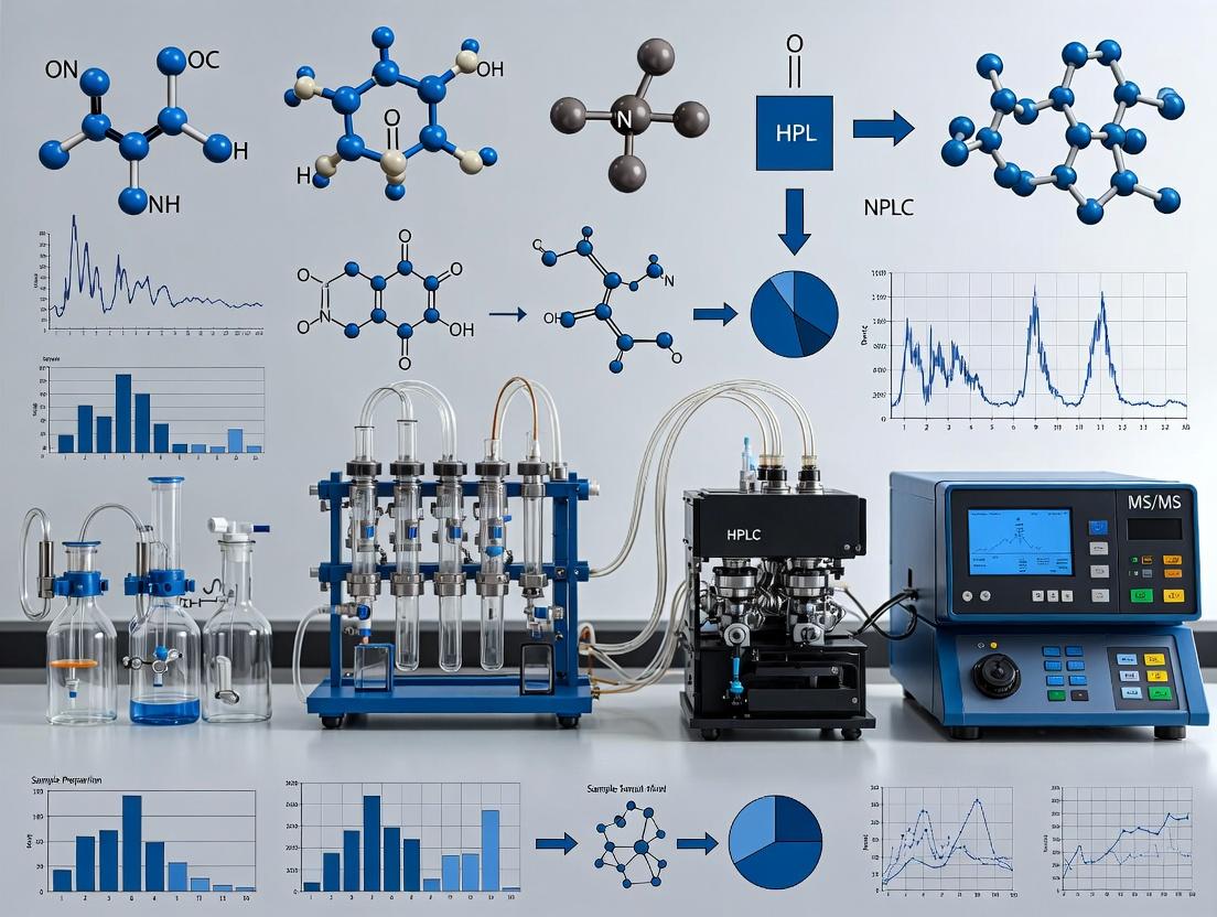
4. Visualization of CSF Analysis Workflow and Pathophysiological Pathways
Title: HPLC-MS/MS CSF Analysis Workflow
Title: CSF Biomarkers in Alzheimer's Disease Pathway
5. The Scientist's Toolkit: Key Research Reagent Solutions
Table 3: Essential Materials for HPLC-MS/MS CSF Research
| Reagent / Material | Function & Importance |
|---|---|
| Stable Isotope-Labeled Internal Standards (SIS) | Critical for absolute quantification by MS. Corrects for matrix effects, ion suppression, and pre-analytical variability. Essential for Aβ, tau, neurotransmitter assays. |
| Immunoaffinity Enrichment Kits (Aβ, Tau, etc.) | Overcome the high dynamic range of CSF protein content. Enrich low-abundance biomarkers prior to MS analysis, dramatically improving sensitivity. |
| Quality Control Pools (Commercial or In-House) | Human CSF pools from healthy/diseased donors. Used for longitudinal assay performance monitoring, precision studies, and batch-to-batch normalization. |
| Specialized LC Columns (e.g., NanoFlow, HILIC) | NanoFlow columns for limited sample proteomics. HILIC columns for polar metabolite retention. Choice directly impacts coverage and sensitivity. |
| Mass Spectrometry Calibrants & Tuning Solutions | Ensure mass accuracy and reproducibility. Vendor-specific solutions for routine instrument calibration and performance verification (e.g., ESI tuning mixes). |
| Certified CSF Collection Kits (Low-Binding Tubes) | Standardize pre-analytical variables. Tubes with low protein-binding properties minimize analyte loss, a major source of variability in biomarker studies. |
Cerebrospinal fluid (CSF) is a uniquely informative biofluid for neurological research, offering a direct biochemical window into the central nervous system (CNS). Its analysis is indispensable for diagnosing, monitoring, and researching neurological diseases. The rationale for its use, particularly in the context of advanced analytical techniques like HPLC-MS/MS, rests on its distinct advantages.
Key Advantages of CSF Analysis
- Proximity to Pathology: CSF bathes the brain and spinal cord, containing proteins, metabolites, and other biomarkers reflective of CNS physiological and pathological states.
- Blood-Brain Barrier (BBB) Filtering: The BBB restricts the passage of many blood-derived proteins into the CSF, resulting in a less complex proteome than serum/plasma and enriching for CNS-specific biomarkers.
- Early Disease Detection: Biochemical changes in CSF often precede clinical symptoms and structural imaging changes (e.g., MRI), enabling earlier diagnosis.
- Quantifiable Target Engagement: For drug development, measuring drug concentrations and pharmacodynamic biomarkers in CSF provides direct evidence of CNS penetration and target modulation.
- Disease Subtyping and Staging: Multi-omics analysis of CSF can stratify heterogeneous diseases (e.g., Alzheimer's, Parkinson's) into distinct molecular subtypes and track progression.
Table 1: Comparative Complexity of CSF and Plasma Proteomes
| Parameter | Cerebrospinal Fluid (CSF) | Plasma/Serum | Implication for Analysis |
|---|---|---|---|
| Total Protein Concentration | 0.15 - 0.45 mg/mL | 60 - 80 mg/mL | CSF requires high-sensitivity methods (e.g., LC-MS/MS). |
| Number of Detectable Proteins | ~3,000 - 5,000 (with deep proteomics) | ~10,000+ (with deep proteomics) | Lower complexity simplifies biomarker discovery. |
| Albumin Contribution | ~50-60% of total protein | ~50-60% of total protein | Similar relative abundance, but absolute mass is ~200x lower in CSF. |
| CNS-Enriched Proteins | High (e.g., Neurofilament Light, S100B) | Very Low | CSF is specific for CNS-derived biomarkers. |
| Dynamic Range | ~7-8 orders of magnitude | ~10-12 orders of magnitude | Less dynamic range facilitates detection of low-abundance CNS biomarkers. |
Key Research Applications in Neurology (HPLC-MS/MS Context)
Neurodegenerative Disease Biomarker Discovery
HPLC-MS/MS is the gold standard for quantifying established and novel biomarkers.
- Alzheimer's Disease (AD): Precise quantification of amyloid-β peptides (Aβ42, Aβ40), tau (total-tau, phosphorylated-tau).
- Parkinson's Disease (PD) & Lewy Body Dementia: Detection of α-synuclein species (total, phosphorylated, oligomeric).
- General Neuroaxonal Damage: Quantification of Neurofilament Light Chain (NfL), a sensitive marker of neuronal injury across multiple diseases (MS, AD, traumatic brain injury).
Clinical Trial Monitoring & Drug Development
CSF analysis is critical for Phase I-III neurological trials.
- Pharmacokinetics (PK): Measurement of drug concentration in CSF to confirm BBB penetration.
- Pharmacodynamics (PD): Quantifying changes in pathogenic proteins (e.g., reduction in Aβ or tau) in response to therapy.
- Target Engagement: Verification that a drug modulates its intended biochemical target within the CNS.
Inborn Errors of Metabolism & Neuroinflammatory Markers
- Metabolomics: HPLC-MS/MS profiles small molecules, neurotransmitters, and metabolites for diagnosing neurometabolic disorders.
- Inflammation: Quantification of cytokines, chemokines, and immunoglobulin indices to diagnose and monitor neuroinflammatory diseases like multiple sclerosis or autoimmune encephalitis.
Experimental Protocols
Protocol: HPLC-MS/MS Quantification of Amyloid-β Peptides (Aβ42/Aβ40) in Human CSF
Objective: To quantitatively measure Aβ42 and Aβ40 peptides in human CSF using immunoaffinity enrichment coupled with HPLC-MS/MS.
Materials (Research Reagent Solutions): Table 2: Essential Reagents and Materials for Aβ MS Assay
| Item | Function | Key Consideration |
|---|---|---|
| Anti-Aβ Monoclonal Antibodies | Immunoaffinity capture of Aβ peptides from CSF. | Must be specific, high-affinity; often used on magnetic beads. |
| Stable Isotope-Labeled (SIL) Aβ Standards (e.g., 15N/13C-Aβ42, Aβ40) | Internal standards for precise quantification. | Correct for pre-analytical and analytical variability. |
| CSF Collection Tubes (Polypropylene) | Pre-analytical collection. | Avoids adsorption of Aβ to tube walls. |
| Mass Spectrometry-Compatible Denaturing Buffer (e.g., with Guanidine HCl) | Dissociates Aβ from binding proteins and prevents aggregation. | Ensures complete recovery and accurate measurement. |
| Reverse-Phase C18 HPLC Column | Chromatographic separation of peptides prior to MS. | Nano-flow or micro-flow columns provide high sensitivity. |
| Triple Quadrupole Mass Spectrometer | Detection and quantification of target peptide fragments. | Operated in Multiple Reaction Monitoring (MRM) mode. |
Procedure:
- CSF Collection & Storage: Collect lumbar CSF in polypropylene tubes. Centrifuge (2,000 x g, 10 min, 4°C) to pellet cells. Aliquot and store at -80°C. Avoid freeze-thaw cycles.
- Sample Preparation: Thaw CSF samples on ice. Mix 200 µL of CSF with 20 µL of SIL internal standard mix and 200 µL of denaturing buffer. Vortex thoroughly.
- Immunoaffinity Enrichment: Add antibody-conjugated magnetic beads to the denatured sample. Incubate with rotation for 2 hours at room temperature. Wash beads 3x with PBS-Tween, then 2x with HPLC-grade water.
- Elution: Elute captured Aβ peptides from beads using 30% acetonitrile in 1% formic acid.
- LC-MS/MS Analysis:
- HPLC: Inject eluate onto a reverse-phase C18 column (e.g., 75µm x 15cm). Use a gradient from 2% to 35% acetonitrile in 0.1% formic acid over 15 minutes.
- MS/MS: Use positive electrospray ionization. Monitor specific precursor-to-product ion transitions for Aβ42, Aβ40, and their SIL counterparts in MRM mode.
- Data Analysis: Integrate peak areas for native and SIL peptides. Generate a calibration curve using standard samples with known concentrations. Calculate endogenous Aβ concentration using the ratio of native/SIL peak areas.
Protocol: Global Proteomic Profiling of CSF by Data-Independent Acquisition (DIA) LC-MS/MS
Objective: To identify and quantify thousands of proteins in CSF for discovery-phase biomarker studies.
Procedure:
- High-Abundance Protein Depletion: Use immunoaffinity columns (e.g., MARS-14) to remove the top 14 abundant proteins (e.g., albumin, immunoglobulins). This step is optional but increases depth.
- Protein Digestion: Reduce (DTT), alkylate (IAA), and digest depleted CSF with trypsin (1:50 enzyme-to-protein ratio, overnight, 37°C).
- Peptide Cleanup: Desalt peptides using C18 solid-phase extraction tips or StageTips.
- LC-MS/MS (DIA):
- HPLC: Separate peptides on a nano-flow C18 column with a 60-120 minute gradient.
- MS/MS: Acquire full MS1 scan followed by sequential, wide isolation window (e.g., 20-25 Da) MS2 scans across the entire m/z range.
- Data Processing: Use spectral library-based software (e.g., Spectronaut, DIA-NN) to query DIA data against a project-specific or public CSF spectral library for protein identification and quantification.
Visualizations
Workflow for Targeted CSF Biomarker Analysis via HPLC-MS/MS
Key Neurological Signaling Pathways Interrogated via CSF Proteomics
The reliability of HPLC-MS/MS data for cerebrospinal fluid (CSF) biomarker discovery and pharmacokinetic studies is fundamentally dependent on rigorous pre-analytical standardization. This document details standardized protocols for CSF collection, stabilization, and storage, framed within a thesis focused on minimizing pre-analytical variation to ensure the integrity of subsequent LC-MS/MS analyses.
CSF Collection Protocols
Patient Preparation and Positioning
Standardized patient positioning (lateral decubitus or sitting) is critical for consistent CSF pressure and protein concentration. Collection should be scheduled to control for diurnal variation in certain biomarkers.
Lumbar Puncture Procedure & Initial Handling
- Needle Type: Use atraumatic (pencil-point) needles (e.g., 22-25G) to reduce the risk of post-lumbar puncture headache and traumatic tap.
- Discard Volume: Discard the first 1-2 mL of CSF to minimize blood contamination from the puncture.
- Collection Tube: Collect directly into low-binding polypropylene tubes. The use of additive-free tubes is standard for most omics studies, though specific protease or phosphatase inhibitor cocktails may be added immediately for targeted analyses (see Stabilization).
- Aliquoting: Gently mix the CSF by inverting the tube 2-3 times. Aliquot into small-volume, low-protein-binding cryovials (e.g., 0.5 mL aliquots) to avoid repeated freeze-thaw cycles.
- Volume: Record the total collection volume.
Contamination Assessment
Immediately assess each sample for blood contamination via visual inspection and erythrocyte count. Hemolyzed samples (>500 RBCs/μL) can confound MS/MS results due to high-abundance plasma protein interference.
Table 1: Contamination Assessment and Action Guidelines
| Contamination Indicator | Acceptance Threshold | Recommended Action for LC-MS/MS |
|---|---|---|
| Erythrocyte Count | < 500 cells/μL | Proceed with analysis. |
| 500 - 10,000 cells/μL | Note contamination; consider centrifugation (2,000 x g, 10 min, 4°C) and supernatant transfer. | |
| > 10,000 cells/μL | Discard or use only for non-proteomic/metabolomic assays. | |
| Visual Inspection | Clear, colorless | Ideal. |
| Xanthochromic | Note; may indicate prior hemorrhage. | |
| Turbid/Cloudy | High likelihood of high cell/protein content; centrifuge before aliquoting. |
Stabilization and Processing
Rapid processing is paramount to halt enzymatic degradation and chemical modification.
Protocol: Immediate Post-Collection Processing
- Time-to-Processing: Begin processing within 30 minutes of collection. Place primary collection tube on wet ice immediately after draw.
- Centrifugation: For cell-free analysis, centrifuge at 2,000 x g for 10 minutes at 4°C to pellet cells and debris.
- Aliquoting: Carefully pipette the supernatant into pre-chilled, low-binding cryovials. Avoid disturbing the pellet.
- Stabilization Additives: For specific analyte classes, add inhibitors immediately after aliquoting:
- Proteomics: Broad-spectrum protease inhibitor cocktails (e.g., containing AEBSF, Aprotinin, Bestatin, etc.).
- Phosphoproteomics: Phosphatase inhibitors (e.g., sodium fluoride, sodium orthovanadate, β-glycerophosphate).
- Metabolomics: Stabilizers like sodium azide to inhibit bacterial growth.
- Snap-Freezing: Immerse aliquots in a dry-ice/ethanol bath or liquid nitrogen for 5 minutes before transfer to -80°C.
Critical Time Variables
Table 2: Maximum Recommended Hold Times Before Freezing
| Processing Step | Room Temp | On Wet Ice (4°C) | Reference |
|---|---|---|---|
| Primary Tube (Unprocessed) | 30 min | 2 hours | Teunissen et al., 2009; later validated |
| Cell-Free Supernatant | Not Recommended | 4 hours | Recent consensus guidelines |
| For Inhibitor Addition | Immediately | N/A | Best practice for targeted assays |
Storage and Thawing Best Practices
Long-Term Storage
- Temperature: Store aliquots at ≤ -80°C in a dedicated, non-frost-free freezer. Monitor temperature continuously.
- Vials: Use screw-cap cryovials with silicone O-rings; avoid internal threads that can trap sample.
- Location: Store vials in the middle of the freezer, away from doors and cooling elements.
- Inventory: Maintain a detailed log with aliquot IDs, collection date, volume, and freeze-thaw history.
Thawing Protocol for HPLC-MS/MS Analysis
- Thaw aliquots rapidly in a 37°C water bath or on a controlled thermal block until just ice-free (~5-10 mins).
- Immediately place on wet ice.
- Gently vortex for 5-10 seconds to ensure homogeneity.
- Centrifuge briefly (e.g., 10,000 x g, 1 min, 4°C) to pellet any potential precipitates before transferring to autosampler vials.
- Critical Rule: Perform a single thaw only. Do not re-freeze unused material.
The Scientist's Toolkit: Essential Research Reagent Solutions
Table 3: Key Materials for CSF Pre-Analytical Processing
| Item | Function & Rationale |
|---|---|
| Atraumatic Spinal Needles (22-25G) | Minimizes trauma, reduces post-LP headache, and lowers risk of blood contamination. |
| Low-Binding Polypropylene Tubes | Minimizes adsorption of low-abundance proteins and peptides to tube walls. |
| Broad-Spectrum Protease Inhibitor Cocktail | Preserves the native proteome/peptidome by inhibiting serine, cysteine, aspartic, and aminopeptidases. |
| Phosphatase Inhibitor Cocktail | Essential for preserving labile phosphorylation states in phosphoproteomic studies. |
| Low-Protein-Binding Cryovials (0.5 mL) | Prevents sample loss during storage; small aliquot size avoids repeated freeze-thaw cycles. |
| Internal Standard Mix (Stable Isotope-Labeled) | Added early in processing (post-thaw) to monitor and correct for losses during sample prep for MS. |
| Protein Precipitation Solvents (e.g., MeOH/ACN) | For deproteinization in metabolomic or targeted peptide assays prior to LC-MS/MS. |
| Solid-Phase Extraction (SPE) Cartridges | For sample clean-up and analyte enrichment to reduce matrix effects in MS ionization. |
Visualized Workflows
Title: End-to-End CSF Processing Workflow for LC-MS/MS
Title: CSF Stabilization Strategy Based on Analyte Class
Application Notes for CSF Analysis in HPLC-MS/MS Research
Within the thesis investigating neurodegenerative disease biomarkers via HPLC-MS/MS analysis of cerebrospinal fluid (CSF), rigorous sample preparation is paramount. The low abundance of protein biomarkers amidst high-abundance proteins and the complex matrix necessitate a multi-step workflow. This protocol details the essential techniques of high-abundance protein depletion, enzymatic digestion, and post-digestion clean-up, optimized for subsequent LC-MS/MS quantification.
High-Abundance Protein Depletion
Application Note: Depletion of highly abundant proteins (e.g., Albumin, IgG) is critical to enhance the detection depth of low-abundance, biologically relevant analytes in CSF. Without depletion, the dynamic range of the mass spectrometer is overwhelmed.
Protocol: Immunoaffinity Column Depletion (Using Commercial Kits)
- Reagent: Commercial spin column kit (e.g., ProteoPrep IgG & Albumin Depletion, MARS-14).
- Procedure:
- Thaw CSF sample on ice and centrifuge at 14,000 x g for 10 min at 4°C to remove particulates.
- Equilibrate the spin column with 500 µL of provided binding/wash buffer by centrifuging at 1000 x g for 1 minute. Discard flow-through.
- Apply 25-50 µL of clarified CSF to the column bed. Incubate at room temperature for 5 minutes with gentle agitation.
- Centrifuge at 1000 x g for 2 minutes. Collect the flow-through (depleted fraction).
- Wash the column with 100 µL of wash buffer, centrifuge, and pool with the initial flow-through.
- Buffer exchange the depleted fraction into 50 mM ammonium bicarbonate using a 5 kDa molecular weight cut-off (MWCO) centrifugal filter. Concentrate to approximately 50 µL.
Quantitative Data on Depletion Efficiency:
| Depletion Method | Target Proteins | % Abundance Reduction (Mean ± SD) | Reported Protein ID Increase |
|---|---|---|---|
| Immunoaffinity (Albumin/IgG) | Albumin, Immunoglobulin G | 95% ± 3% (Albumin), 92% ± 4% (IgG) | ~20-30% |
| Multi-Affinity (MARS-14) | 14 High-Abundance Proteins | >85% for each target | ~40-50% |
| Ultracentrifugation | Lipoproteins, >50 kDa proteins | Variable (Method-dependent) | ~10-15% |
Enzymatic Digestion
Application Note: Digestion converts proteins into predictable peptides amenable to LC-MS/MS analysis. The efficiency and reproducibility of digestion directly impact peptide yield and quantitative accuracy.
Protocol: In-Solution Trypsin Digestion
- Reagent: Sequencing-grade modified trypsin.
- Procedure:
- Measure the protein concentration of the depleted CSF sample using a colorimetric assay (e.g., BCA).
- Transfer 20 µg of protein into a low-protein-binding tube. Adjust volume to 50 µL with 50 mM ammonium bicarbonate.
- Add 5 µL of 45 mM dithiothreitol (DTT). Incubate at 56°C for 30 minutes to reduce disulfide bonds.
- Cool to room temperature. Add 5 µL of 100 mM iodoacetamide (IAA). Incubate in the dark for 30 minutes for alkylation.
- Quench excess IAA by adding 5 µL of 45 mM DTT.
- Add trypsin at a 1:50 (enzyme:protein) mass ratio. Incubate at 37°C for 16-18 hours in a thermomixer.
- Stop digestion by acidifying with formic acid to a final concentration of 1% (v/v).
Quantitative Data on Digestion Parameters:
| Digestion Parameter | Standard Condition | Optimized for CSF (Thesis Context) | Impact on Peptide Yield |
|---|---|---|---|
| Enzyme:Substrate Ratio | 1:50 | 1:50 | Standard yield. 1:100 may be used for cost-saving. |
| Time | 4-6 hours | 16-18 hours (Overnight) | Increases yield by ~15-25% for complex samples. |
| Buffer | 50 mM ABC | 50 mM ABC with 0.01% ProteaseMax | Surfactant can increase yield by ~10-15%. |
| Temperature | 37°C | 37°C | Standard. |
Post-Digestion Clean-up
Application Note: Clean-up removes salts, polymers, and detergents from the peptide mixture that can cause ion suppression and chromatographic interference in LC-MS/MS.
Protocol: Solid-Phase Extraction (SPE) using C18 Tips
- Reagent: C18 StageTips or commercial C18 pipette tips.
- Procedure:
- Condition the C18 tip with 100 µL of 100% acetonitrile (ACN). Centrifuge at 1500 x g for 2 min.
- Equilibrate with 100 µL of 0.1% trifluoroacetic acid (TFA) in water. Centrifuge.
- Load the acidified digest onto the tip. Centrifuge slowly (800 x g) to pass sample through.
- Wash with 100 µL of 0.1% TFA in 5% ACN. Centrifuge.
- Elute peptides with 60 µL of 0.1% TFA in 60% ACN into a fresh tube.
- Dry the eluate completely in a vacuum concentrator. Reconstitute in 20 µL of 0.1% formic acid for MS analysis.
Quantitative Data on Clean-up Efficiency:
| Clean-up Method | Primary Function | Peptide Recovery Rate | Salt/Lipid Removal Efficacy |
|---|---|---|---|
| C18 SPE (Tip/Column) | Desalting & Concentration | >85% | Excellent (Removes TFA, ABC) |
| Strong Cation Exchange | Fractionation & Desalting | ~70-80% per fraction | Excellent |
| Protein Precipitation (Pre-Digest) | Remove Lipids, Salts | Protein loss can be high | Good for lipids |
Visualization of Workflows and Pathways
CSF Prep Workflow for LC-MS/MS
The Scientist's Toolkit: Research Reagent Solutions
| Reagent/Material | Function in Protocol | Key Consideration for CSF |
|---|---|---|
| Immunoaffinity Depletion Spin Columns | Selective removal of top 2-20 abundant proteins. | Kit choice balances depth (more proteins) vs. cost & sample loss. |
| Sequencing-Grade Modified Trypsin | Highly specific proteolytic enzyme cleaves at Lys/Arg. | Low autolysis rate is critical for clean background in MS. |
| Dithiothreitol (DTT) | Reducing agent for breaking protein disulfide bonds. | Fresh preparation required to maintain efficacy. |
| Iodoacetamide (IAA) | Alkylating agent for capping reduced cysteine residues. | Light-sensitive; must be used in dark. |
| C18 Solid-Phase Extraction Tips | Desalting and concentration of peptide mixtures. | Low-binding tips maximize recovery of low-abundance peptides. |
| Ammonium Bicarbonate (ABC) | Volatile buffer for digestion, easily removed in MS. | Preferred over non-volatile buffers (e.g., Tris) for MS compatibility. |
| Mass Spectrometry Grade Solvents | Water, Acetonitrile, Formic Acid for LC-MS. | Purity essential to minimize chemical noise and background ions. |
| 0.22 µm Ultrafiltration Units | Sterile filtration and buffer exchange of depleted CSF. | Removes particulates and potential clogging agents for LC. |
Core Principles of HPLC and Tandem Mass Spectrometry Relevant to CSF Analysis
High-Performance Liquid Chromatography (HPLC) coupled with tandem mass spectrometry (MS/MS) is the gold standard for the sensitive, specific, and multiplexed analysis of endogenous and exogenous compounds in cerebrospinal fluid (CSF). The analysis of CSF presents unique challenges due to its low protein content, limited sample volume, and the low abundance of many neurochemical targets. This article details the core principles, application notes, and protocols for HPLC-MS/MS in CSF research, framed within a broader thesis investigating neurodegenerative disease biomarkers and neuropharmacokinetics.
Core HPLC Principles for CSF:
- High Resolution Separation: Reversed-phase chromatography (C18 columns) is predominant, separating analytes based on hydrophobicity. Ultra-High-Performance Liquid Chromatography (UHPLC) with sub-2µm particles provides superior resolution and speed, critical for complex CSF matrices.
- Minimized Extra-Column Volume: To prevent peak broadening, especially with low-volume CSF injections (typically 1-10 µL), all system tubing is of minimal internal diameter (e.g., 0.005").
- Aqueous-Compatible Mobile Phases: Gradients often start with a high aqueous content (water with 0.1% formic acid) to retain polar metabolites and peptides, moving to organic solvents (acetonitrile or methanol).
Core Tandem MS Principles for CSF:
- Selective Reaction Monitoring (SRM)/Multiple Reaction Monitoring (MRM): The cornerstone of CSF quantitation. The first quadrupole (Q1) selects the precursor ion (often [M+H]⁺). The collision cell (q2) fragments it, and the third quadrupole (Q3) selects a specific product ion. This two-stage selection yields exceptional specificity against matrix background.
- High Sensitivity: Modern triple quadrupole MS systems are essential to detect pg/mL to ng/mL level analytes in small CSF volumes.
- Ion Source Considerations: Electrospray Ionization (ESI) is most common. Heated ESI sources enhance sensitivity for lower flow rates typical of UHPLC.
Application Notes & Quantitative Data
Table 1: Typical HPLC-MS/MS Parameters for CSF Analysis of Small Molecules (e.g., Neurotransmitters, Drugs)
| Parameter | Typical Setting/Value | Rationale for CSF Analysis |
|---|---|---|
| Column | C18, 2.1 x 50-100 mm, 1.7-1.8 µm | Optimal balance of resolution, speed, and backpressure for UHPLC. |
| Injection Volume | 1-10 µL | Conserves precious CSF sample; volumes >10 µL may cause matrix effects. |
| Flow Rate | 0.2-0.4 mL/min | Compatible with ESI sensitivity and UHPLC column dimensions. |
| Gradient Time | 5-10 minutes | Enables high-throughput analysis of batch samples. |
| MS Scan Type | SRM/MRM | Provides highest sensitivity and selectivity for target quantitation. |
| Ion Source | H-ESI or ESI | Robust ionization for a broad range of compounds. |
| Source Temp. | 300-350°C | Aids desolvation, improving signal-to-noise. |
Table 2: Example Quantitative Panel for CSF Neurotransmitter Metabolites
| Analytic (Biomarker Class) | Typical Conc. in Healthy CSF (Quantitative Range) | Relevant Pathological Context (e.g., Alzheimer's, Parkinson's) | Common Internal Standard |
|---|---|---|---|
| Amyloid-β 1-42 (Peptide) | ~500-1000 pg/mL | ↓ in Alzheimer's Disease | ¹⁵N-labeled or ¹³C-labeled Aβ 1-42 |
| Phospho-Tau (p-Tau181) (Protein) | ~20-50 pg/mL | ↑ in Alzheimer's Disease | Synthetic p-Tau181 peptide with stable isotopes |
| 5-Hydroxyindoleacetic acid (5-HIAA) (Monoamine metabolite) | 20-40 ng/mL | ↓ in Depression | d5-5-HIAA |
| Homovanillic acid (HVA) (Dopamine metabolite) | 40-80 ng/mL | ↓ in Parkinson's Disease | d5-HVA |
| Neurofilament Light Chain (NfL) (Protein) | <380 pg/mL | ↑ in Neurodegeneration & Neuroinflammation | Recombinant ¹⁵N-labeled NfL |
Experimental Protocols
Protocol 1: Sample Preparation for Targeted CSF Metabolomics (SPE-based)
Aim: To clean, concentrate, and stabilize small molecule analytes from 100 µL of human CSF. Materials: See "The Scientist's Toolkit" below. Steps:
- Thawing & Aliquoting: Thaw CSF samples slowly on ice. Vortex gently and aliquot 100 µL into a low-protein-binding microcentrifuge tube.
- Protein Precipitation: Add 300 µL of ice-cold methanol containing a cocktail of isotope-labeled internal standards (IS). Vortex vigorously for 30 seconds.
- Centrifugation: Centrifuge at 14,000 x g for 15 minutes at 4°C to pellet proteins.
- Solid-Phase Extraction (SPE): Load the supernatant onto a pre-conditioned (1 mL methanol, then 1 mL water) mixed-mode cation-exchange SPE cartridge (e.g., Oasis MCX).
- Washing & Elution: Wash with 1 mL of 2% formic acid in water. Elute analytes with 1 mL of 5% ammonium hydroxide in methanol.
- Concentration & Reconstitution: Evaporate the eluent to complete dryness under a gentle stream of nitrogen. Reconstitute the dried extract in 50 µL of initial mobile phase (e.g., 0.1% formic acid in water).
- Analysis: Vortex, transfer to an HPLC vial with insert, and inject 5 µL onto the HPLC-MS/MS system.
Protocol 2: LC-MS/MS Analysis of Amyloid-β Peptides in CSF
Aim: To quantify Aβ 1-40 and Aβ 1-42 via immunoprecipitation (IP) coupled with HPLC-MS/MS. Materials: See "The Scientist's Toolkit" below. Steps:
- Immunoaffinity Enrichment: Incubate 500 µL of CSF with magnetic beads conjugated to anti-Aβ monoclonal antibodies (targeting mid-domain epitopes) for 2 hours at room temperature with gentle mixing.
- Bead Washing: Isolate beads on a magnetic rack. Wash 3x with PBS-Tween 20.
- Elution: Elute bound Aβ peptides with 50 µL of 1% formic acid.
- Digestion (Optional): For peptide mapping, digest with trypsin. For intact analysis, proceed directly.
- LC-MS/MS Analysis:
- Column: C4 or C8 column (2.1 x 50 mm, 3.5 µm) for intact protein/peptide separation.
- Gradient: 20-80% B over 8 min (A: 0.1% FA in water; B: 0.1% FA in acetonitrile).
- MS: SRM transition monitoring for specific charge states of Aβ 1-40 and Aβ 1-42, and their corresponding isotope-labeled IS.
Diagrams
Title: Core HPLC-MS/MS Workflow for CSF Analysis
Title: SRM/MRM Principle in Tandem MS
The Scientist's Toolkit: Research Reagent Solutions
Table 3: Essential Materials for HPLC-MS/MS Analysis of CSF
| Item/Category | Specific Example/Type | Function in CSF Analysis |
|---|---|---|
| CSF Collection System | Sterile, polypropylene tubes; atraumatic spinal needles | Minimizes blood contamination and adsorptive losses to tube walls. |
| Protease/Phosphatase Inhibitor Cocktail | Broad-spectrum, EDTA-free cocktails | Preserves labile protein/peptide biomarkers (e.g., Aβ, tau) during collection and storage. |
| Stable Isotope-Labeled Internal Standards (SIL-IS) | ¹³C, ¹⁵N-labeled versions of target analytes (Aβ, neurotransmitters, drugs) | Corrects for matrix effects, ionization efficiency variance, and sample preparation losses. Critical for absolute quantitation. |
| Solid-Phase Extraction (SPE) Cartridges | Mixed-mode (e.g., Oasis MCX, WCX), HLB | Clean-up and concentrate analytes from complex CSF matrix, improving sensitivity and LC column lifetime. |
| UHPLC Columns | Reversed-phase C18 (1.7-1.8 µm, 2.1 mm ID) | Provides high-resolution separation of CSF components with minimal peak broadening. |
| LC Vials & Inserts | Polypropylene vials with low-volume glass inserts (e.g., 100-200 µL) | Prevents adsorptive losses of low-abundance analytes and accommodates small sample volumes. |
| Mass Spectrometry Calibrants | ESI tuning mix (e.g., from Agilent, Waters) | Ensures mass accuracy and optimal instrument performance before sample batch analysis. |
| Immunoaffinity Beads | Magnetic beads coated with specific antibodies (e.g., for Aβ, tau) | Enables high-specificity enrichment of low-abundance protein biomarkers prior to MS analysis (IP-MS). |
Step-by-Step HPLC-MS/MS Protocols for CSF Proteomics, Metabolomics, and Targeted Assays
Cerebrospinal fluid (CSF) is a prime biospecimen for discovering biomarkers of neurological disorders due to its proximity to the brain and spinal cord. Within the context of a broader thesis on HPLC-MS/MS analysis of CSF, this document outlines the integrated application of bottom-up (shotgun) and top-down proteomic approaches. This dual-strategy enables comprehensive protein profiling, from identifying thousands of proteins and their post-translational modifications (PTMs) to characterizing proteoforms with intact mass analysis, thereby accelerating biomarker discovery for conditions like Alzheimer's disease, multiple sclerosis, and brain tumors.
Table 1: Key Characteristics of Bottom-Up and Top-Down Proteomic Approaches for CSF Analysis
| Feature | Bottom-Up (Shotgun) Proteomics | Top-Down Proteomics |
|---|---|---|
| Analytical Target | Peptides from digested proteins | Intact proteins/proteoforms |
| Typical Workflow | Protein extraction → Enzymatic digestion → LC-MS/MS of peptides | Protein extraction → Intact protein separation → LC-MS/MS of proteins |
| Primary Instrumentation | High-flow or nanoflow HPLC coupled to high-resolution tandem MS (Q-Exactive, timsTOF) | Nanoflow HPLC coupled to high-resolution/FT-based tandem MS (Orbitrap Eclipse, FT-ICR) |
| Key Metrics | Protein IDs, sequence coverage, PTM site localization | Proteoform IDs, intact mass, combinatorial PTM characterization |
| Typical CSF Depth | 1,500 – 3,000+ proteins identified | 100 – 500 proteoforms characterized |
| PTM Analysis | Site-specific but inferred from peptides | Direct observation of intact PTM patterns |
| Primary Challenge | Inference of proteoforms from peptides; sample complexity | Low abundance; inefficient fragmentation; data analysis complexity |
| Throughput | High | Medium to Low |
Table 2: Summary of Quantitative Data from Recent Integrated CSF Proteomics Studies (2023-2024)
| Study Focus | Bottom-Up Findings | Top-Down Findings | Integrated Biomarker Outcome |
|---|---|---|---|
| Alzheimer's Disease (AD) | Quantified 2,345 proteins; 112 significantly altered (p<0.01). Aβ precursor protein (APP) peptides increased 2.5-fold. | Identified 15 unique proteoforms of Amyloid-beta (Aβ) with varying truncations. Aβ-42/Aβ-40 ratio shift confirmed. | Combined data increased diagnostic specificity to 94% vs. 88% for Aβ42 alone. |
| Multiple Sclerosis (MS) | 1,890 proteins quantified. 45 immunomodulatory proteins dysregulated. C1QB complement protein increased 3.1-fold. | Characterized 8 proteoforms of Myelin Basic Protein (MBP) with distinct citrullination and phosphorylation states. | Citrullinated MBP proteoform C8 correlated strongly with radiographic disease activity (r=0.78). |
| Glioblastoma | 2,150 proteins quantified. Chitinase-3-like-1 (CHI3L1) increased 4.7-fold vs. controls. | Discovered novel glycosylated proteoforms of YKL-40 (CHI3L1) not detectable via bottom-up. | Specific glyco-proteoform YKL-40-G2 predicted survival with HR of 2.4 (95% CI: 1.5-3.8). |
Experimental Protocols
Protocol 1: Bottom-Up Proteomics for Deep CSF Profiling
Objective: To identify and quantify the global CSF proteome using tryptic digestion and nanoLC-MS/MS.
Materials: See "Research Reagent Solutions" table. Procedure:
- CSF Preparation: Thaw aliquots (typically 100-200 µL) on ice. Centrifuge at 16,000 x g for 10 min at 4°C to remove any insoluble debris. Transfer supernatant to a fresh LoBind tube.
- Protein Precipitation & Quantification: Add 4 volumes of ice-cold acetone. Vortex and incubate at -20°C for 2 hours. Pellet proteins by centrifugation at 15,000 x g for 15 min at 4°C. Air-dry the pellet. Resuspend in 50 µL of 8M urea in 50mM TEAB, pH 8.5. Quantify using a microBCA assay.
- Reduction, Alkylation, and Digestion: Reduce with 5mM dithiothreitol (DTT) at 37°C for 45 min. Alkylate with 15mM iodoacetamide (IAA) at room temp in the dark for 30 min. Dilute urea concentration to <2M with 50mM TEAB. Add trypsin at a 1:50 (enzyme:protein) ratio. Incubate at 37°C for 16 hours. Quench with 1% formic acid (FA).
- Peptide Cleanup: Desalt peptides using C18 StageTips. Elute peptides with 60% acetonitrile (ACN)/0.1% FA. Dry in a vacuum concentrator.
- LC-MS/MS Analysis: Reconstitute in 2% ACN/0.1% FA. Load 1-2 µg onto a 50cm C18 column (75µm id, 2µm beads). Perform a 120-min gradient from 5% to 30% Buffer B (0.1% FA in ACN) at 300 nL/min. Acquire data on a timsTOF Pro 2 or Orbitrap Eclipse in DIA-PASEF or data-independent acquisition (DIA) mode.
- Data Analysis: Process DIA data using Spectronaut or DIA-NN against the Human UniProt database. For LFQ, use MaxQuant or FragPipe.
Protocol 2: Top-Down Proteomics for CSF Proteoform Characterization
Objective: To separate and analyze intact CSF proteins and their proteoforms using nanoLC coupled to high-resolution tandem MS.
Procedure:
- CSF Preparation for Intact Analysis: Deplete high-abundance proteins (e.g., albumin, immunoglobulins) using a multiple affinity removal spin cartridge. Desalt the flow-through using a 10kDa MWCO filter. Reconstitute in 20 µL of top-down lysis buffer (2% SDS, 50mM TEAB).
- Intact Protein Separation (LC-MS): Load 5-10 µL onto a PLRP-S column (300Å, 5µm, 150mm x 0.3mm). Use a 60-min gradient from 20% to 60% Buffer B (0.1% FA in ACN) at 5 µL/min, coupled directly to an ESI source.
- Intact Mass Acquisition: Acquire full MS scans on an Orbitrap Eclipse (with UVPD or ETD) or FT-ICR MS at a resolution of 120,000 (at m/z 200) over m/z 600-2000.
- Targeted Tandem MS for Proteoforms: Isolate precursor ions of interest with a 10-20 m/z window. Fragment using Electron-Transfer/Higher-Energy Collisional Dissociation (EThcD) or Ultraviolet Photodissociation (UVPD). Acquire fragment spectra at 60,000 resolution.
- Data Analysis: Deconvolute intact masses using Xtract or UniDec. Process MS/MS data with ProSight PD or TopPIC for proteoform identification and PTM localization.
Visualizations
CSF Proteomics Dual Workflow
Biomarker Discovery & Validation Pipeline
The Scientist's Toolkit
Table 3: Key Research Reagent Solutions for CSF Proteomics
| Item | Function & Rationale |
|---|---|
| Human 14 High-Abundance Protein Depletion Spin Cartridge | Removes ~95% of abundant proteins (e.g., albumin) to deepen coverage of low-abundance, CNS-derived proteins in both bottom-up and top-down workflows. |
| Mass Spectrometry-Grade Trypsin/Lys-C Mix | Provides specific, efficient digestion for bottom-up proteomics, maximizing peptide yield and minimizing missed cleavages for confident IDs. |
| C18 StageTips (Empore) | Micro-scale solid-phase extraction for desalting and concentrating peptide digests prior to LC-MS/MS, improving sensitivity and reproducibility. |
| Nanoflow UHPLC System (e.g., Dionex Ultimate 3000 RSLChano) | Delivers ultra-low flow rates (200-300 nL/min) for high-resolution peptide separation, directly coupled to the MS for maximum sensitivity. |
| PLRP-S Column (300Å, 5µm, 150mm x 0.3mm) | Polymeric reversed-phase column optimized for separating intact proteins and large polypeptides in top-down proteomics. |
| High-Resolution Tandem Mass Spectrometer (Orbitrap Eclipse/timsTOF Pro 2) | Provides the mass accuracy, resolution, and advanced fragmentation (HCD, ETD, UVPD) required for both peptide sequencing and intact proteoform analysis. |
| Tris(2-carboxyethyl)phosphine (TCEP) | A non-thiol, MS-compatible reducing agent superior to DTT for stabilizing reduced cysteine residues in top-down sample prep. |
| Proteomics Data Analysis Suite (e.g., FragPipe, Spectronaut, ProSight PD) | Software platforms essential for processing complex DIA/DDA data, database searching, quantitation, and proteoform characterization. |
Within the broader context of a thesis on HPLC-MS/MS analysis of cerebrospinal fluid (CSF), this document details application notes and protocols for targeted peptide and neuropeptide analysis. CSF presents a complex matrix for biomarker discovery and pharmacokinetic studies in neurological diseases and drug development. This work focuses on overcoming challenges of low analyte abundance, high matrix interference, and structural diversity through optimized sample preparation, chromatographic separation, and MS/MS quantification.
Key Methodological Challenges & Solutions
- Low Abundance: Neuropeptides often exist in the low pg/mL to fg/mL range in CSF. Required solution: Immunoaffinity enrichment and nano-flow LC-MS/MS.
- Matrix Complexity: High salt and protein content. Required solution: Robust solid-phase extraction (SPE) and immunoaffinity depletion.
- Structural Heterogeneity: Presence of isoforms, post-translational modifications (PTMs), and precursors. Required solution: High-resolution separations (UPLC) and selective fragmentation.
- Dynamic Range: Need to quantify both high-abundance proteins and trace neuropeptides. Required solution: Scheduled MRM and stable isotope-labeled internal standards (SIL IS).
Table 1: Representative Neuropeptides in CSF: Typical Concentrations and MRM Parameters
| Neuropeptide | Precursor (m/z) | Product (m/z) | CE (V) | Typical CSF Concentration (pg/mL) | Biological Relevance |
|---|---|---|---|---|---|
| Substance P | 674.9 | 112.1 | 22 | 5 - 50 | Pain transmission, neuroinflammation |
| β-Endorphin | 694.8 | 130.1 | 28 | 10 - 200 | Analgesia, stress response |
| Neurotensin | 558.3 | 187.1 | 20 | 2 - 30 | Dopamine modulation, hypothermia |
| Orexin A | 937.5 | 249.2 | 35 | 1 - 10 | Sleep/wake regulation |
| Vasopressin | 543.8 | 120.1 | 18 | 1 - 20 | Social behavior, water balance |
| SIL IS (Avg.) | -- | -- | -- | Spiked at 100 pg/mL | Quantification control |
Table 2: Comparison of Sample Preparation Methods for CSF Peptidomics
| Method | Principle | Recovery (%) | Key Advantage | Key Limitation | Best For |
|---|---|---|---|---|---|
| SPE (C18) | Hydrophobic interaction | 60-85 | Broad applicability, high capacity | Co-elution of salts, less selective | General peptidome profiling |
| Immunoaffinity | Antigen-antibody binding | >90 | Exceptional specificity & enrichment | High cost, target-specific | Ultra-trace single analyte |
| Ultrafiltration | Molecular weight cutoff | 40-70 | Simple, rapid, preserves PTMs | Low recovery, membrane adsorption | Large peptide/protein removal |
| Acetonitrile PPT | Solvent precipitation | 50-80 | Fast, removes most proteins | Poor for small, hydrophilic peptides | Fast sample clean-up |
Detailed Experimental Protocols
Protocol 1: Immunoaffinity SPE coupled with nanoLC-MS/MS for Substance P Quantification
I. Objective: Quantify Substance P in 500 µL of human CSF with a target LLOQ of 1 pg/mL.
II. Materials & Reagents:
- CSF samples (aliquoted, stored at -80°C).
- Stable Isotope-Labeled Substance P internal standard (SIL-SP).
- Anti-Substance P antibody-coupled magnetic beads.
- Phosphate Buffered Saline (PBS), pH 7.4.
- Low-binding microcentrifuge tubes and tips.
- Elution buffer: 1% Formic Acid in 30% ACN/H₂O.
- Reconstitution solvent: 0.1% FA in 3% ACN/H₂O.
- Nano-flow HPLC system coupled to QTRAP 6500+.
III. Procedure:
- Sample Thawing & Internal Standard Addition:
- Thaw CSF samples on wet ice.
- Centrifuge at 14,000 x g for 10 min at 4°C.
- Transfer 500 µL of supernatant to a low-binding tube.
- Spike with 25 µL of SIL-SP solution (final conc. 100 pg/mL).
Immunoaffinity Enrichment:
- Add 50 µL of antibody-bead suspension to the sample.
- Incubate with end-over-end mixing for 2 hours at 4°C.
- Place tube on a magnetic rack for 2 min. Discard supernatant.
- Wash beads twice with 500 µL of ice-cold PBS with brief vortexing.
Elution & Reconstitution:
- Elute bound peptides with 2 x 50 µL of elution buffer. Pool eluates.
- Dry eluates in a vacuum concentrator at 45°C for 45 min.
- Reconstitute dried residue in 20 µL of reconstitution solvent. Vortex for 1 min.
nanoLC-MS/MS Analysis:
- Column: PepMap C18, 75 µm x 25 cm, 2 µm.
- Gradient: 3% to 35% B over 30 min (A: 0.1% FA in H₂O, B: 0.1% FA in ACN).
- Flow: 300 nL/min.
- MS: Positive ion mode, ESI voltage 2400V.
- Detection: Scheduled MRM for Substance P (674.9/112.1) and SIL-SP.
Data Analysis:
- Use peak area ratio (Analyte/SIL IS) for quantification.
- Generate calibration curve (1-500 pg/mL) using blank CSF spiked with analyte and constant SIL IS.
Protocol 2: Global Peptidome Profiling Using C18 SPE and microLC-MS/MS
I. Objective: Perform untargeted profiling and semi-quantitation of peptides in CSF (MW < 10 kDa).
II. Procedure:
- Depletion & Desalting:
- Process 1 mL CSF using 10kDa MWCO centrifugal filters.
- Acidify filtrate with TFA to 0.1%.
- Condition and equilibrate a C18 SPE cartridge.
- Load acidified filtrate. Wash with 0.1% TFA.
- Elute peptides with 60% ACN, 0.1% FA.
- Dry and reconstitute in 50 µL of 0.1% FA.
microLC-MS/MS Analysis:
- Column: BEH C18, 1.0 µm x 150 mm.
- Gradient: 2% to 40% B in 60 min.
- Flow: 50 µL/min.
- MS: Data-Dependent Acquisition (DDA). Full scan (350-1500 m/z) followed by top 20 MS/MS scans.
Data Processing:
- Use software (e.g., Peaks, MaxQuant) for database searching against human proteome.
- Perform label-free quantification based on precursor ion intensity.
Visualizations
CSF Peptide Analysis Core Workflow
Neuropeptide Signaling Pathway
The Scientist's Toolkit: Research Reagent Solutions
Table 3: Essential Materials for Targeted CSF Peptide Analysis
| Item | Function & Rationale |
|---|---|
| Stable Isotope-Labeled (SIL) Peptide Standards | Gold-standard internal standards for absolute quantification; corrects for MS ionization variability and sample preparation losses. |
| Low-Binding Consumables (Tubes/Tips) | Minimizes adsorptive losses of hydrophobic peptides to plastic surfaces, critical for recovery. |
| Immunoaffinity Beads (Magnetic/Resin) | Enables highly selective pre-concentration of target analytes from complex CSF, improving sensitivity by 100-1000 fold. |
| Mass Spectrometry-Compatible Buffers | Volatile buffers (e.g., Formic Acid, Ammonium Bicarbonate) are essential for efficient LC-MS analysis and ion source cleanliness. |
| High-Recovery SPE Cartridges | Designed for low-abundance analytes; removes salts and proteins while maximizing peptide yield. |
| Nano/Micro LC Columns & Systems | Provide superior sensitivity for limited sample volumes by reducing flow rates, increasing ionization efficiency. |
| Scheduled MRM Assay Kits | Pre-optimized mass transitions and chromatography parameters for specific peptide panels, accelerating method development. |
This application note details protocols for the comprehensive analysis of cerebrospinal fluid (CSF) small molecules, executed within the broader methodological thesis of HPLC-MS/MS in biofluid research. The low abundance and high complexity of the CSF metabolome and lipidome present unique analytical challenges. This document provides validated workflows for untargeted discovery and targeted quantification, focusing on robustness and reproducibility essential for identifying disease-specific phenotypic signatures in neurological disorders.
Experimental Protocols
Protocol 1: CSF Sample Preparation for Untargeted Metabolomics and Lipidomics Objective: To deproteinize and extract a broad range of small molecules and lipids from CSF with minimal bias. Materials: See "Research Reagent Solutions" table. Steps:
- Thaw CSF aliquots (typically 50-100 µL) on ice.
- Add 400 µL of ice-cold methanol:acetonitrile (1:1, v/v) to 100 µL of CSF. Vortex vigorously for 30 seconds.
- Incubate at -20°C for 60 minutes to precipitate proteins.
- Centrifuge at 18,000 × g for 20 minutes at 4°C.
- Carefully transfer 450 µL of supernatant to a fresh LC-MS vial.
- Dry under a gentle stream of nitrogen at room temperature.
- Reconstitute in 100 µL of initial mobile phase (e.g., 95% Water, 5% Acetonitrile with 0.1% Formic Acid for positive mode; or 95% Water, 5% Acetonitrile with 10 mM Ammonium Acetate for negative mode). Vortex for 60 seconds.
- Centrifuge at 18,000 × g for 10 minutes at 4°C before transferring to an LC-MS vial insert.
Protocol 2: Targeted LC-MS/MS Quantification of Neuroactive Metabolites Objective: To accurately quantify a panel of 15 key neurotransmitters and related metabolites (e.g., serotonin, dopamine, glutamate, GABA, tryptophan, kynurenine). Chromatography:
- Column: HILIC column (e.g., 2.1 x 100 mm, 1.7 µm).
- Mobile Phase A: 10 mM Ammonium Formate in Water, pH 3.0.
- Mobile Phase B: Acetonitrile.
- Gradient: 90% B (0-1 min), to 40% B over 8 min, hold for 2 min, re-equilibrate for 4 min.
- Flow Rate: 0.4 mL/min. Temperature: 40°C. MS/MS Detection:
- Ionization: ESI positive/negative switching.
- MRM transitions optimized for each analyte using certified standards.
- Dwell time: 20-50 ms per transition. Data Analysis: Quantify using external calibration curves (1-1000 ng/mL) with isotopically labeled internal standards for each analyte to correct for matrix effects.
Data Presentation
Table 1: Quantitative Summary of Key CSF Metabolite Alterations in Neurodegenerative Diseases
| Metabolite Class | Example Metabolite | Alzheimer's Disease (AD) vs. Control (Fold Change) | Parkinson's Disease (PD) vs. Control (Fold Change) | Amyotrophic Lateral Sclerosis (ALS) vs. Control (Fold Change) | Primary Analytical Method |
|---|---|---|---|---|---|
| TCA Cycle | Citrate | ↓ 0.65 | HILIC-MS/MS (Neg) | ||
| Neurotransmitter | Glutamate | ↑ 1.8 | ↓ 0.7 | HILIC-MS/MS (Pos) | |
| Tryptophan Pathway | Quinolinic Acid | ↑ 2.5 | ↑ 1.9 | ↑ 3.1 | RPLC-MS/MS (Neg) |
| Lipid - PC | PC(36:4) | ↓ 0.5 | ↓ 0.6 | RPLC-MS/MS (Pos) | |
| Lipid - SM | SM(d18:1/16:0) | ↓ 0.7 | RPLC-MS/MS (Pos) | ||
| Polyamine | Spermidine | ↑ 1.6 | ↑ 2.0 | HILIC-MS/MS (Pos) |
Legend: ↑/↓ indicates significant increase/decrease (p<0.05, FDR-corrected); indicates no significant change. Data synthesized from recent cohort studies (2022-2024).
Table 2: Key Research Reagent Solutions
| Item | Function & Specification |
|---|---|
| HybridSPE-Phospholipid 96-well Plate | Efficient removal of phospholipids to reduce ion suppression in MS. Critical for lipidomics. |
| Deuterated / 13C-Labeled Internal Standards | Enables correction for matrix effects & analyte loss. Essential for absolute quantification (e.g., d4-Glutamate, 13C6-Choline). |
| LC-MS Grade Solvents (MeOH, ACN, Water) | Minimizes background chemical noise, ensuring high signal-to-noise ratios. |
| Ammonium Formate & Acetic Acid (LC-MS Grade) | Volatile buffers for mobile phases, compatible with ESI-MS. |
| Synthetic SPLASH LIPIDOMIX Mass Spec Standard | Quantitative standard mixture for lipid identification and semi-quantification across lipid classes. |
| Porous Graphitic Carbon (PGC) Column | Orthogonal separation for polar metabolites and isomers poorly retained by RPLC/HILIC. |
Visualization
Diagram 1: Integrated CSF Omics Workflow for Disease Phenotyping
Diagram 2: Key Metabolic Pathways in CSF Neurodegeneration Research
Quantitative Analysis of Neurotransmitters, Drugs, and Their Metabolites in CSF
This document provides detailed application notes and protocols for the quantitative analysis of endogenous neurotransmitters, xenobiotic drugs, and their metabolites in cerebrospinal fluid (CSF) using high-performance liquid chromatography coupled with tandem mass spectrometry (HPLC-MS/MS). The analysis of CSF provides a direct window into the biochemical environment of the central nervous system (CNS), making it a critical matrix for neuroscience research, biomarker discovery, and CNS drug development. This work is framed within a broader thesis investigating the optimization of HPLC-MS/MS methodologies for complex biofluids, with a focus on overcoming challenges specific to CSF, such as low sample volumes, low analyte concentrations, and high salt content.
Key Quantitative Data from Recent Studies
Table 1: Representative HPLC-MS/MS Analytical Parameters for Selected Neurotransmitters in CSF
| Analytic (Class) | Typical CSF Concentration Range | LLOQ (pmol/mL) | Internal Standard | Reference Method (Year) |
|---|---|---|---|---|
| Dopamine (Monoamine) | 0.1 - 0.5 nM | 0.05 | Dopamine-d₄ | HILIC-MS/MS (2023) |
| Serotonin (5-HT) (Monoamine) | 1 - 10 nM | 0.1 | Serotonin-d₄ | RP-MS/MS with derivatization (2024) |
| Norepinephrine (Monoamine) | 0.5 - 2 nM | 0.2 | Norepinephrine-d₆ | Ion-pairing LC-MS/MS (2023) |
| Glutamate (Amino Acid) | 1 - 10 µM | 50 | Glutamate-d₅ | HILIC-MS/MS (2024) |
| GABA (Amino Acid) | 50 - 500 nM | 5 | GABA-d₆ | Derivatization (AccQ-Tag) & RP-MS/MS (2023) |
| Glycine (Amino Acid) | 2 - 15 µM | 100 | Glycine-d₅ | Underivatized HILIC-MS/MS (2024) |
| Tryptophan (Precursor) | 1 - 3 µM | 20 | Tryptophan-d₅ | RP-MS/MS (2023) |
| 5-HIAA (Metabolite) | 50 - 150 nM | 2 | 5-HIAA-d₆ | RP-MS/MS (2024) |
| HVA (Metabolite) | 100 - 500 nM | 5 | HVA-d₅ | RP-MS/MS (2023) |
Table 2: Quantitative Data for Selected CNS Drugs and Metabolites in CSF
| Drug (Therapeutic Class) | Active Metabolite(s) | Typical CSF/Plasma Ratio (%) | Target CSF Conc. Range (Therapeutic) | Common IS | Key Sample Prep Consideration |
|---|---|---|---|---|---|
| Levodopa (Anti-Parkinson) | Dopamine, 3-OMD | ~10% | 100 - 500 ng/mL | Levodopa-d₃ | Acidification to prevent degradation |
| Carbamazepine (Anticonvulsant) | Carbamazepine-10,11-epoxide | 20-30% | 1 - 5 µg/mL | Carbamazepine-d₁₀ | Simple protein precipitation |
| Citalopram (SSRI) | Desmethylcitalopram, didesmethylcitalopram | ~50% | 10 - 100 ng/mL | Citalopram-d₆ | Alkalinization for efficient extraction |
| Morphine (Opioid) | Morphine-3-glucuronide, M6G | <10% (for parent) | 1 - 50 ng/mL | Morphine-d₃ | Enzymatic hydrolysis for conjugates |
| Methotrexate (Chemotherapy) | 7-hydroxymethotrexate | Variable | 0.1 - 10 µM (high-dose) | Methotrexate-d₃ | Requires folate degradation blocker |
Detailed Experimental Protocols
Protocol 1: Targeted Quantification of Monoamine Neurotransmitters and Metabolites
A. Sample Preparation (Derivatization with Propionic Anhydride)
- Thawing: Thaw CSF samples on wet ice. Centrifuge at 10,000 x g for 10 minutes at 4°C to pellet any particulates.
- Aliquoting: Transfer 100 µL of clear CSF supernatant to a low-binding microcentrifuge tube.
- Internal Standard Addition: Add 10 µL of a deuterated internal standard mix (containing DA-d₄, 5-HT-d₄, NE-d₆, HVA-d₅, 5-HIAA-d₆ at 10 ng/mL in 0.1% formic acid).
- Protein Precipitation: Add 300 µL of ice-cold methanol containing 0.1% formic acid. Vortex vigorously for 60 seconds.
- Centrifugation: Centrifuge at 15,000 x g for 15 minutes at 4°C.
- Derivatization: Transfer 300 µL of supernatant to a clean vial. Add 50 µL of 2M sodium carbonate buffer (pH 10) and 50 µL of propionic anhydride. Vortex and incubate at 60°C for 15 minutes.
- Reaction Quenching & Extraction: Cool to room temperature. Add 100 µL of 10% formic acid to quench. Load the mixture onto a pre-conditioned (with methanol then water) Oasis MCX mixed-mode cation-exchange SPE cartridge.
- SPE Clean-up: Wash with 1 mL 2% formic acid, then 1 mL methanol. Elute analytes with 1 mL of 5% ammonium hydroxide in methanol. Evaporate eluent to dryness under a gentle nitrogen stream at 40°C.
- Reconstitution: Reconstitute the dry residue in 100 µL of mobile phase A (0.1% formic acid in water). Vortex and centrifuge before injection.
B. HPLC-MS/MS Conditions
- HPLC System: UHPLC with a C18 column (2.1 x 100 mm, 1.7 µm).
- Mobile Phase: A: 0.1% Formic acid in water. B: 0.1% Formic acid in acetonitrile.
- Gradient: 2% B to 30% B over 8 min, then to 95% B at 8.1 min, hold for 2 min, re-equilibrate.
- Flow Rate: 0.35 mL/min. Column Temp: 40°C. Injection Volume: 5 µL.
- MS/MS: Triple quadrupole with ESI+ mode. Source Temp: 350°C. Capillary Voltage: 3.5 kV.
- Detection: Multiple Reaction Monitoring (MRM). Example transition: Dopamine derivative: 294.2 -> 136.1 (CE 18 V).
Protocol 2: Simultaneous Analysis of Amino Acid Neurotransmitters via HILIC-MS/MS
A. Sample Preparation (Protein Precipitation)
- To 50 µL of centrifuged CSF, add 10 µL of deuterated amino acid IS mix.
- Add 150 µL of ice-cold acetonitrile. Vortex for 60 sec.
- Centrifuge at 18,000 x g for 15 min at 4°C.
- Transfer 150 µL of supernatant to an HPLC vial with insert. Evaporate under nitrogen to ~20 µL.
- Add 80 µL of acetonitrile (final acetonitrile content >85%), vortex, and centrifuge. Inject.
B. HILIC-MS/MS Conditions
- Column: Atlantis Premier BEH HILIC Silica, 2.1 x 150 mm, 1.7 µm.
- Mobile Phase: A: 10 mM ammonium formate, pH 3.0 in water. B: 10 mM ammonium formate, pH 3.0 in 90% acetonitrile/10% water.
- Gradient: 95% B to 60% B over 7 min. Re-equilibrate at 95% B for 4 min.
- Flow Rate: 0.4 mL/min. Column Temp: 40°C.
- MS/MS: ESI+ for most (Glu, Gln, Asp, Gly), ESI- for GABA. MRM detection.
Visualizations
Title: Key Neurotransmitter Synthesis and Metabolism Pathways
Diagram 2: CSF Analysis Workflow from Collection to Quantification
Title: End-to-End CSF HPLC-MS/MS Analysis Workflow
The Scientist's Toolkit
Table 3: Essential Research Reagent Solutions for CSF HPLC-MS/MS Analysis
| Item | Function & Rationale |
|---|---|
| Deuterated Internal Standards (IS) | Correct for variability in sample prep, ionization efficiency, and matrix effects. Essential for accurate quantification (stable isotope dilution). |
| Mass Spectrometry Grade Solvents (Acetonitrile, Methanol, Water) | Minimize chemical noise and background ions, ensuring high sensitivity and reproducibility. |
| Low-Binding Microtubes & Pipette Tips | Prevent adsorptive loss of analytes, especially critical for low-concentration monoamines. |
| Oasis MCX / WCX SPE Cartridges | Mixed-mode solid-phase extraction for selective clean-up of ionic analytes from complex CSF matrix. |
| Derivatization Reagents (e.g., Propionic Anhydride, AccQ-Tag) | Enhance chromatographic retention (for polar compounds) and MS ionization efficiency (ESI+). |
| Ion-Pairing Reagents (e.g., Heptafluorobutyric Acid - HFBA) | Improve retention of very polar, acidic neurotransmitters (like amino acids) on reverse-phase columns. |
| Enzymatic Inhibitor Cocktails (in collection tubes) | Preserve labile analytes (e.g., peptides, phosphorylated species) immediately upon CSF sampling. |
| UHPLC Column (C18, HILIC, PFP) | Provide high-resolution separation tailored to analyte polarity (HILIC for polar, C18 for mid-polar, PFP for isomers). |
| Artificial CSF (aCSF) | Used as a surrogate matrix for preparing calibration standards to mimic the salt/protein background of real CSF. |
In the context of HPLC-MS/MS analysis of cerebrospinal fluid (CSF) for biomarker discovery in neurological diseases, the journey from raw instrument output to biological insight is a critical, multi-step process. This pipeline directly impacts the reliability and translatability of findings for drug development. The inherent complexity and low protein concentration of CSF demand a robust, standardized computational workflow to manage data volume, ensure quality, and extract statistically valid biological signatures.
Experimental Protocols
Protocol 2.1: Raw Spectra Processing and Feature Detection (DDA Mode)
- Software: MSConvert (ProteoWizard), DIA-NN, or MaxQuant.
- Procedure:
- File Conversion: Convert raw
.raw(Thermo) or.d(Agilent) files to an open format (.mzML) using MSConvert with peak picking and demultiplexing enabled. - Database Search (for DDA): For Data-Dependent Acquisition (DDA) data, configure search engine (e.g., Andromeda in MaxQuant).
- Parameters: Set precursor mass tolerance to 10 ppm, fragment ion tolerance to 0.02 Da. Specify fixed modification: Carbamidomethylation (C). Variable modifications: Oxidation (M), Acetyl (Protein N-term). Use a UniProt human reference proteome FASTA file.
- Identification: Set False Discovery Rate (FDR) thresholds to ≤1% at both peptide and protein levels.
- Label-Free Quantification (LFQ): Enable the LFQ algorithm in MaxQuant with a minimum ratio count of 2.
- File Conversion: Convert raw
Protocol 2.2: Differential Abundance Analysis
- Software: Perseus, R (limma, DEP packages), or Python (scikit-learn).
- Procedure:
- Data Filtering: Remove proteins identified only by site, reverse database hits, and potential contaminants. Filter for proteins present in ≥70% of samples in at least one experimental group (e.g., Alzheimer's disease vs. control).
- Imputation: Replace missing values using a method appropriate for likely missing-not-at-random data (e.g., imputation from a normal distribution, width=0.3, downshift=1.8 in Perseus).
- Normalization: Apply variance stabilizing normalization (VSN) or quantile normalization to correct for technical variation.
- Statistical Testing: Perform a two-sample t-test (or ANOVA for >2 groups) with permutation-based FDR correction (e.g., 250 permutations, FDR=0.05). A significant protein must satisfy: p-adjusted < 0.05 and |log2(fold-change)| > 0.585 (≈1.5-fold).
Protocol 2.3: Functional Enrichment & Pathway Analysis
- Software: ClusterProfiler (R), Metascape, or IPA.
- Procedure:
- Input: Upload the list of statistically significant protein IDs (UniProt or Gene Symbol) and their log2(fold-change) values.
- Enrichment Analysis: Run over-representation analysis (ORA) or gene set enrichment analysis (GSEA) against databases: Gene Ontology (Biological Process, Cellular Component, Molecular Function), KEGG, Reactome.
- Parameters: Set minimum overlap to 3, p-value cutoff to 0.01, FDR cutoff to 0.05.
- Network Integration: Submit proteins to the STRING database to generate protein-protein interaction networks with a confidence score >0.7. Visualize and cluster the resulting network.
Data Presentation
Table 1: Summary of Key Quantitative Metrics from a Representative CSF Proteomics Pipeline
| Pipeline Stage | Key Metric | Typical Value (CSF DDA-MS) | Acceptance Threshold / Note | ||
|---|---|---|---|---|---|
| Raw Data | Total MS/MS Spectra | 80,000 - 120,000 per sample | Instrument-dependent | ||
| Identification | Protein Groups Identified | 800 - 1,500 | FDR ≤ 1% | ||
| Peptide Sequences Identified | 6,000 - 10,000 | FDR ≤ 1% | |||
| Quantification | Proteins Quantified (LFQ) | 700 - 1,200 | Present in ≥70% replicates | ||
| Differential Analysis | Significant Hits (AD vs. Control) | 50 - 150 proteins | p-adjusted < 0.05, | FC | > 1.5 |
| Enrichment | Top Pathway (e.g., in AD) | Complement Activation | FDR < 0.01, Count=15 |
Mandatory Visualizations
Title: LC-MS/MS Data Processing Workflow
Title: From Protein Lists to Biological Hypothesis
The Scientist's Toolkit: Research Reagent Solutions
| Item / Solution | Function in Pipeline |
|---|---|
| LC-MS Grade Solvents (Water, Acetonitrile) | Ensure minimal background noise and ion suppression in mobile phases for optimal MS sensitivity. |
| Trypsin (Sequencing Grade) | Proteolytic enzyme for digesting CSF proteins into peptides for LC-MS/MS analysis. Standardized activity is critical for reproducibility. |
| Stable Isotope-Labeled Standard (SIS) Peptides | Internal standards for absolute quantification or quality control in targeted MS (MRM/SRM) assays. |
| Protease/Phosphatase Inhibitor Cocktails | Added immediately upon CSF collection to preserve the native proteome/phosphoproteome and prevent artifactual degradation. |
| High-Affinity Depletion Columns (e.g., MARS-14) | Remove high-abundance proteins (e.g., albumin, IgG) from CSF to enhance detection of low-abundance, clinically relevant biomarkers. |
| Quality Control (QC) Reference Pool | A pooled sample from all study aliquots, injected repeatedly throughout the run to monitor instrument stability and for normalization. |
| Commercial Human Proteome Database | Curated, non-redundant FASTA files for accurate and standardized protein identification searches. |
Solving Common Challenges: Troubleshooting and Optimizing Your CSF HPLC-MS/MS Workflow
Mitigating Matrix Effects and Ion Suppression in the Complex CSF Background
1. Introduction Within the broader thesis on HPLC-MS/MS analysis of cerebrospinal fluid (CSF) for biomarker and neuropharmacokinetic research, addressing matrix effects is paramount. CSF, while less complex than plasma, contains salts, proteins, peptides, and endogenous metabolites that can cause significant ion suppression or enhancement, compromising assay accuracy, precision, and sensitivity. This document outlines application notes and detailed protocols for identifying and mitigating these effects to ensure robust quantitative results.
2. Quantifying Matrix Effects: The Post-Column Infusion Experiment A critical first step is to empirically evaluate matrix effects across the chromatographic run.
2.1. Protocol: Post-Column Infusion for Matrix Effect Mapping
- Materials: HPLC-MS/MS system, syringe pump, post-column T-connector, pooled blank CSF (from at least 6 individual sources), mobile phase, standard solution of a stable analyte (e.g., 50-100 ng/mL in mobile phase).
- Procedure:
- Connect the syringe pump via the T-connector between the HPLC column outlet and the MS/MS source.
- Infuse the standard solution at a constant rate (e.g., 5-10 µL/min).
- Inject a blank CSF sample (e.g., 10-20 µL) onto the column and start the analytical gradient.
- In the MS/MS, set the detector to monitor a selected reaction monitoring (SRM) transition for the infused analyte.
- The resulting chromatogram visualizes ion suppression/enhancement. A stable signal indicates no matrix effect; a dip indicates suppression; a peak indicates enhancement.
2.2. Data Interpretation The output is a qualitative map. Key quantitative metrics are derived from a separate experiment (see Section 3).
3. Core Mitigation Strategies: Protocol & Data Three primary strategies are employed, often in combination.
3.1. Strategy 1: Advanced Sample Preparation
- Protocol: Hybrid Solid-Phase Extraction (SPE)
- Condition a mixed-mode cation-exchange SPE cartridge (e.g., Oasis MCX) with 1 mL methanol, then 1 mL water.
- Acidify 500 µL of CSF sample with 50 µL of 2% formic acid. Load onto the cartridge.
- Wash with 1 mL of 2% formic acid in water, then 1 mL methanol.
- Elute with 1 mL of 5% ammonium hydroxide in methanol.
- Evaporate the eluent to dryness under a gentle nitrogen stream at 40°C.
- Reconstitute in 100 µL of initial mobile phase and inject.
3.2. Strategy 2: Chromatographic Resolution
- Protocol: Optimized Shallow Gradient Elution
- Column: HSS T3 or CSH C18 (2.1 x 100 mm, 1.8 µm) for polar analyte retention.
- Mobile Phase A: 0.1% Formic Acid in Water.
- Mobile Phase B: 0.1% Formic Acid in Acetonitrile.
- Gradient: 2% B to 40% B over 8 minutes, followed by a wash and re-equilibration.
- Flow Rate: 0.4 mL/min.
- Column Temp: 45°C.
- Rationale: Slows elution of potentially interfering, highly polar matrix components, separating them temporally from analytes.
3.3. Strategy 3: Internal Standard Calibration
- Protocol: Use of Stable Isotope-Labeled Internal Standards (SIL-IS)
- Add a known, constant amount of SIL-IS (e.g., analyte labeled with ¹³C, ¹⁵N) to every sample, calibration standard, and QC before sample preparation.
- The SIL-IS co-elutes with the native analyte and experiences identical matrix effects and recovery losses.
- Quantify using the response ratio (analyte peak area / SIL-IS peak area) vs. concentration.
4. Quantitative Comparison of Mitigation Strategies The effectiveness of strategies is quantified by the Matrix Factor (MF) and Processed Sample Accuracy.
Table 1: Efficacy of Mitigation Strategies for a Model Neuropeptide in CSF
| Strategy | Matrix Factor (MF)* | %CV of MF (n=6 lots) | Processed Sample Accuracy (%) |
|---|---|---|---|
| Protein Precipitation Only | 0.45 (Severe Suppression) | 25.3 | 72 |
| Hybrid SPE | 0.85 (Mild Suppression) | 8.7 | 95 |
| SPE + SIL-IS | 1.00 (Corrected) | 3.5 | 101 |
| Shallow Gradient + SIL-IS | 1.02 (Corrected) | 4.1 | 99 |
*MF = Peak area in presence of matrix / Peak area in neat solution. MF=1 indicates no effect.
5. The Scientist's Toolkit: Research Reagent Solutions Table 2: Essential Materials for CSF HPLC-MS/MS Method Development
| Item | Function & Rationale |
|---|---|
| Pooled Blank CSF (≥6 donors) | Provides a representative matrix for developing and evaluating methods, capturing biological variability. |
| Stable Isotope-Labeled IS (SIL-IS) | Gold standard for correcting variability in ionization efficiency and sample preparation recovery. |
| Mixed-Mode SPE Cartridges (e.g., MCX, WCX) | Remove a broader range of ionic and non-ionic interferences compared to reverse-phase-only sorbents. |
| Low-Binding Microtubes/Pipette Tips | Minimize adsorptive losses of hydrophobic or protein-binding analytes. |
| Mass Spectrometry-Compatible Buffers | Volatile additives (Formic Acid, Ammonium Acetate) prevent source contamination and ion suppression. |
| High-Purity Water & Solvents (LC-MS Grade) | Reduces chemical noise and background ions that contribute to baseline interference. |
6. Visualized Workflows
Diagram 1: CSF HPLC-MS/MS Workflow & Mitigation Points
Diagram 2: Cause & Mitigation of Ion Suppression
Within the context of cerebrospinal fluid (CSF) research using HPLC-MS/MS, the reliable detection of low-abundance biomarkers (e.g., neurofilament light chain, amyloid-β peptides, synaptic proteins) is a significant challenge. The complexity of the CSF matrix and the often picomolar-to-femtomolar concentrations of target analytes demand robust technical strategies to enhance both sensitivity and specificity. This application note details current methodologies and protocols designed to overcome these barriers, enabling more accurate biomarker quantification for neurological disease diagnosis and drug development.
Key Technical Solutions
Advanced Sample Preparation
The cornerstone of low-abundance biomarker analysis is efficient and reproducible sample cleanup and preconcentration.
Protocol 1.1: Immunoaffinity Depletion and Enrichment
- Objective: Remove high-abundance proteins (e.g., albumin, IgG) and simultaneously enrich target low-abundance biomarkers.
- Materials: Commercial spin-column or bead-based immunoaffinity depletion kits (e.g., for Top 14 CSF proteins); target-specific antibody-coated magnetic beads.
- Procedure:
- Dilute 100-200 µL of CSF sample with the provided binding buffer.
- Apply sample to the depletion column/beads and incubate with gentle mixing for 15 minutes at room temperature.
- Collect flow-through (depleted fraction). For enrichment, transfer flow-through to target-specific antibody beads.
- Wash beads 3x with PBS-Tween (0.05%).
- Elute bound biomarkers using 50-100 µL of a low-pH glycine buffer (0.1 M, pH 2.5) or a gentle acid (0.5% formic acid). Immediately neutralize with Tris-HCl buffer (1 M, pH 8.0).
- Outcome: Up to a 95% reduction in high-abundance protein mass, enabling a 10-100x effective concentration of low-abundance species.
Protocol 1.2: Micro-Solid Phase Extraction (µ-SPE) and Derivatization
- Objective: Desalt, concentrate, and chemically modify analytes to improve ionization efficiency.
- Procedure:
- Acidify depleted CSF sample with 1% formic acid.
- Load onto a C18 µ-SPE plate/cartridge (conditioned with methanol then water).
- Wash with 5% methanol/0.1% formic acid.
- Elute with 50 µL of 80% acetonitrile/0.1% formic acid. Dry under vacuum.
- Reconstitute in derivatization reagent (e.g., tandem mass tags -TMT- for multiplexing, or propionic anhydride for amine-containing peptides). Incubate at 55°C for 1 hour.
- Quench the reaction and clean up via a second µ-SPE step.
Nanoflow HPLC-MS/MS Optimization
Moving from conventional to nanoflow chromatography dramatically increases ionization efficiency and sensitivity.
Protocol 2.1: NanoLC Method for CSF Biomarkers
- Column: C18, 75 µm ID x 25 cm, 2 µm particle size.
- Mobile Phases: A: 0.1% Formic acid in water; B: 0.1% Formic acid in acetonitrile.
- Gradient: 2-35% B over 60 minutes, following a 10-minute loading/washing period at 2% B.
- Flow Rate: 300 nL/min.
- MS Instrument: State-of-the-art triple quadrupole or high-resolution Q-TOF/tribrid instrument.
- Source Conditions: Nanospray voltage: 2.0 kV; Capillary temperature: 275°C.
Targeted Acquisition Modes
Protocol 3.1: Parallel Reaction Monitoring (PRM) on a High-Resolution Mass Spectrometer
- Objective: Achieve high specificity and confirmatory fragment ion data in complex matrices.
- Setup:
- Isolate the precursor ion of interest with a 1-2 m/z window.
- Fragment using higher-energy collisional dissociation (HCD) at a normalized collision energy optimized for the analyte (typically 25-35).
- Acquire all fragment ions in the orbitrap or time-of-flight analyzer at high resolution (>30,000 FWHM).
- Quantify by integrating the area under the curve for 3-5 unique, high-intensity fragment ions.
Protocol 3.2: Immunoaffinity-LC-SRM (Stable Reaction Monitoring)
- Objective: Ultimate sensitivity for single, critical biomarkers.
- Procedure: Perform target enrichment via Protocol 1.1. Use a triple quadrupole MS in SRM mode. Optimize collision energy for 2-3 specific transitions per analyte. Use a stable isotope-labeled internal standard (SIL-IS) for absolute quantification.
Data Presentation
Table 1: Comparative Performance of Sample Preparation Techniques for CSF Amyloid-β 1-42
| Technique | Sample Volume (µL) | Preconcentration Factor | LOD (pg/mL) | %CV (Intra-day) | Key Benefit |
|---|---|---|---|---|---|
| Direct Dilution & Injection | 20 | 1x | 500 | 15-20 | Simple, fast |
| Protein Precipitation | 100 | 5x | 150 | 10-12 | Broad applicability |
| µ-SPE | 200 | 10x | 50 | 8-10 | Good cleanup & concentration |
| Immunoaffinity Enrichment (Beads) | 500 | 50-100x | 2 | 5-7 | Highest sensitivity |
Table 2: Impact of LC Configuration on MS Signal for Neurofilament Light Chain (NfL) Peptides
| LC Configuration | Column ID | Flow Rate | Injection Volume | Peak Area (Counts) | Peak Width (sec) | Signal-to-Noise Ratio |
|---|---|---|---|---|---|---|
| Conventional HPLC | 2.1 mm | 200 µL/min | 10 µL | 5.2e4 | 12 | 45 |
| MicroLC | 1.0 mm | 50 µL/min | 10 µL | 1.8e5 | 8 | 150 |
| NanoLC | 75 µm | 300 nL/min | 10 µL | 1.1e6 | 6 | 950 |
The Scientist's Toolkit
Table 3: Research Reagent Solutions for CSF Biomarker Analysis
| Item | Function | Example/Supplier |
|---|---|---|
| Immunoaffinity Depletion Column | Removes top abundant proteins to reduce dynamic range and spectral interference. | Seppro Human 14, MARS-14 |
| Anti-target Magnetic Beads | Enriches specific biomarker(s) from complex matrix via antibody-antigen binding. | Custom from Proteintech, Bio-Rad |
| Stable Isotope-Labeled Peptides (SIL) | Internal standards for absolute quantification, correcting for recovery and ion suppression. | JPT Peptide Technologies, Thermo Scientific |
| Low-Binding Microtubes/Pipette Tips | Minimizes adsorptive losses of low-abundance proteins/peptides to plastic surfaces. | Protein LoBind Tubes (Eppendorf) |
| Mass Spectrometry-Compatible Surfactant | Efficient protein solubilization and digestion without MS signal interference. | RapiGest SF (Waters) |
| Derivatization Reagents (TMT/iTRAQ) | Enhances ionization, enables multiplexed quantitative comparisons across samples. | TMTpro 16plex (Thermo Scientific) |
| NanoLC Column (C18, 2 µm) | Provides high-resolution separation of peptides at low flow rates for optimal ionization. | PepMap Neo (Thermo), IonOpticks Aurora |
| LC Trap Column | Desalts and concentrates samples online before analytical separation. | µ-Precolumn Cartridge (Thermo) |
Visualizations
CSF Sample Preparation Workflow
NanoLC-HRMS Analysis Workflow
Within the context of an HPLC-MS/MS thesis focused on quantifying neuropharmaceuticals and biomarkers in cerebrospinal fluid (CSF), maintaining chromatographic integrity is paramount. This application note details systematic protocols to diagnose and resolve three pervasive issues: peak tailing, carryover, and retention time shifts. These challenges are particularly acute in CSF analysis due to the matrix's low protein content, potential for high salt concentrations, and the trace levels of target analytes.
Table 1: Root Causes and Diagnostic Parameters for Chromatographic Issues in CSF Analysis
| Issue | Primary Indicators (CSF Context) | Typical Impact on Data (HPLC-MS/MS) | Key Diagnostic Parameter to Check |
|---|---|---|---|
| Peak Tailing | Asymmetry Factor (As) > 1.5 for early-eluting analytes. | Reduced sensitivity, inaccurate integration, poor resolution from matrix interferences. | Peak Asymmetry (As) at 10% peak height. |
| Carryover | Peak area in blank injection > 0.1% of peak area in preceding high-concentration sample. | Overestimation of analyte concentration, compromised calibration curve accuracy. | % Carryover = (Blank Peak Area / Sample Peak Area) x 100. |
| Retention Time Shift | RT change > ±0.1 min over a batch, or > ±2% for a given analyte. | Misidentification of peaks, failed MRM scheduling, inaccurate quantification. | Standard Deviation of RT for QC samples. |
Table 2: Optimized Research Reagent Solutions for CSF HPLC-MS/MS
| Item | Function in CSF Analysis | Recommended Specification / Notes |
|---|---|---|
| LC-MS Grade Water | Mobile phase A; sample reconstitution. | Resistivity >18 MΩ-cm, TOC <5 ppb. Minimizes background noise. |
| LC-MS Grade Acetonitrile | Mobile phase B; protein precipitation. | Low UV absorbance, acidic and polymeric impurities controlled. |
| Ammonium Formate | Volatile buffer salt for mobile phase (e.g., 2-10 mM). | Preferred over phosphate for MS compatibility. pH adjust with formic acid. |
| Formic Acid (Optima Grade) | Mobile phase additive (0.1% typical) for positive ionization. | Enhances [M+H]+ ion formation. Provides low-pH for cation-exchange suppression. |
| Ammonium Hydroxide | Mobile phase additive for negative ionization mode. | Used to elevate pH for anion suppression or [M-H]- formation. |
| CSF Surrogate Matrix | For preparing calibration standards. | Artificial CSF or charcoal-stripped, analyte-free human CSF. |
| Silanized Glass Vials | Sample storage and injection. | Prevents adsorption of hydrophobic analytes to glass surfaces. |
Experimental Protocols for Diagnosis and Resolution
Protocol 2.1: Diagnosing and Remedying Peak Tailing
Objective: Identify source of peak tailing and implement corrective action. Materials: Test mixture (analytes in surrogate CSF matrix), analytical column, guard column, mobile phases.
- Initial Diagnosis: Inject test mix. Calculate Asymmetry Factor (As) = B/A, where A is distance from peak front to RT at 10% height, B is distance from RT to tailing edge.
- Check Column Inlet: Examine for voids or contamination. If As > 2.0, consider replacing guard column or analytical column.
- Test Secondary Interactions (Silanol Activity): For basic drugs in CSF, add 0.1% triethylamine to mobile phase. A reduction in As indicates silanol interaction.
- Optimize Mobile Phase pH: Adjust pH 1.0 unit below analyte pKa for acids, or 1.0 unit above for bases, to ensure charged state and reduce interaction with residual silanols.
- Evaluate Sample Solvent Strength: Ensure injection solvent strength ≤ mobile phase initial strength. Reconstitute dried CSF extracts in initial mobile phase composition.
Protocol 2.2: Quantifying and Eliminating Carryover
Objective: Measure and reduce carryover to <0.01%. Materials: High-concentration standard (100x upper limit of quantification), blank solvent (mobile phase A/B mix), needle wash solution.
- Measurement: Inject high-concentration standard in triplicate, followed by three blank injections. Quantify target analyte in each blank.
- Calculation: Average the blank peak areas and divide by the average peak area of the high standard. Express as percentage.
- Source Identification:
- Autosampler: Prime and flush needle and injection port with strong wash (e.g., 50:50 acetonitrile:water with 0.5% formic acid). Ensure wash solvent is compatible with MS flow path.
- Column: Inject a strong-wash blank (e.g., 80% B) after each sample. If carryover persists, backflush the column if permitted by manufacturer.
- MS Ion Source: Check for contamination in the ESI source or skimmer cones.
- Implement Solution: Incorporate an extended, multi-solvent needle wash (weak wash → strong wash → weak wash) into the sequence method.
Protocol 2.3: Correcting Retention Time Shifts
Objective: Stabilize retention times within an analytical batch. Materials: Quality Control (QC) samples in surrogate CSF matrix, column heater, degassed mobile phases.
- Monitoring: Embed QC samples at start, middle, and end of batch. Plot RT of internal standard vs. injection number.
- Check for Mobile Phase Degradation/Evaporation: Prepare fresh mobile phase buffers daily. Use tightly sealed solvent reservoirs. For aqueous buffers >10mM, do not store >48 hours.
- Verify Temperature Control: Ensure column oven temperature is stable (±0.5°C). Increase temperature can decrease RT.
- Assess Column Equilibration: After gradient return, allow ≥5 column volumes for re-equilibration. Monitor pressure stability.
- Check for Column Deterioration: Compare RT and pressure of early-eluting vs. late-eluting compounds. Increasing pressure with RT shortening indicates blockage at column inlet.
Visualized Workflows and Relationships
Title: Troubleshooting Decision Tree for HPLC Issues
Title: CSF HPLC-MS/MS Workflow with Trouble Points
Optimizing MS Parameters for Complex Mixtures and Improving Data Quality
Application Notes: HPLC-MS/MS Analysis of Cerebrospinal Fluid
Cerebrospinal fluid (CSF) presents a unique analytical challenge due to its low protein concentration, high salt content, and the presence of endogenous compounds that can cause ion suppression. Optimizing MS parameters is critical for detecting low-abundance biomarkers in neurodegenerative disease research and CNS drug development. This protocol details a systematic approach to method development for complex CSF mixtures.
Key Quantitative Data for CSF Analysis
Table 1: Optimized Electrospray Ionization (ESI) Source Parameters for CSF Analysis
| Parameter | Value/Range | Rationale |
|---|---|---|
| Capillary Voltage | 2.8 - 3.2 kV | Balances ion yield with in-source fragmentation for fragile analytes. |
| Source Temperature | 300 - 350 °C | Ensures desolvation of aqueous CSF matrix without thermal degradation. |
| Desolvation Gas Flow | 800 - 1000 L/hr | Removes solvent effectively; higher flows reduce cluster formation. |
| Cone Gas Flow | 50 - 150 L/hr | Optimized for CSF's lower viscosity compared to plasma. |
| Nebulizer Gas Pressure | 6 - 7 Bar | Creates stable spray for consistent droplet formation. |
Table 2: Recommended Mass Spectrometer Tuning Parameters for Triple Quadrupole MS/MS
| Parameter | Precursor Scan | Product Ion Scan | Dwell Time |
|---|---|---|---|
| Resolution (Q1 & Q3) | Unit (0.7 Da) | Unit (0.7 Da) | -- |
| Collision Energy | -- | Compound-specific* (15-40 eV) | -- |
| Collision Gas Pressure | -- | 2.0 - 3.5 mTorr (Argon) | -- |
| Dwell Time per MRM Transition | -- | -- | 15 - 50 ms |
*Must be optimized via direct infusion for each analyte.
Table 3: Impact of LC Gradient on Signal-to-Noise (S/N) for CSF Peptides
| Gradient Duration (min) | Average Peak Width (s) | Average S/N (n=10 peptides) | Throughput (Samples/day) |
|---|---|---|---|
| 10 | 12 ± 2 | 45 ± 15 | High (~120) |
| 30 | 22 ± 4 | 120 ± 30 | Medium (~40) |
| 60 | 35 ± 6 | 195 ± 45 | Low (~20) |
Experimental Protocols
Protocol 1: Systematic Optimization of Collision Energy (CE)
Objective: To determine the optimal CE for each MRM transition to maximize product ion signal.
- Sample Preparation: Prepare a standard solution of the target analyte at 100 ng/mL in a surrogate matrix (0.1% BSA in PBS).
- Infusion: Use a syringe pump to directly infuse the standard into the MS at a flow rate of 5-10 µL/min.
- MS Method: Set the instrument to Product Ion Scan mode. Fix the precursor ion.
- CE Ramp: Program a series of experiments where the CE is ramped from 5 eV to 50 eV in increments of 2-5 eV.
- Data Analysis: Plot the intensity of the top 2-3 product ions vs. CE. The optimal CE is typically at the apex of the curve for the most intense transition.
Protocol 2: Assessing and Mitigating Matrix Effects in CSF
Objective: To quantify ion suppression/enhancement and validate method robustness.
- Post-Column Infusion Setup: Connect a tee-piece between the HPLC column outlet and the MS source. Maintain LC flow (e.g., 0.3 mL/min).
- Infusion Solution: Infuse a mixture of target analytes at a constant rate (e.g., 5 µL/min of a 500 ng/mL solution) via the tee-piece.
- LC Injection: Inject a blank matrix sample (pooled, processed CSF from control subjects).
- MRM Monitoring: Monitor the MRM channels for the infused analytes throughout the LC run.
- Analysis: A stable signal indicates no matrix effect. A dip or rise in the baseline indicates suppression or enhancement, respectively, at that chromatographic retention time. Adjust LC conditions (gradient, column chemistry) to shift analyte retention away from suppression zones.
Protocol 3: Optimizing Dwell Time for Large MRM Panels
Objective: To balance sensitivity and sufficient data points across a peak for reliable quantification in multi-analyte panels.
- Determine Peak Width: Run a representative sample and calculate the average baseline peak width (W) in seconds for a set of early, middle, and late-eluting compounds.
- Calculate Minimum Points per Peak: For reliable quantification, aim for 12-15 data points across the peak.
- Calculate Maximum Dwell Time: Maximum dwell time (ms) = (Peak Width W in seconds * 1000) / (Minimum Points per Peak).
- Cycle Time Check: Calculate total cycle time: Sum of (Dwell Time + Inter-Channel Delay) for all MRM transitions in a scheduled window. Ensure the cycle time is less than ~1/3 of the narrowest peak width to maintain points per peak.
- Iterate: If cycle time is too long, reduce dwell times for high-abundance analytes or implement intelligent MRM scheduling.
Visualizations
Diagram 1: CSF Metabolomics/Proteomics Workflow (98 chars)
Diagram 2: MS Parameter Optimization Hierarchy (78 chars)
The Scientist's Toolkit: Research Reagent Solutions
Table 4: Essential Materials for HPLC-MS/MS CSF Analysis
| Item | Function & Rationale |
|---|---|
| Stable Isotope-Labeled Internal Standards (SIL-IS) | Corrects for variability in sample prep, ionization efficiency, and matrix effects. Critical for absolute quantification. |
| High-Affinity Depletion Columns (e.g., MARS-14, IgY) | Removes high-abundance proteins (e.g., albumin, IgG) to enhance detection of low-abundance biomarkers. |
| Solid-Phase Extraction (SPE) Plates (C18, Mixed-Mode) | Desalts and concentrates analytes, improving sensitivity and LC column longevity. |
| LC Columns: Fused-Core C18 (2.7 µm, 150 mm) | Provides high separation efficiency with moderate backpressure, ideal for complex CSF separations. |
| Calibration Mixtures in Artificial CSF | Mimics the ionic strength and pH of real CSF for accurate calibration curves, avoiding the use of precious pooled biological matrix. |
| Quality Control (QC) Pools from Study Samples | Monitors instrumental performance and data reproducibility throughout long analytical batches. |
Addressing Sample Throughput and Reproducibility Issues in Clinical Research Settings
Application Note: Automated SPE-HPLC-MS/MS for High-Throughput CSF Biomarker Analysis
This application note details a robust protocol for the quantitative analysis of neurodegenerative disease biomarkers (e.g., amyloid-β peptides, tau species) in human cerebrospinal fluid (CSF) using automated solid-phase extraction (SPE) coupled with HPLC-MS/MS. The system addresses critical bottlenecks in clinical research by standardizing sample preparation and chromatographic separation, enhancing throughput and reproducibility.
Table 1: Comparison of Manual vs. Automated CSF Sample Preparation Protocols
| Parameter | Manual SPE Protocol | Automated SPE Protocol (Detailed Below) |
|---|---|---|
| Sample Processing Time (per 96-well plate) | ~10 hours | ~3 hours |
| Hands-on Technician Time | High (~8 hours) | Low (<1 hour for setup) |
| Inter-batch CV (for Aβ42) | 12-18% | 5-8% |
| Intra-batch CV (for Aβ42) | 8-12% | 3-5% |
| Potential for Sample Mix-up | Higher | Minimal (tracked by plate barcode) |
| Solvent Consumption (per sample) | Higher | Reduced by ~30% |
| Scalability | Low | High (parallel processing of multiple plates) |
Detailed Experimental Protocols
Protocol 1: Automated SPE of CSF Samples
Objective: To reproducibly isolate and concentrate target analytes from CSF while removing salts, proteins, and phospholipids.
Materials & Reagents:
- CSF Samples: Aliquoted and stored at -80°C. Thaw on wet ice.
- Internal Standard (IS) Solution: Stable isotope-labeled analogs of target analytes (e.g., 15N-Aβ42) in 0.1% formic acid.
- SPE Cartridge: 96-well protein precipitation/phospholipid removal plate (e.g., Ostro).
- Equipment: Positive pressure SPE workstation or robotic liquid handler.
Procedure:
- Sample Thawing & IS Addition: Thaw CSF samples on wet ice. Centrifuge at 10,000 x g for 10 minutes at 4°C to pellet any particulates.
- Aliquot 200 µL of clear CSF supernatant into a designated well of a 96-well collection plate.
- Spike with 20 µL of appropriate Internal Standard (IS) solution. Vortex mix gently for 1 minute.
- Condition SPE plate: Apply 300 µL of methanol to each well, followed by 300 µL of water. Do not let wells dry.
- Load Sample: Transfer the entire 220 µL CSF+IS mixture to the corresponding well of the conditioned SPE plate.
- Wash: Apply 400 µL of 5% methanol in water containing 0.1% formic acid.
- Elute: Apply 2 x 75 µL of elution solvent (80:20 acetonitrile:water with 0.1% formic acid) to each well. Collect eluate into a clean 96-well collection plate.
- Evaporation & Reconstitution: Evaporate eluate to dryness under a gentle stream of nitrogen at 37°C. Reconstitute in 50 µL of initial mobile phase (95% water, 5% acetonitrile, 0.1% formic acid). Seal plate, vortex for 3 minutes, and centrifuge prior to LC-MS/MS injection.
Protocol 2: HPLC-MS/MS Analysis of CSF Extracts
Objective: To achieve high-resolution separation and sensitive, specific quantification of target biomarkers.
Chromatography Conditions:
- Column: C18, 2.1 x 100 mm, 1.7 µm particle size.
- Mobile Phase A: Water with 0.1% Formic Acid.
- Mobile Phase B: Acetonitrile with 0.1% Formic Acid.
- Gradient: 5% B to 40% B over 10 min, then to 95% B in 0.5 min, hold for 2 min, re-equilibrate.
- Flow Rate: 0.4 mL/min.
- Column Temp: 45°C.
- Injection Volume: 10 µL.
Mass Spectrometry Conditions (Triple Quadrupole):
- Ionization Mode: Positive Electrospray Ionization (ESI+).
- Source Temp: 350°C.
- Ion Spray Voltage: 5500 V.
- Detection Mode: Scheduled Multiple Reaction Monitoring (sMRM).
- Dwell Time: ≥ 20 ms per transition.
Quantification:
- Calibration curves (1-1000 pg/mL) prepared in artificial CSF. Peak area ratios (analyte/IS) are plotted against concentration using a 1/x² weighted linear regression model.
Visualizations
Title: Automated CSF Biomarker Analysis Workflow
Title: Causes & Solutions for CSF Assay Reproducibility
The Scientist's Toolkit: Key Research Reagent Solutions
Table 2: Essential Materials for Reproducible CSF HPLC-MS/MS
| Item | Function & Criticality |
|---|---|
| Stable Isotope-Labeled Internal Standards (SIL-IS) | Compensates for losses during sample prep and ion suppression during MS analysis. Critical for accurate quantification. |
| Artificial Cerebrospinal Fluid (aCSF) | Matrix for preparing calibration standards, ensuring matrix-matched quantification and minimizing background interference. |
| Phospholipid Removal / Hybrid SPE Plates | Selectively removes major ion-suppressing phospholipids from CSF, enhancing signal stability and instrument uptime. |
| LC-MS Grade Solvents & Additives | Minimizes background chemical noise, prevents system contamination, and ensures consistent ionization efficiency. |
| Barcoded, Pre-Silanized Vials & Plates | Prevents analyte adsorption to surfaces and enables reliable sample tracking from freezer to data file. |
| Multiplexed Quality Control (QC) Pools | Low, Mid, High concentration QC samples made from pooled donor CSF; used to monitor inter-batch performance. |
Table 3: Representative sMRM Transitions for Key CSF Biomarkers
| Analyte | Precursor Ion (m/z) | Product Ion 1 (Quantifier) | Product Ion 2 (Qualifier) | Collision Energy (V) |
|---|---|---|---|---|
| Aβ 1-42 | 1084.5 [M+5H]⁵⁺ | 1105.5 (y₉₇⁺) | 1203.6 (y₁₀₈⁺) | 28 |
| [¹⁵N]-Aβ 1-42 (IS) | 1094.5 [M+5H]⁵⁺ | 1115.5 | 1213.6 | 28 |
| Phospho-Tau 181 | 1002.8 [M+3H]³⁺ | 1123.4 (y₁₀²⁺) | 1297.5 (y₁₂²⁺) | 22 |
| Total Tau | 998.1 [M+3H]³⁺ | 1107.4 (y₁₀²⁺) | 1281.5 (y₁₂²⁺) | 22 |
Conclusion: The integration of automated, standardized sample preparation with optimized, multi-analyte HPLC-MS/MS sMRM methods directly addresses the dual challenges of sample throughput and reproducibility in clinical CSF research. This systematic approach ensures the generation of high-quality, reliable data suitable for robust biomarker discovery and validation studies.
Ensuring Reliability: Method Validation, Cross-Platform Comparison, and Clinical Translation
Introduction and Regulatory Context Within the scope of thesis research on HPLC-MS/MS analysis of cerebrospinal fluid (CSF), rigorous method validation is paramount. This document synthesizes application notes and protocols aligned with two primary regulatory and guidance frameworks: the U.S. Food and Drug Administration (FDA) guidance for bioanalytical method validation and the Clinical and Laboratory Standards Institute (CLSI) guideline C62-A. CSF presents unique matrices challenges, including low protein content, low analyte concentrations, and limited sample volume, necessitating tailored validation approaches.
Summary of Key Validation Parameters and Acceptance Criteria The following table consolidates quantitative validation parameters and typical acceptance criteria from FDA and CLSI guidelines, adapted for CSF assays.
Table 1: Core Validation Parameters for Quantitative CSF HPLC-MS/MS Assays
| Parameter | FDA Guidance (Bioanalytical Method Validation) | CLSI C62-A (Liquid Chromatography-Mass Spectrometry Methods) | CSF-Specific Considerations |
|---|---|---|---|
| Accuracy & Precision | Within ±15% of nominal (±20% at LLOQ). Precision (RSD) ≤15% (≤20% at LLOQ). | Similar tiers: within-run and total precision. Bias within ±15%. | May require tighter criteria for endogenous analytes or near LLOQ due to low basal levels. |
| Calibration Curve | Minimum of 6 non-zero standards. Correlation coefficient (r) ≥0.99. Use simplest appropriate model. | 6-8 calibrators, excluding blank. Statistically justified weighting (e.g., 1/x, 1/x²). | Linear range must cover expected pathological concentrations; often sub-nanogram per mL. |
| Lower Limit of Quantification (LLOQ) | Signal-to-noise ≥5. Accuracy & Precision within ±20%. | Concentration where imprecision (CV) ≤20%. Established via precision profile. | Critical parameter due to low analyte levels. Confirm with ≤25% deviation in incurred sample reanalysis. |
| Selectivity & Specificity | No significant interference ≥20% of LLOQ analyte response or ≥5% of IS response. | Assess with ≥10 individual matrix sources. Evaluate for isobaric/interfering species. | Use >10 individual CSF samples. Assess from normal and diseased states (e.g., high protein). |
| Matrix Effect & Recovery | Assess via post-column infusion and post-extraction addition. IS normalization required. | Quantify via matrix factor (MF). IS-normalized MF CV should be ≤15%. | CSF matrix effects can be highly variable; lot-to-lot consistency of surrogate matrix must be validated. |
| Carryover | Should not exceed 20% of LLOQ and 5% of IS. | Contamination ≤20% of LLOQ. Assess after high-concentration sample. | Critical due to wide dynamic range and limited sample volume for re-injection. |
| Stability | Bench-top, processed, freeze-thaw, long-term. Within ±15% of nominal. | Similar criteria. Include stability in injector (autosampler). | Assess stability at low concentrations relevant to CSF. Validate in whole CSF, not just processed. |
| Dilution Integrity | Demonstrate accuracy and precision for samples diluted >2-fold to be within ±15%. | Not explicitly detailed. Follow FDA principle. | Often required for research samples with concentrations above ULOQ. |
Detailed Experimental Protocols
Protocol 1: Establishment of Selectivity and Matrix Effect Using Individual CSF Lots Objective: To demonstrate assay specificity and quantify ionization suppression/enhancement. Procedure:
- Obtain at least 10 individual sources of blank human CSF.
- Prepare two sets of low and high QC samples:
- Set A (Post-extraction spike): Extract blank CSF from each source. Spike analyte and internal standard into the cleaned extract.
- Set B (Neat solution): Prepare analyte and IS in mobile phase at identical concentrations.
- Prepare unextracted standards in mobile phase for calibration.
- Inject all samples in a single batch. Calculate the Matrix Factor (MF) for each CSF lot and each QC level: MF = (Peak Response Set A / Peak Response Set B).
- Calculate the Internal Standard Normalized MF: IS-normalized MF = (MF Analyte / MF IS).
- The CV% of the IS-normalized MF across the 10 individual lots should be ≤15% to demonstrate consistent matrix effect compensation.
Protocol 2: Partial Volume and Micro-Sample Analysis for CSF Objective: To validate assay accuracy and precision when analyzing sub-100 µL volumes of CSF. Procedure:
- Prepare a calibration curve and QC samples (Low, Mid, High) using a surrogate matrix (e.g., artificial CSF or 0.1% BSA in saline) at the final scaled volume (e.g., 50 µL).
- Spike analytes and IS directly into the low-volume CSF/surrogate, then proceed with protein precipitation or solid-phase extraction.
- For accuracy, compare the measured concentration of surrogate matrix QCs to their nominal value (acceptance: ±15%).
- For precision, analyze six replicates of each surrogate matrix QC level in a single run.
- Conduct a parallelism test: Spike analyte into 5 individual pools of blank human CSF at low and high concentrations. Dilute each pool 2-fold and 4-fold with its corresponding blank CSF. Analyze all samples. The measured concentrations, when corrected for dilution, should be within ±15% of the expected value of the undiluted sample to demonstrate no dilution-mediated matrix effects.
The Scientist's Toolkit: Essential Research Reagent Solutions
| Item | Function in CSF HPLC-MS/MS Analysis |
|---|---|
| Artificial CSF (aCSF) | Surrogate matrix for preparing calibration standards, minimizing use of precious pooled human CSF. Must mimic ion composition. |
| Stable Isotope-Labeled Internal Standard (SIL-IS) | Corrects for variability in extraction recovery, matrix effects, and ionization efficiency. Essential for reliable quantification. |
| Phospholipid Removal Plate (e.g., HybridSPE, Ostro) | Selectively removes phospholipids during sample prep, a major source of ion suppression and long-term system contamination. |
| Low-Binding Microcentrifuge Tubes & Pipette Tips | Minimizes adsorptive loss of hydrophobic or protein-bound analytes to container surfaces, critical for low-concentration analytes. |
| Mass Spectrometry Grade Water & Solvents | Reduces background noise and prevents source contamination, improving sensitivity and signal stability for trace-level detection. |
| Charcoal-Stripped CSF or Serum | Used in selectivity experiments to generate a "true" analyte-free matrix or as a base for surrogate matrix when analyte is endogenous. |
Visualization of Workflows and Relationships
CSF HPLC-MS/MS Analysis and Validation Workflow
Regulatory Convergence on CSF-Specific Challenges
Within the framework of a thesis on HPLC-MS/MS analysis of cerebrospinal fluid (CSF) for neurodegenerative disease biomarker discovery and pharmacokinetic studies, selecting the optimal analytical platform is paramount. CSF presents unique challenges: low analyte concentrations, limited sample volume, and a complex matrix. This document provides a comparative analysis of four key technologies—HPLC-MS/MS, Immunoassays, GC-MS, and Capillary Electrophoresis (CE)—detailing their applications, protocols, and suitability for CSF research.
Comparative Performance Data
Table 1: Quantitative Comparison of Analytical Platforms for CSF Analysis
| Parameter | HPLC-MS/MS | Immunoassays (e.g., ELISA) | GC-MS | Capillary Electrophoresis |
|---|---|---|---|---|
| Typical Sensitivity | 0.1-1 pg/mL (MRM) | 1-10 pg/mL | 0.1-1 ng/mL (after derivatization) | 1-10 ng/mL (UV detection) |
| Dynamic Range | 3-5 orders of magnitude | 2-3 orders of magnitude | 3-4 orders of magnitude | 2-3 orders of magnitude |
| Multiplexing Capacity | High (100s of analytes) | Low to Moderate (1-10) | Moderate (10s of analytes) | Moderate (10s of analytes) |
| Sample Throughput | Moderate (10-40 samples/day) | High (96+ samples/day) | Low (5-15 samples/day) | Moderate-High (20-60 samples/day) |
| Sample Volume Required | 10-100 µL | 50-200 µL | 50-1000 µL | 1-50 nL (injection) |
| Key Strength | Untargeted/targeted, specificity, multiplex | High throughput, ease of use | Volatile/small molecule resolution | High efficiency, minimal solvent use |
| Key Limitation for CSF | Matrix effects, cost, expertise | Cross-reactivity, limited multiplex | Need for derivatization, low throughput | Lower sensitivity for complex samples |
Detailed Application Notes & Protocols
HPLC-MS/MS for Neurotransmitter Metabolites in CSF
Application Note: Targeted quantification of dopamine, serotonin, and their metabolites (HVA, 5-HIAA) to assess monoaminergic dysfunction.
Protocol:
- Sample Preparation: Thaw CSF on ice. Aliquot 100 µL into a microtube. Add 20 µL of isotopically labeled internal standard mix (d4-HVA, d4-5-HIAA). Precipitate proteins with 300 µL of cold methanol. Vortex for 1 min, centrifuge at 14,000 x g for 15 min at 4°C. Transfer supernatant to a clean vial and evaporate to dryness under nitrogen. Reconstitute in 50 µL of 0.1% formic acid in water.
- Chromatography: Column: C18 (2.1 x 100 mm, 1.7 µm). Mobile Phase A: 0.1% Formic acid in H₂O. B: 0.1% Formic acid in MeOH. Gradient: 5% B to 95% B over 8 min. Flow: 0.3 mL/min.
- MS/MS Detection: ESI positive mode. MRM transitions: HVA: 181>137, d4-HVA: 185>141; 5-HIAA: 191>146. Source Temp: 150°C, Desolvation Temp: 500°C.
- Data Analysis: Quantify using peak area ratios (analyte/IS) against a 6-point calibration curve prepared in artificial CSF.
Title: HPLC-MS/MS CSF Sample Analysis Workflow
Immunoassay for Amyloid-β 42/40 Ratio
Application Note: High-throughput screening of Alzheimer's disease biomarker ratio using validated ELISA kits.
Protocol:
- Kit & Plate Preparation: Use commercial colorimetric ELISA kits for Aβ42 and Aβ40. Bring all reagents to room temperature. Pre-wet wells with 300 µL Wash Buffer.
- Sample & Standard Addition: Dilute CSF sample 1:2 with Standard Diluent. Load 100 µL of standards (0-200 pg/mL), controls, and diluted samples in duplicate.
- Incubation: Add 50 µL of detection antibody. Seal plate, incubate 3 hours at room temperature on a plate shaker (300 rpm). Wash 4x with 300 µL Wash Buffer.
- Detection: Add 100 µL of Streptavidin-HRP. Incubate 30 min. Wash 4x. Add 100 µL TMB substrate, incubate 15 min in the dark. Stop reaction with 100 µL Stop Solution.
- Readout: Measure absorbance at 450 nm (reference 620 nm) within 30 min. Calculate concentration from standard curve. Compute Aβ42/Aβ40 ratio.
GC-MS for Steroid Hormone Profiling in CSF
Application Note: Analysis of neuroactive steroids (e.g., allopregnanolone) requiring high volatility.
Protocol:
- Derivatization: To 500 µL of CSF, add deuterated internal standard. Perform liquid-liquid extraction with 2 mL ethyl acetate. Evaporate organic layer. Derivatize dry extract with 50 µL MSTFA (N-Methyl-N-(trimethylsilyl)trifluoroacetamide) + 1% TMCS at 60°C for 30 min.
- GC-MS Analysis: Column: 30m x 0.25mm ID, 0.25µm film thickness (5% phenyl methyl polysiloxane). Carrier Gas: Helium, 1.2 mL/min. Oven Program: 100°C hold 2 min, ramp 15°C/min to 300°C, hold 5 min. Injection: Splitless at 280°C.
- Detection: Electron Impact (EI) source at 70 eV. Operate in Selected Ion Monitoring (SIM) mode for target analyte and IS fragments.
Capillary Electrophoresis for Protein Charge Variant Analysis
Application Note: Monitoring charge heterogeneity of therapeutic antibodies in CSF after intrathecal delivery.
Protocol:
- Capillary Conditioning: Flush new fused-silica capillary (50 µm ID, 40 cm effective length) with 1M NaOH for 30 min, H₂O for 10 min, and running buffer for 15 min.
- Sample Preparation: Dilute CSF sample 1:5 with running buffer (100 mM phosphoric acid, adjusted to pH 8.0 with Tris). Filter through 0.22 µm centrifugal filter.
- Run Conditions: Hydrodynamic injection: 50 mbar for 10 s. Separation Voltage: +20 kV. Temperature: 25°C. Detection: UV at 214 nm.
- Data Analysis: Identify peaks by migration time compared to spiked standards. Calculate relative peak areas for variant quantification.
Pathway and Decision Logic
Title: Analytical Platform Selection Logic for CSF
The Scientist's Toolkit: Key Research Reagent Solutions
Table 2: Essential Materials for HPLC-MS/MS CSF Biomarker Research
| Item | Function/Justification |
|---|---|
| Stable Isotope-Labeled Internal Standards (SIL-IS) | Critical for compensating for matrix effects and losses during sample prep; enables accurate quantification. |
| Artificial Cerebrospinal Fluid (aCSF) | Used as a surrogate matrix for preparing calibration standards, avoiding analyte interference from pooled human CSF. |
| SPE Cartridges (e.g., Mixed-Mode C8/SCX) | For selective cleanup and enrichment of low-abundance analytes from complex CSF matrix. |
| LC-MS Grade Solvents & Additives | Minimize background noise and ion suppression; essential for reproducible chromatography and MS response. |
| Phospholipid Removal Plates | Specifically reduce phospholipids, a major source of ion suppression in biological MS. |
| Stable, Low-Binding Microtubes/Vials | Prevent adsorptive losses of sticky analytes (e.g., amyloid peptides) onto plastic surfaces. |
| Certified Mass Spectrometry Calibrants | For regular instrument calibration ensuring mass accuracy and reproducibility over long studies. |
| High-Purity Enzyme/Protein Standards | For system suitability tests and verifying assay performance for protein/peptide analyses. |
Establishing Robust Quality Control (QC) and Assurance (QA) Measures for Longitudinal Studies
In HPLC-MS/MS analysis of cerebrospinal fluid (CSF) for biomarker discovery or pharmacokinetic studies, longitudinal designs introduce unique challenges. Robust QC/QA measures are critical to distinguish biological variation from technical drift, ensuring data integrity across weeks or months of sample collection and analysis.
Application Notes
1. Longitudinal QC Design: Implement a tiered QC system consisting of:
- Process QCs: Blank samples and zero-analyte matrices analyzed with each batch to monitor carryover and background.
- Within-Batch QCs: Prepared at low, medium, and high concentrations from a separate analyte stock, dispersed throughout the analytical run (e.g., at start, after every 6-10 study samples, and at end).
- Longitudinal QC (LQC) Pool: A large, homogeneous pool of surrogate matrix or pooled study sample aliquots, stored identically to study samples. Multiple LQC aliquots are analyzed with every batch to monitor long-term system stability.
2. Acceptance Criteria for Longitudinal Batches:
- Precision: ≤15% CV for LQCs across all batches, with ≤20% CV at the lower limit of quantitation (LLOQ).
- Accuracy: LQC mean concentrations within ±15% of nominal value (±20% at LLOQ).
- Signal Drift: Internal Standard (IS) response CV ≤ 20-25% across the batch.
- Calibration: Standard curve R² ≥ 0.99, with ≥75% of standards (including LLOQ and ULOQ) within ±15% accuracy.
Protocols
Protocol 1: Preparation and Use of Longitudinal QC (LQC) Material
Objective: Create a stable, representative QC material to monitor inter-batch performance. Materials: Surrogate artificial CSF or pooled donor CSF (void of target analytes), analyte stock solutions, low-protein-binding tubes. Procedure:
- Prepare a large volume (e.g., 50-100 mL) of pooled matrix. Spike with analytes at low (3x LLOQ), medium (mid-range), and high (80% ULOQ) levels.
- Vortex mix thoroughly for 30 minutes. Centrifuge at 10,000 x g for 10 min to remove particulates.
- Aliquot immediately into single-use volumes (e.g., 100 µL) in pre-labeled cryovials.
- Store all aliquots at -80°C in a dedicated, non-frost-free freezer.
- With each analytical batch, thaw one aliquot per QC level. Process and analyze identically to unknown samples.
Protocol 2: In-Batch QC Placement and System Suitability Test
Objective: Ensure batch acceptability and detect intra-batch drift. Procedure:
- Sequence: Inject in the following order: 1) System Blank, 2) Calibration Standards (from low to high), 3) LQC Level 1, 4-*) Unknown samples (randomized), with Within-Batch QCs (L/M/H) inserted after every 6-10 unknowns, 5) LQC Level 2 & 3, 6) Calibration Standard (mid-point) as a continuing calibration verification.
- System Suitability: Prior to batch analysis, perform six consecutive injections of a mid-level QC. The %CV of the analyte/IS response must be ≤15%.
- Evaluation: Plot the IS response area for each injection in the sequence to visually assess signal drift.
Data Presentation
Table 1: Summary of Key Longitudinal QC Metrics and Acceptance Criteria
| QC Tier | Purpose | Frequency | Acceptance Criteria | Corrective Action if Failed |
|---|---|---|---|---|
| Longitudinal QC Pool | Monitor inter-batch precision & accuracy | Every batch, all levels | Mean conc. ±15% of nominal; CV ≤15% | Investigate storage, prep; re-inject previous LQC to assess drift. |
| Within-Batch QC | Assess intra-batch precision & accuracy | Min. 5 per batch per level | ≥67% (4 of 6) within ±15% of nominal | Re-inject failing QC; if persists, re-prep and re-analyze batch from start. |
| Calibration Curve | Define quantitative range | Every batch | R² ≥ 0.99; ≥75% standards within ±15% | Prepare fresh standards; check dilution series and instrument calibration. |
| Internal Standard Response | Monitor instrument stability & injection consistency | Every injection | CV across batch ≤ 20-25% | Check IS degradation, pump performance, or ionization source contamination. |
Visualizations
Title: Longitudinal QC Workflow from Collection to Data Acceptance
Title: Recommended QC Placement in an Analytical Batch Sequence
The Scientist's Toolkit
Table 2: Essential Research Reagent Solutions for CSF HPLC-MS/MS QC
| Item | Function in QC/QA | Example/Notes |
|---|---|---|
| Artificial Cerebrospinal Fluid (aCSF) | Matrix for preparing calibration standards and QCs when analyte-free biological CSF is unavailable. | Mimics ion composition; must be validated for lack of interference. |
| Stable Isotope-Labeled Internal Standards (SIL-IS) | Corrects for matrix effects, recovery variability, and instrument fluctuation. Critical for longitudinal precision. | Ideally, ( ^{13}C )- or ( ^{15}N )-labeled analog for each target analyte. |
| Commercially Prepared QC Material | Provides an independent, third-party verification of method accuracy and long-term performance. | Available for some neurotransmitters, amyloid-β, tau peptides. |
| Low-Protein/Low-Bind Tubes & Tips | Minimizes adsorptive loss of hydrophobic or protein-bound analytes during aliquoting and processing. | Polypropylene tubes with polymer additive. |
| Quality Control Charting Software | Tracks performance of LQCs and WBQCs over time via Levey-Jennings plots, triggering alerts for rule violations. | Enables statistical process control (SPC) for objective drift detection. |
| Characterized, Large-Volume CSF Pool | The optimal matrix for preparing biologically relevant LQC pools for endogenous biomarker assays. | Requires ethical sourcing; must screen for target analyte baseline. |
Statistical Considerations for Biomarker Verification and Validation in CSF
Within a broader thesis on HPLC-MS/MS analysis of cerebrospinal fluid (CSF), the statistical rigor applied to biomarker verification and validation is paramount. This phase translates initial discovery findings into robust, clinically actionable assays. The low-abundance, high-dimensional nature of CSF proteomics demands specialized statistical approaches to mitigate false positives and ensure reliability.
Key Statistical Considerations & Application Notes
1. Experimental Design & Power Analysis Prior to verification, a formal power analysis is required to determine the minimum sample size. This minimizes the risk of Type II errors (false negatives). For CSF studies, effect size estimates from discovery-phase data should be used, with adjustments for the anticipated greater variance in the independent cohort.
2. Correction for Multiple Testing Verification panels often involve tens to hundreds of candidates. Controlling the family-wise error rate (FWER) or false discovery rate (FDR) is essential.
- Benjamini-Hochberg (BH) Procedure: Recommended for verification as it controls the FDR, offering a balance between discovery of true positives and limitation of false positives.
- Bonferroni Correction: More stringent, suitable for final validation of a very small, pre-selected panel, but increases Type II error risk.
3. Data Normalization & Standardization CSF analyte concentration can vary due to pre-analytical factors. Normalization is critical before statistical testing.
- Common Approaches: Normalization to total protein content, use of exogenous internal standards (stable isotope-labeled peptides/proteins), or endogenous "housekeeping" proteins validated for stability in the specific disease context.
- Statistical Quality Control: Use of quality control (QC) samples to monitor batch effects and correct them using algorithms like Combat or linear mixed-effects models.
4. Performance Metrics for Validation Upon verification, validated assays require rigorous performance evaluation.
Table 1: Key Statistical Metrics for Biomarker Assay Validation
| Metric | Formula/Description | Acceptance Benchmark (Typical) |
|---|---|---|
| Accuracy | (Mean Observed Concentration) / (True Concentration) x 100% | 85-115% |
| Precision (Repeatability) | Coefficient of Variation (CV%) within a run | CV < 20% (lower for abundant analytes) |
| Precision (Intermediate Precision) | CV% across operators, days, instruments | CV < 25% |
| Lower Limit of Quantification (LLOQ) | Lowest concentration with Accuracy 80-120% and Precision CV < 20% | Must be below clinically relevant threshold |
| Linearity | Coefficient of determination (R²) over assay range | R² > 0.99 |
| Receiver Operating Characteristic (ROC) Analysis | Area Under the Curve (AUC) evaluating diagnostic performance | AUC > 0.8 for useful discrimination |
Detailed Experimental Protocols
Protocol 1: Verification of Candidate Biomarkers Using Targeted HPLC-MS/MS (MRM/SRM) Objective: To verify the differential expression of a panel of 50 candidate biomarkers in an independent set of CSF samples. Materials: CSF samples (e.g., 50 AD cases, 50 controls), stable isotope-labeled peptide standards (SIS), trypsin, LC-MS/MS system. Procedure:
- Sample Preparation: Thaw CSF aliquots on ice. Dilute 50 µL CSF 1:2 with digestion buffer. Add SIS mixture.
- Digestion: Reduce with 10mM DTT (30 min, 60°C), alkylate with 20mM IAA (30 min, RT, dark). Digest with trypsin (1:50 w/w, 37°C, 16h). Quench with 1% formic acid.
- HPLC-MS/MS Analysis: Inject 10 µL onto a reversed-phase nanoLC column coupled to a triple quadrupole MS. Use scheduled MRM mode with optimized transitions for each native and SIS peptide.
- Data Processing: Integrate peaks using Skyline or equivalent. Calculate native-to-SIS peak area ratios.
- Statistical Analysis:
- Normalize ratios to median global standard signal.
- Perform Shapiro-Wilk test for normality.
- Apply Mann-Whitney U test (non-parametric) for case-control comparison for each candidate.
- Correct p-values using the Benjamini-Hochberg method (FDR set at 10%).
- Candidates with FDR-adjusted p-value < 0.1 proceed to validation.
Protocol 2: Analytical Validation of a Single Biomarker Assay Objective: To validate the analytical performance of a verified biomarker (e.g., Phosphorylated Tau-181). Materials: Calibrators (recombinant protein in artificial CSF), QC samples (low, mid, high), individual CSF samples, SIS. Procedure:
- Calibration Curve: Prepare and analyze six non-zero calibrators across expected range (e.g., 10-2000 pg/mL) in duplicate over three separate runs.
- Precision & Accuracy: Analyze QC samples at three concentrations (n=5 per run) across three runs. Calculate intra- and inter-run CV% (Precision) and % deviation from nominal value (Accuracy).
- LLOQ Determination: Analyze dilutions of the lowest calibrator. LLOQ is the lowest with CV < 20% and Accuracy 80-120%.
- Linearity: Assess via linear regression of observed vs. expected calibrator concentrations. Report R² and slope confidence interval.
- Statistical Reporting: Compile all data into a validation summary report following FDA/EMA bioanalytical method validation guidelines.
The Scientist's Toolkit
Table 2: Key Research Reagent Solutions for CSF Biomarker LC-MS/MS
| Reagent/Material | Function in Workflow | Critical Note |
|---|---|---|
| Stable Isotope-Labeled Peptide/Protein Standards (SIS) | Internal standards for absolute or relative quantification; corrects for sample prep and ionization variability. | Must be added as early as possible (pre-digestion for protein SIS). |
| Artificial CSF Matrix | Used for preparing calibration standards to mimic sample matrix without endogenous analyte. | Essential for accurate quantification. |
| Immobilized Trypsin | Provides rapid, consistent digestion with minimal autolysis. | Reduces sample handling and improves reproducibility. |
| SPE Plates (C18, Mixed-Mode) | For sample clean-up, desalting, and enrichment of target analytes from complex CSF. | Low-binding plates are essential to prevent analyte loss. |
| LC Column (C18, 75µm x 25cm, 2µm) | High-resolution separation of peptides prior to MS detection. | Nanoflow configurations maximize sensitivity for low-abundance biomarkers. |
| Quality Control (QC) Pooled CSF Sample | Injected intermittently throughout analytical batch to monitor system stability and correct for drift. | Should be aliquoted and stored at -80°C to ensure long-term supply. |
Visualizations
Title: Statistical Workflow for CSF Biomarker Verification
Title: Link Between Analytical and Clinical Validation
Application Notes
The integration of HPLC-MS/MS in cerebrospinal fluid (CSF) analysis has moved from a purely research-oriented tool to a cornerstone of clinical diagnostics and therapeutic monitoring. The following application notes detail key translational successes.
1. Early and Specific Diagnosis of Neurodegenerative Diseases The quantification of CSF amyloid-β (Aβ42), total tau (t-tau), and phosphorylated tau (p-tau181) by HPLC-MS/MS has provided a biochemical diagnosis for Alzheimer’s disease (AD) with superior accuracy over traditional immunoassays. MS-based assays minimize matrix effects and antibody cross-reactivity, leading to improved diagnostic specificity. This has been critical for patient stratification in clinical trials for anti-amyloid therapies.
2. Monitoring Inborn Errors of Metabolism HPLC-MS/MS panels for neurotransmitters (e.g., pterins, monoamine metabolites) and amino acids in CSF are now first-line tests for diagnosing pediatric neurotransmitter disorders, such as aromatic L-amino acid decarboxylase deficiency and cerebral folate deficiency. The ability to quantify multiple low-abundance analytes simultaneously allows for rapid diagnosis and initiation of targeted treatments like levodopa or folinic acid.
3. Therapeutic Drug Monitoring (TDM) in CNS Infections For antifungals like voriconazole in cryptococcal meningitis, CSF penetration is variable and critical for outcome. HPLC-MS/MS enables precise, rapid quantification of drug levels in CSF, allowing for real-time dose adjustment to achieve therapeutic targets while minimizing toxicity, directly impacting patient survival.
4. Biomarker Verification for Drug Development In clinical trials for neurological drugs, candidate biomarkers discovered in proteomics studies are transitioned to robust, quantitative HPLC-MS/MS assays (e.g., using multiplexed selected reaction monitoring). This verifies biomarker changes in response to treatment, providing pharmacodynamic evidence of target engagement.
Quantitative Data Summary of Key CSF HPLC-MS/MS Clinical Assays Table 1: Summary of Validated Clinical HPLC-MS/MS Assays for CSF
| Analytic Class | Specific Analytes (Examples) | Clinical Application | Typical Concordance vs. Immunoassay | Key Advantage of MS |
|---|---|---|---|---|
| AD Biomarkers | Aβ42, Aβ40, t-tau, p-tau181 | Alzheimer’s Diagnosis | 85-95% | Distinguishes Aβ isoforms; absolute quantification. |
| Neurotransmitters | 5-HIAA, HVA, MHPG, 5-MTHF | IEM Diagnosis, Psychiatric Disorders | N/A (MS is gold standard) | Multiplexing of chemically diverse metabolites. |
| Therapeutic Drugs | Voriconazole, Posaconazole, Methotrexate | TDM for CNS Infections/Cancer | >98% (more precise) | High precision at low concentrations; no antibody interference. |
| Proteomic Panels | Neurofilament Light (NfL), Glial Fibrillary Acidic Protein (GFAP) | Neuroaxonal Injury, Astrocytopathy | 90-98% | Linear dynamic range; species-specific peptide targets. |
Experimental Protocols
Protocol 1: Quantitative Analysis of CSF AD Biomarkers (Aβ42, t-tau, p-tau181) via HPLC-MS/MS
1. Sample Preparation:
- CSF Aliquot: Use 200 µL of clear CSF (centrifuged at 16,000 x g, 10 min, 4°C).
- Denaturation/Reduction: Add 20 µL of 5% RapiGest SF (Waters) in 50 mM ammonium bicarbonate and 5 µL of 200 mM dithiothreitol (DTT). Vortex, incubate at 60°C for 30 min.
- Alkylation: Add 5 µL of 400 mM iodoacetamide (IAA). Vortex, incubate in dark at room temp for 30 min.
- Enzymatic Digestion: Add 2 µg of sequencing-grade trypsin (Promega) in 50 mM ammonium bicarbonate. Vortex, incubate at 37°C for 4 hours.
- Acidification: Add 10 µL of 10% formic acid (FA) to cleave RapiGest and stop digestion. Incubate at 37°C for 30 min, then centrifuge at 16,000 x g for 10 min.
- Solid-Phase Extraction (SPE): Load supernatant onto Oasis HLB µElution Plate (Waters). Wash with 0.1% FA, elute with 50 µL of 50% acetonitrile (ACN)/0.1% FA. Dry in a vacuum concentrator and reconstitute in 50 µL of 3% ACN/0.1% FA containing stable isotope-labeled (SIL) internal standard peptides.
2. HPLC-MS/MS Analysis:
- HPLC System: Nanoflow or microflow UHPLC system.
- Column: C18 reversed-phase column (e.g., 75 µm x 250 mm, 1.7 µm particle size).
- Mobile Phase: A: 0.1% FA in water; B: 0.1% FA in ACN.
- Gradient: 3% B to 35% B over 25 min, then to 95% B in 2 min, hold for 5 min.
- MS System: Triple quadrupole MS operated in positive ion, Selected Reaction Monitoring (SRM) mode.
- Key SRM Transitions: For each target peptide (e.g., VAEVDK for t-tau) and its SIL counterpart, monitor 2-3 precursor→product ion transitions.
- Data Analysis: Use peak area ratios (analyte/SIL) for quantification against a 6-point external calibration curve prepared in artificial CSF.
Protocol 2: Multiplexed CSF Neurotransmitter Metabolite Analysis
1. Sample Preparation:
- CSF Aliquot: Use 50 µL of CSF.
- Protein Precipitation: Add 200 µL of ice-cold methanol containing deuterated internal standards (e.g., d4-HVA, d3-5-HIAA). Vortex vigorously for 1 min.
- Centrifugation: Centrifuge at 18,000 x g for 15 min at 4°C.
- Supernatant Transfer: Transfer 200 µL of clear supernatant to a clean HPLC vial.
- Evaporation & Reconstitution: Dry under a gentle nitrogen stream at 40°C. Reconstitute in 50 µL of 0.1% formic acid in water, vortex.
2. HPLC-MS/MS Analysis:
- HPLC System: HILIC or charged surface hybrid (CSH) C18 column.
- Column: (e.g., 2.1 x 100 mm, 1.7 µm).
- Mobile Phase: A: 10 mM ammonium formate, 0.1% formic acid in water; B: 0.1% formic acid in ACN.
- Gradient: 95% B to 50% B over 5 min, hold, then re-equilibrate.
- MS System: Triple quadrupole MS with electrospray ionization (ESI), negative ion mode for acidic metabolites.
- Detection: SRM mode for transitions of HVA, 5-HIAA, MHPG, 5-MTHF, etc., against their deuterated IS.
Visualizations
Title: Translational Workflow from CSF Sample to Clinical Application
Title: AD Pathway and CSF Biomarkers Measured by MS
The Scientist's Toolkit: Research Reagent Solutions
Table 2: Essential Materials for CSF HPLC-MS/MS Clinical Assay Development
| Item | Function & Rationale |
|---|---|
| Artificial CSF | Matrix-matched solution for preparing calibration standards, critical for accurate quantification and compensating for ionization suppression/enhancement. |
| Stable Isotope-Labeled (SIL) Peptides/Analytes | Internal standards for absolute quantification. They correct for variability in sample preparation, injection, and MS ionization efficiency. |
| Anti-Proteolytic Cocktail | Added immediately after CSF collection to prevent degradation of labile proteins/peptides (e.g., Aβ) prior to analysis. |
| RapiGest SF or Similar Surfactant | Acid-cleavable detergent for efficient protein denaturation and solubilization prior to enzymatic digestion, improving digestion efficiency and reproducibility. |
| Sequencing-Grade Modified Trypsin | High-purity enzyme for reproducible and complete proteolytic digestion of CSF proteins into measurable peptides. |
| Oasis HLB SPE Plates (µElution Format) | For efficient desalting and concentration of peptide digests or small molecules, removing interfering salts and improving MS sensitivity. |
| UHPLC-QqQ Mass Spectrometer | The core analytical platform. Triple quadrupole (QqQ) configuration is optimal for sensitive, specific, and robust SRM-based quantification. |
| Validated Bioinformatic Pipeline (e.g., Skyline) | Open-source software for designing SRM assays, processing raw MS data (peak integration), and calculating quantitative results. |
Conclusion
HPLC-MS/MS has cemented its role as an indispensable tool for the detailed molecular characterization of cerebrospinal fluid, offering unparalleled specificity, sensitivity, and multiplexing capability. This comprehensive guide has traversed the journey from understanding CSF's fundamental biology and implementing robust methodologies to overcoming analytical hurdles and ensuring rigorous validation. The integration of optimized sample preparation, advanced instrumentation, and rigorous bioinformatics is key to unlocking CSF's diagnostic and mechanistic potential. Future directions point toward increased automation, higher sensitivity for trace analytes, single-cell proteomics from limited volumes, and the integration of multi-omics data from CSF with neuroimaging and digital biomarkers. As these technologies mature, HPLC-MS/MS-based CSF analysis will continue to drive breakthroughs in understanding neurodegenerative diseases, neuro-oncology, and psychiatric disorders, accelerating the development of targeted therapies and personalized medicine approaches in neurology.