FTIR Spectroscopy for Fiber Analysis: A Comprehensive Comparative Guide for Biomedical and Material Researchers
This article provides a detailed comparative analysis of natural and synthetic fibers using Fourier Transform Infrared (FTIR) spectroscopy, tailored for researchers, scientists, and drug development professionals.
FTIR Spectroscopy for Fiber Analysis: A Comprehensive Comparative Guide for Biomedical and Material Researchers
Abstract
This article provides a detailed comparative analysis of natural and synthetic fibers using Fourier Transform Infrared (FTIR) spectroscopy, tailored for researchers, scientists, and drug development professionals. It explores the foundational principles of fiber composition and their distinct FTIR spectral fingerprints. The scope extends to advanced methodological applications, including ATR-FTIR and chemometric analysis, for quality control and material characterization in biomedical contexts. It addresses troubleshooting for complex samples and validates FTIR's efficacy against other analytical techniques. By synthesizing current trends and research, this review serves as a critical resource for applying FTIR spectroscopy in advanced material development and biomedical research, highlighting its growing role in process analytical technology (PAT) and personalized medicine.
Molecular Foundations: Decoding the FTIR Spectral Fingerprints of Natural and Synthetic Fibers
In the evolving landscape of sustainable materials, natural plant fibers have garnered significant attention as viable, eco-friendly alternatives to synthetic fibers in diverse applications ranging from textile manufacturing to composite material reinforcement [1] [2]. The intrinsic properties of these fibers—such as biodegradability, low density, and specific strength—are predominantly governed by their chemical constitution [1] [3]. The primary components of natural fibers include cellulose, hemicellulose, lignin, and pectin, each contributing distinctly to the fiber's overall characteristics [4] [1]. Within this context, Fourier Transform Infrared (FT-IR) spectroscopy emerges as a powerful analytical technique, enabling researchers to conduct a precise comparative analysis of natural and synthetic fibers based on their unique molecular fingerprints [5] [6]. This guide provides a structured overview of the chemical composition of key natural fibers, supported by experimental data and detailed FT-IR methodologies, to serve as a reference for professionals engaged in fiber research and application development.
Chemical Composition of Selected Natural Fibers
The properties of a natural fiber are a direct consequence of the proportion and arrangement of its constituent polymers. The table below summarizes the typical chemical composition of several important natural fibers, highlighting their variable nature.
Table 1: Chemical Composition of Common Natural Fibers
| Fiber Source | Cellulose (wt%) | Hemicellulose (wt%) | Lignin (wt%) | Pectin (wt%) | Waxes & Fats (wt%) | References |
|---|---|---|---|---|---|---|
| Corypha Taliera Fruit Fiber | 55.1 | 22.4 | 19.3 | - | 2.9 | [3] |
| Flax (variety: MODRAN) | 70.2 | 16.1 | 4.2 | 6.3 | 1.9 | [1] |
| Flax (variety: NIKE) | 70.8 | 16.8 | 3.8 | 6.0 | 1.6 | [1] |
| Hemp (variety: BENIKO) | 73.5 | 16.6 | 3.9 | 4.3 | 1.1 | [1] |
| Hemp (variety: TYGRA) | 74.1 | 16.3 | 3.7 | 4.5 | 1.2 | [1] |
| Tinospora Cordifolia Fiber (Raw) | ~60 (Holocellulose) | (Part of Holocellulose) | ~19 | - | Removed on treatment | [2] |
Functional Roles of Fiber Components
- Cellulose: This is a linear polymer of glucose and the primary structural component of the plant cell wall. The semi-crystalline microfibrils of cellulose are responsible for the high tensile strength and stiffness of the fiber [7] [3]. A higher cellulose content and crystallinity generally correlate with enhanced mechanical properties [3].
- Hemicellulose: Unlike cellulose, hemicellulose is a branched, amorphous polymer composed of various sugars. It is highly hydrophilic and acts as a molecular linkage between cellulose and lignin, contributing to the fiber's moisture absorption and biodegradability [1] [2].
- Lignin: As a complex, cross-linked aromatic polymer, lignin provides structural rigidity and compressive strength to the plant. It is chemically inert and hydrophobic, offering resistance to biological decay [1]. However, its presence can sometimes hinder the effective bonding between the fiber and a polymer matrix in composites.
- Pectin: These acidic polysaccharides act as a "glue" that binds plant cells together. Like hemicellulose, pectins are hydrophilic and can influence the moisture sorption behavior of the fiber [4] [1].
- Waxes and Fats: Located on the fiber's surface, these non-polar compounds contribute to the hydrophobic nature of the raw fiber, but they can be removed or reduced through processing to improve surface adhesion for composite applications [4] [1] [2].
Experimental Protocols for FT-IR Analysis of Fibers
Fourier Transform Infrared (FT-IR) spectroscopy is a versatile technique for identifying the chemical functional groups in fibers based on their absorption of infrared light. Below are detailed methodologies for two common modes of analysis.
Attenuated Total Reflectance FT-IR (ATR-FT-IR)
ATR-FT-IR is a widely used method for the rapid and direct analysis of fiber samples with minimal preparation [4] [6].
- Sample Preparation: A small piece of the textile fiber or a single filament is placed directly on the diamond or germanium crystal of the ATR accessory. Pressure is applied to ensure good contact between the sample and the crystal [6].
- Instrumentation & Parameters:
- Spectrometer: Thermo Scientific Nicolet 6700 FT-IR spectrometer with a Smart Orbit micro-ATR accessory (diamond crystal) [6].
- Microspectrometer: Thermo Scientific Nicolet iN10 MX FT-IR microscope with a Slide-On Germanium (Ge) ATR crystal [6].
- Spectral Range: 600–4000 cm⁻¹ [6].
- Resolution: 4 cm⁻¹ [6].
- Number of Scans: 64 to 128 scans are co-added to improve the signal-to-noise ratio [5] [6].
- Data Collection: The infrared spectrum is collected, showing absorption peaks corresponding to the molecular vibrations of the fiber's components. The ATR technique is particularly effective for differentiating between natural and modified cellulosic fibers, such as viscose rayon [4].
Reflectance FT-IR (r-FT-IR) Spectroscopy
r-FT-IR is a non-invasive alternative suitable for analyzing valuable or delicate samples where physical contact must be avoided [6].
- Sample Preparation: The fiber sample is placed on a gold-plated surface or another reflective substrate without any compression. This method is ideal for analyzing historic textiles or forensic evidence without causing damage [6].
- Instrumentation & Parameters:
- Instrument: FT-IR microspectrometer (e.g., Thermo Scientific Nicolet iN10 MX) used in reflectance mode [6].
- Detector: Mercury Cadmium Telluride (MCT) cooled with liquid nitrogen [6].
- Aperture Size: Adjustable, typically from 25x25 μm to 150x150 μm, allowing for the analysis of single fibers or specific regions [6].
- Spectral Range: 600–4000 cm⁻¹ [6].
- Resolution: 4 cm⁻¹ [6].
- Number of Scans: 64 [6].
- Data Collection: The instrument measures the infrared light reflected from the sample surface. While the resulting spectra can be influenced by scattering effects, pathlength correction algorithms like Standard Normal Variate (SNV) are often applied during data processing to yield high-quality, identifiable spectra [6].
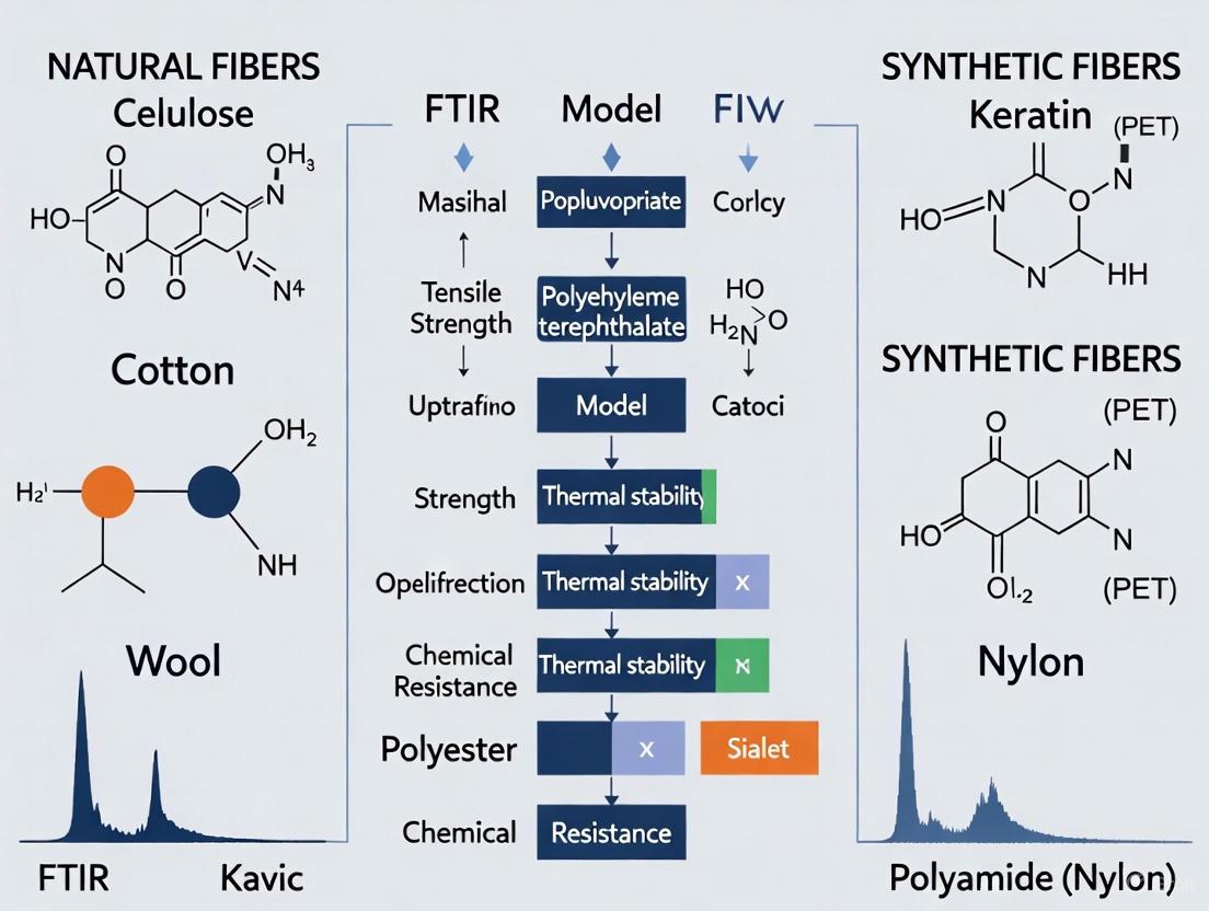
FT-IR Spectral Workflow for Fiber Identification
The following diagram illustrates the logical workflow for identifying and comparing natural fibers using FT-IR spectroscopy, from sample preparation to final classification.
The Scientist's Toolkit: Key Reagents & Materials for Fiber FT-IR Analysis
Table 2: Essential Research Reagents and Materials
| Item | Function in Experiment | Specific Example / Note |
|---|---|---|
| FT-IR Spectrometer with Microscope | Enables high-sensitivity analysis of single fibers or small samples. | Thermo Scientific Nicolet iN10 MX with MCT detector [6]. |
| ATR Accessory | Allows direct, minimal-preparation analysis of solid fiber samples. | Germanium or diamond crystal tip (e.g., Slide-On MicroTip Ge ATR) [4] [6]. |
| Potassium Bromide (KBr) | Used to prepare pellets for transmission FT-IR analysis of powdered fiber samples. | Fibers are crushed and blended with transparent KBr [3]. |
| Reference Gold Plate | Serves as a reflective background for non-invasive r-FT-IR measurements. | Used to acquire background and sample spectra in reflectance mode [6]. |
| Cleaning Solvent (e.g., Ethanol) | Essential for decontaminating the ATR crystal between samples to prevent cross-contamination. | Applied after each sample analysis [5]. |
| Chemometrics Software | Used for advanced spectral data processing, classification, and modeling. | Aspen Unscrambler, TQ Analyst, or in-house Python scripts with sklearn library [5] [6]. |
The comparative analysis of natural fibers hinges on a deep understanding of their chemical composition, particularly the varying proportions of cellulose, hemicellulose, lignin, and pectin. These components collectively determine the physical, mechanical, and hydrophilic properties of the fiber, influencing their suitability for different applications. As demonstrated, FT-IR spectroscopy, in both ATR and reflectance modes, is an indispensable tool in this analytical process. It provides a reliable and efficient means to obtain a chemical fingerprint of fibers, differentiate between fiber types, and even assess the effects of chemical treatments. The experimental protocols and data interpretation frameworks outlined in this guide provide a foundation for researchers to systematically characterize and compare natural fibers, thereby accelerating the development of advanced, sustainable materials.
The comparative analysis of natural and synthetic fibers represents a critical area of research in material science, forensic investigation, and industrial quality control. Within this field, Fourier-transform infrared (FTIR) spectroscopy has emerged as a powerful analytical tool for elucidating polymer structures and enabling precise fiber identification [8]. This technique provides a molecular fingerprint of fiber composition by detecting characteristic vibrational modes of chemical functional groups, allowing researchers to distinguish between chemically similar materials with high accuracy [9] [5].
The synthetic fibers polyester, nylon, and acrylic dominate global textile markets due to their durability, versatility, and cost-effectiveness [10] [11]. Despite their widespread application, these fibers present significant analytical challenges in both research and practical settings. Traditional methods for fiber identification, including microscopy, often fail to distinguish between synthetic fibers with similar physical properties [6]. FTIR spectroscopy overcomes this limitation by probing the fundamental chemical composition of these polymers, providing a non-destructive and highly reliable method for fiber characterization [5] [6].
This review provides a comprehensive comparison of the polymer structures of polyester, nylon, and acrylic through the lens of FTIR spectroscopy. By examining their distinctive spectral features and presenting standardized experimental protocols, this work aims to establish a reference framework for researchers engaged in fiber analysis across diverse scientific disciplines.
Polymer Structures and Characteristic FTIR Bands
The chemical composition of synthetic fibers directly determines their physical properties and analytical signatures. Polyester, primarily composed of polyethylene terephthalate (PET), features an ester functional group (-COO-) linking aromatic rings [12]. Nylon, a polyamide, is characterized by amide groups (-CONH-) within its molecular structure, forming strong intermolecular hydrogen bonds [12] [13]. Acrylic consists of polymerized acrylonitrile units with a nitrile group (-C≡N) as its distinguishing feature [10] [11].
These fundamental chemical differences manifest as distinctive absorption patterns in FTIR spectra, enabling clear differentiation between fiber types. The following table summarizes the characteristic FTIR bands for each synthetic fiber:
Table 1: Characteristic FTIR Absorption Bands for Synthetic Fibers
| Fiber Type | Polymer Composition | Characteristic FTIR Bands (cm⁻¹) | Functional Group Assignment |
|---|---|---|---|
| Polyester | Polyethylene terephthalate (PET) | 1710-1725 | C=O stretch, ester |
| 1240-1270 | C-O-C stretch, aromatic ester | ||
| 730-870 | C-H bend, aromatic ring | ||
| Nylon | Polyamide | 1630-1640 | C=O stretch, amide I |
| 1540-1550 | N-H bend, amide II | ||
| 3300 | N-H stretch | ||
| Acrylic | Polyacrylonitrile | 2240-2245 | C≡N stretch, nitrile |
| 1450 | CH₂ bend | ||
| 1250, 1350-1500 | Methylene deformation |
The spectral differences between these fibers are substantial. Polyester exhibits a strong carbonyl stretch around 1710-1725 cm⁻¹, while nylon shows amide signatures at 1630-1640 cm⁻¹ (amide I) and 1540-1550 cm⁻¹ (amide II) [5]. Acrylic is readily identified by its sharp nitrile stretch at 2240-2245 cm⁻¹, a band absent in both polyester and nylon spectra [10].
Experimental Protocols for FTIR Analysis of Synthetic Fibers
Sample Preparation
For ATR-FTIR analysis, small fiber samples (approximately 2-5 mm in length) can be analyzed directly without extensive preparation [5] [6]. Ensure the ATR crystal is clean before analysis by wiping with ethanol and running a background spectrum. Place the fiber sample on the diamond crystal and apply consistent pressure using the instrument's pressure arm to ensure good contact [6]. For reflectance FT-IR (r-FT-IR), position the fiber on a gold plate or similar reflective surface without applying pressure, making this method suitable for delicate or valuable samples [6].
Instrumental Parameters
Standardized instrumental parameters are essential for reproducible FTIR analysis of synthetic fibers:
- Spectral Range: 4000-600 cm⁻¹ [5] [6]
- Resolution: 4 cm⁻¹ [5] [6]
- Number of Scans: 64-128 scans to improve signal-to-noise ratio [5] [6]
- Detector: Mercury Cadmium Telluride (MCT) cooled with liquid nitrogen or DLaTGS [6]
Data Collection and Preprocessing
Collect background spectra regularly under identical conditions. For ATR-FTIR, apply pressure of 60-75% with a germanium or diamond crystal [6]. For each fiber sample, collect multiple spectra from different areas to assess homogeneity. Preprocess spectra using standard techniques including Savitzky-Golay derivative (e.g., first derivative) and Standard Normal Variate (SNV) to smooth spectra and minimize scattering effects [5].
Spectral Analysis and Classification
Following data collection, employ chemometric methods for robust fiber classification. Principal Component Analysis (PCA) can be built to observe unique patterns and cluster samples by fiber type [5]. Classification models such as Soft Independent Modeling by Class Analogy (SIMCA) have demonstrated 97.1% correct classification of synthetic fibers at a 5% significance level [5]. Alternative methods including Random Forest classification and discriminant analysis have also proven effective for fiber identification [6].
Comparative Performance Analysis
Spectral Differentiation Capabilities
FTIR spectroscopy provides exceptional capability to differentiate between synthetic fiber types based on their polymer structures. Research demonstrates that ATR-FTIR combined with chemometric analysis can achieve 97.1% correct classification of synthetic fibers including nylon, polyester, and acrylic [5]. The differentiation power stems from fundamental chemical differences in these polymers:
Nylon's amide groups produce distinctive N-H stretching at 3300 cm⁻¹ and amide I/II bands, while polyester shows strong ester carbonyl stretching and aromatic signatures [5]. Acrylic is uniquely identified by its sharp nitrile absorption, which doesn't overlap with major bands from other synthetic fibers [10].
Quantitative Spectral Data
The following table presents quantitative FTIR spectral data for the three synthetic fibers, highlighting key differentiating absorption bands:
Table 2: Quantitative FTIR Spectral Data for Synthetic Fiber Identification
| Spectral Parameter | Polyester | Nylon | Acrylic |
|---|---|---|---|
| Major Identification Band (cm⁻¹) | 1710-1725 (C=O stretch) | 1630-1640 (Amide I) | 2240-2245 (C≡N stretch) |
| Secondary Identification Bands (cm⁻¹) | 1240-1270 (C-O-C stretch)730-870 (C-H aromatic) | 1540-1550 (Amide II)3300 (N-H stretch) | 1450 (CH₂ bend)1250, 1350-1500 (CH deformation) |
| Spectral Range (cm⁻¹) | 600-4000 | 600-4000 | 600-4000 |
| Classification Accuracy | >97% [5] | >97% [5] | >97% [5] |
Method Performance Comparison
Different FTIR sampling techniques offer varying advantages for synthetic fiber analysis:
Table 3: Comparison of FTIR Techniques for Synthetic Fiber Analysis
| Parameter | ATR-FT-IR | Reflectance FT-IR |
|---|---|---|
| Sensitivity | High | Moderate to High |
| Sample Preparation | Minimal | Minimal |
| Sample Damage | Potential deformation from pressure | Non-destructive |
| Spectral Quality | Excellent | Very Good |
| Suitability for Delicate Samples | Limited | Excellent |
| Differentiation of Amide Fibers | Good | Better [6] |
Reflectance FT-IR has demonstrated particular effectiveness for differentiating between amide-based fibers like nylon, wool, and silk, sometimes outperforming ATR-FT-IR in classification accuracy for these materials [6].
The Scientist's Toolkit: Essential Research Reagents and Materials
Successful FTIR analysis of synthetic fibers requires specific instrumentation, software, and analytical materials. The following table details essential components for establishing a robust fiber analysis workflow:
Table 4: Essential Research Reagents and Materials for FTIR Fiber Analysis
| Item | Specification/Function | Application Notes |
|---|---|---|
| FT-IR Spectrometer | With ATR and reflectance capabilities | Essential for flexible sampling approaches [6] |
| ATR Crystal | Diamond or Germanium crystal | Diamond provides durability; Germanium offers higher refractive index [6] |
| Microscope Attachment | FT-IR microspectrometer for small samples | Enables analysis of single fibers or small samples [6] |
| Cleaning Solvent | Ethanol (≥95%) | For cleaning ATR crystal between samples to prevent cross-contamination [5] |
| Background Material | Gold plate or reflective surface | For reflectance FT-IR measurements [6] |
| Chemometrics Software | ASPEN Unscrambler, TQ Analyst, or Python with sklearn | For multivariate analysis including PCA and classification models [5] [6] |
| Standard Reference Fibers | Certified nylon, polyester, and acrylic samples | For method validation and calibration [5] |
| Pressure Calibration Standards | Polystyrene film | For verifying instrument performance and wavelength accuracy [5] |
The integration of appropriate chemometric tools is particularly crucial for modern fiber analysis. Software packages such as ASPEN Unscrambler and Python with sklearn libraries provide powerful platforms for implementing classification algorithms including SIMCA and Random Forest, which have demonstrated exceptional accuracy in synthetic fiber identification [5] [6].
FTIR spectroscopy provides an powerful analytical framework for differentiating synthetic fibers based on their fundamental polymer structures. The characteristic chemical functional groups of polyester (ester), nylon (amide), and acrylic (nitrile) produce distinctive spectral signatures that enable highly accurate identification and classification. Through standardized ATR-FTIR and reflectance FT-IR protocols, combined with multivariate statistical analysis, researchers can achieve classification accuracy exceeding 97% [5].
The comparative analysis presented herein establishes that FTIR spectroscopy surpasses traditional microscopic methods for synthetic fiber identification, particularly for distinguishing between chemically similar synthetic polymers. Furthermore, the non-destructive nature of reflectance FT-IR makes it particularly valuable for analyzing delicate or historically significant textile samples where preservation is paramount [6].
As synthetic fibers continue to evolve through material innovations and sustainability initiatives, FTIR spectroscopy remains an indispensable tool for researchers across disciplines including forensic science, materials characterization, and quality control. The protocols and reference data presented in this review provide a foundation for standardized fiber analysis, facilitating more accurate and reproducible research in the comparative analysis of natural and synthetic fibers.
Fourier Transform Infrared (FT-IR) spectroscopy is a powerful analytical technique that takes advantage of the interaction between infrared light and matter to create a unique "chemical fingerprint" of a sample [14]. In the context of forensic and materials science, this technique is invaluable for the comparative analysis of natural and synthetic fibers. When textile fibers are analyzed properly, they can help establish crucial linkages between suspects, victims, and crime scenes [5]. The fundamental principle underpinning this application is that different covalent bonds characterizing various functional groups have distinct characteristic absorption frequencies, allowing researchers to identify specific molecular structures present in fiber samples [15]. This guide provides a structured comparison of characteristic IR absorption bands, with particular emphasis on the diagnostically crucial 1800–800 cm⁻¹ region, to enable researchers to objectively differentiate between fiber types commonly encountered in forensic investigations.
The infrared spectrum is conventionally divided into specific regions that provide different types of structural information. The region from 4000 to 1500 cm⁻¹ is typically considered the functional group region, where characteristic stretches of common functional groups appear. In contrast, the region below 1500 cm⁻¹, extending down to approximately 600 cm⁻¹, is known as the fingerprint region [16] [15]. This fingerprint region contains a complex pattern of absorption bands resulting from a variety of C-C, C-O, C-N, and C-X single-bond vibrations, as well as bending motions. Much like a human fingerprint, the pattern in this region is unique to every molecule, allowing for positive identification of compounds by comparison to known standards [16]. For fiber analysis, this specificity enables discrimination even between fibers belonging to the same generic class [5].
Characteristic Absorption Bands in the 1800–800 cm⁻¹ Region
Comparative Table of Key Absorption Bands
The following table summarizes the characteristic infrared absorption frequencies for functional groups most relevant to fiber analysis in the 1800–800 cm⁻¹ range:
Table 1: Characteristic IR Absorption Bands in the 1800–800 cm⁻¹ Region for Common Fiber Components
| Functional Group/Bond | Compound Class | Absorption Range (cm⁻¹) | Band Intensity | Notes/Specific Characteristics |
|---|---|---|---|---|
| C=O stretching | Carbonyl groups | 1650–1750 | Strong | Carboxylic acids: 1700–1725; key band for synthetic polymers like polyesters [15]. |
| C=C stretching | Aromatic rings | ~1600 & 1500–1430 | Strong to weak | Two sharp bands characteristic of benzene rings [15]. |
| C=C stretching | Alkenes | 1620–1680 | Weak | Present in some natural fibers and modification sites [15]. |
| C-N stretching | Amines/Nylons | 1180–1360 | Medium | Multiple bands often present in fingerprint region [16]. |
| C-O stretching | Esters, Alcohols | 1000–1300 | Strong | Prominent in cellulose-based natural fibers and polyesters [16] [15]. |
| C-C stretching | Various skeletons | 800–1200 | Variable | Multiple contributions in fingerprint region [15]. |
| C-H bending | Aromatic compounds | 900–700 | Medium | Characteristic out-of-plane bending vibrations [16]. |
The Fingerprint Region (1500–800 cm⁻¹)
The fingerprint region, spanning approximately 1500 to 800 cm⁻¹, is where the most subtle distinctions between similar fibers can be observed. While functional groups above 1500 cm⁻¹ provide initial classification clues, the complex pattern of the fingerprint region allows for definitive identification. A recent forensic study demonstrated this capability by analyzing 138 synthetic textile fibers (nylon, polyester, acrylic, and rayon) using ATR–FT-IR spectroscopy combined with chemometric methods [5]. The researchers utilized the entire spectral range, but particularly emphasized the fingerprint region's unique patterns to build classification models that achieved 97.1% correct classification at a 5% significance level using the Soft Independent Modeling by Class Analogy (SIMCA) method [5].
Table 2: Forensic Discrimination of Synthetic Fibers via FT-IR (Adapted from [5])
| Fiber Type | Total Samples | Key Discriminating Absorptions | Chemometric Classification Success | Forensic Utility |
|---|---|---|---|---|
| Nylon | 48 | Amide I & II bands (~1640, ~1550 cm⁻¹) | 97.1% overall correct classification | High discrimination potential |
| Polyester | 52 | Ester C=O (~1710 cm⁻¹), C-O (~1240, ~1090 cm⁻¹) | High separation distance in SIMCA | Excellent for comparative analysis |
| Acrylic | 26 | Nitrile C≡N (~2240 cm⁻¹), ester groups | Correct classification at 5% significance | Distinguishes within generic class |
| Rayon | 12 | Complex C-O, O-H patterns in fingerprint region | Effective clustering in PCA models | Useful for regenerated cellulose identification |
Experimental Protocols for Fiber Analysis by FT-IR
Standard Methodology for Forensic Fiber Comparison
The following experimental workflow details the optimized protocol for comparative analysis of natural and synthetic fibers using FT-IR spectroscopy, based on established forensic methodologies [5]:
Sample Preparation:
- For synthetic fibers, direct placement on ATR–FT-IR crystal is sufficient
- For natural fibers, minimal preparation is required; ensure clean, undamaged fiber segments
- Mount samples to ensure good contact with ATR crystal without deformation
- For transmission FT-IR (less common for fibers), potassium bromide (KBr) pellet preparation is necessary [14]
Instrumentation Parameters:
- Utilize FT-IR Microscope with diamond crystal ATR accessory
- Set spectral range to 4000–400 cm⁻¹ to capture both functional group and fingerprint regions
- Employ resolution of 4 cm⁻¹ for optimal detail and recording time
- Collect 100 scans per spectrum to ensure adequate signal-to-noise ratio
- Perform background (air) measurement before each sample session
- Clean ATR crystal with ethanol between samples to prevent cross-contamination
Data Collection:
- Analyze each sample in triplicate to ensure reproducibility
- Use polystyrene film as a standard to verify instrument performance
- Apply automatic smoothing functions in instrument software (e.g., OPUS) to enhance spectrum quality
- Export spectra in compatible formats (e.g., OPUS, CSV) for multivariate analysis
Experimental Workflow for Forensic Fiber Analysis by FT-IR
Chemometric Analysis Protocol
For objective comparison and classification of fibers, the following chemometric protocol should be implemented [5]:
Data Preprocessing:
- Apply Savitzky–Golay first derivative method to smooth spectra and enhance resolution
- Utilize Standard Normal Variate (SNV) method to minimize scattering effects
- Mean-center the data to improve multivariate model performance
Multivariate Model Development:
- Perform Principal Component Analysis (PCA) to observe natural clustering and patterns
- Develop Soft Independent Modeling by Class Analogy (SIMCA) models for classification
- Set significance level to 5% for class boundaries
- Validate models using cross-validation techniques
Data Interpretation:
- Examine score plots to visualize clustering of different fiber types
- Analyze loading plots to identify wavenumbers responsible for class separation
- Establish classification rules based on statistical distance measures
The Scientist's Toolkit: Essential Research Reagents and Materials
Table 3: Essential Research Reagents and Materials for FT-IR Fiber Analysis
| Item/Reagent | Function/Purpose | Application Notes |
|---|---|---|
| FT-IR Microscope with ATR | Spectral acquisition of fiber samples | Diamond crystal preferred for durability and spectral range [5] |
| Synthetic Fiber Standards | Reference materials for classification | Nylon, polyester, acrylic, rayon with known provenance [5] |
| Natural Fiber Standards | Reference materials for comparison | Cotton, wool, silk with documented origin |
| Absolute Ethanol | Cleaning ATR crystal between measurements | Prevents cross-contamination; high purity recommended [5] |
| Polystyrene Film | Instrument performance verification | Validates wavenumber accuracy and resolution [5] |
| Potassium Bromide (KBr) | Pellet preparation for transmission mode | Required only if transmission FT-IR is performed [14] |
| Multivariate Analysis Software | Chemometric data processing | Aspen Unscrambler or equivalent for PCA and SIMCA modeling [5] |
Comparative Data Interpretation Strategies
Distinguishing Natural and Synthetic Fibers
The comparative analysis of natural and synthetic fibers relies on recognizing characteristic band patterns in both the functional group and fingerprint regions. Natural cellulosic fibers like cotton and rayon exhibit prominent O-H stretching around 3300 cm⁻¹, C-H stretching around 2900 cm⁻¹, and a complex pattern of C-O and C-C vibrations in the 1200–1000 cm⁻¹ region [15]. In contrast, synthetic fibers display more specific functional group signatures: polyesters show strong carbonyl stretching around 1710 cm⁻¹ and C-O-C stretching around 1100–1250 cm⁻¹; nylons exhibit characteristic amide I (~1640 cm⁻¹) and amide II (~1550 cm⁻¹) bands; and acrylics show a distinctive nitrile absorption at ~2240 cm⁻¹ [5].
The power of modern FT-IR analysis in fiber comparison lies in combining traditional band assignment with multivariate statistical methods. In the referenced forensic study, researchers were able to achieve 97.1% correct classification of synthetic fibers by applying PCA and SIMCA models to the full spectral data [5]. This approach moves beyond simple visual comparison of spectra to objective, statistical classification that can withstand forensic scrutiny. The fingerprint region proves particularly valuable in these models, as it contains the subtle spectral variations that differentiate structurally similar polymers.
Spectral Interpretation Strategy for Fiber Identification
The comparative analysis of natural and synthetic fibers through FT-IR spectroscopy provides a powerful tool for researchers and forensic scientists. The characteristic absorption bands in the 1800–800 cm⁻¹ region, particularly the complex patterns in the fingerprint region below 1500 cm⁻¹, enable both preliminary classification and definitive identification of fiber types. When combined with multivariate statistical methods such as PCA and SIMCA, FT-IR spectroscopy achieves exceptional discrimination power, with recent studies demonstrating 97.1% correct classification of synthetic fibers [5]. This objective, data-driven approach transforms FT-IR from a simple identification technique into a robust comparative methodology capable of supporting forensic conclusions and material science investigations with statistical confidence.
Within the broader thesis on the comparative analysis of natural and synthetic fibers via FTIR research, the critical role of spectral libraries emerges as a foundational element. Spectral library searching represents the most common approach for compound annotation in untargeted analyses, operating on the principle that molecules produce reproducible fragmentation "fingerprints" [17]. For fiber identification, this translates to leveraging unique infrared absorption patterns to differentiate between material classes. The growth of publicly accessible spectral libraries, which have expanded more than 60-fold in the past eight years, now provides an essential knowledge base for interpreting analytical data, from forensics to cultural heritage [17]. This guide objectively compares the performance of FTIR spectroscopy and its associated library tools in differentiating natural and synthetic fibers, providing the experimental data and protocols necessary to implement these techniques effectively.
Key Spectral Peaks for Fiber Differentiation
Fourier Transform Infrared (FT-IR) spectroscopy identifies materials by detecting characteristic molecular vibrations, represented as absorption peaks at specific wavenumbers (cm⁻¹). The following tables consolidate the key diagnostic peaks for major natural and synthetic fiber classes, serving as a primary reference for spectral library searches.
Natural Cellulosic Fibers
Table 1: Characteristic FT-IR Peaks of Common Natural Cellulosic Fibers
| Fiber Type | Key Diagnostic Peaks (cm⁻¹) | Chemical Assignment & Notes |
|---|---|---|
| Cotton | 1105 (vs), 3335 (s,b), 2900 (m), 1430 (m), 1370 (m), 1160 (m), 1050-1000 (s) [18] [4] | The band at 1105 cm⁻¹ is a strong, sharp peak for cellulose-Iβ (natural cellulose). High cellulose content (67-78%) [19] [4]. |
| Linen (Flax) | Similar to cotton, but with detectable peaks for pectin and lignin (e.g., ~1730 cm⁻¹, ~1508 cm⁻¹) [4] | A bast fiber. Differentiated from cotton by the presence of non-cellulosic compounds like pectin and lignin [4]. |
| Jute | 1508 (m), 1450 (w), 1270 (m), 1230 (m) in addition to cellulose peaks [4] | A bast fiber with high lignin content (~12-13%) [19]. The band at 1508 cm⁻¹ (aromatic C=C in lignin) is a key differentiator from other natural celluloses [4]. |
| Hemp | Spectrum similar to linen; requires differentiation based on relative peak intensities or complementary methods [4] | A bast fiber. Contains ~70-74% cellulose and ~3-6% lignin [19]. |
| Sisal | 1735 (w), 1508 (m) in addition to cellulose peaks [4] | A leaf fiber. Contains ~67-78% cellulose and ~8-11% lignin [19]. |
| Silk | 3280 (s, Amide A), 1620 (s, Amide I), 1515 (s, Amide II), 1260 (s, Amide III), 1160 (w), 1060 (w) [4] | A protein-based fiber (β-sheet conformation). Identified by the classic "amide" bands [4]. |
| Wool | 3280 (s, Amide A), 1620 (s, Amide I), 1515 (s, Amide II), 1230 (s, Amide III) [4] | A protein-based fiber (α-helix conformation). Differentiated from silk by the relative intensities and shapes of the amide bands [4]. |
Abbreviations: vs = very strong, s = strong, m = medium, w = weak, b = broad.
Semi-Synthetic and Synthetic Fibers
Table 2: Characteristic FT-IR Peaks of Semi-Synthetic and Synthetic Fibers
| Fiber Type | Key Diagnostic Peaks (cm⁻¹) | Chemical Assignment & Notes |
|---|---|---|
| Viscose Rayon | 1105 (w, broad), 890 (s), 1429 (w) [18] [4] | A regenerated cellulose (cellulose-II). The band at 1105 cm⁻¹ is weak and broad, distinct from its strong, sharp counterpart in natural celluloses. The strong peak at 890 cm⁻¹ is characteristic [18] [4]. |
| Cellulose Acetate | 1735-1750 (s, C=O), 1365-1375 (s, CH₃), 1220-1240 (s, C-O), 1030-1050 (s, C-O) [18] [4] | A semi-synthetic fiber. The intense carbonyl (C=O) stretch at ~1740 cm⁻¹ is the primary differentiator from natural and other regenerated celluloses [18] [4]. |
| Polyester (PET) | 1700-1725 (s, C=O), 1250-1270 (s, C-O), 1090-1120 (s, C-O), 720-730 (m) [6] | A synthetic polymer. The sharp carbonyl stretch is diagnostic [6]. |
| Polyamide (Nylon) | 3280 (m, N-H), 3050 (w), 2930 (m), 2860 (m), 1630 (s, Amide I), 1530 (s, Amide II), 1265 (s), 1200-700 (m) [6] | A synthetic polymer with amide linkages. The amide bands can be distinguished from natural proteins like silk and wool by their spectral context and lack of other biological components [6]. |
| Polyacrylic | 2240-2242 (m, C≡N), 1730-1740 (s, C=O ester), 1440-1470 (m) [6] | A synthetic polymer. The nitrile (C≡N) stretch near 2242 cm⁻¹ is a key identifier [6]. |
| Polypropylene | 2950 (s), 2915 (s), 2835 (s), 1450 (s), 1375 (s) [6] | A synthetic polyolefin. Lacks oxygen-containing functional groups, showing only C-H and C-C stretches [6]. |
Abbreviations: vs = very strong, s = strong, m = medium, w = weak, b = broad.
Experimental Protocols for Fiber Analysis
The reliability of spectral identification hinges on strict adherence to validated experimental protocols. The following methodologies are cited from research studies that have successfully differentiated natural and synthetic fibers.
Protocol for ATR-FT-IR Spectroscopy
ATR-FT-IR is a widely acknowledged technique for fiber analysis due to its minimal sample preparation and high-quality spectra [6].
- Instrumentation: Use an FT-IR spectrometer equipped with a single-bounce diamond ATR accessory (e.g., Thermo Scientific Nicolet 6700) [6].
- Parameters:
- Procedure:
- Clean the ATR crystal with isopropyl alcohol and acquire a background spectrum.
- Place a single fiber or small snippet of fabric directly onto the crystal.
- Apply consistent, firm pressure using the instrument's pressure arm to ensure good contact between the sample and the crystal.
- Collect the spectrum.
- Repeat the measurement on different areas of the sample to ensure homogeneity and representativeness.
Protocol for Non-Invasive Reflectance FT-IR (r-FT-IR) Spectroscopy
For valuable or fragile samples where contact is undesirable, r-FT-IR provides a viable non-invasive alternative [6].
- Instrumentation: Use an FT-IR microspectrometer equipped for reflectance measurements (e.g., Thermo Scientific Nicolet iN10 MX) [6].
- Parameters:
- Procedure:
- Place the textile sample on a gold plate, which also serves as the background reference.
- Select an analysis area using the microscope.
- Adjust the aperture to isolate the region of interest.
- Collect the reflectance spectrum without applying any pressure to the sample.
- Collect multiple spectra from different areas to assess sample homogeneity.
Protocol for Discriminating Natural and Semi-Synthetic Celluloses
Differentiating between natural (e.g., cotton) and semi-synthetic (e.g., viscose) cellulosic fibers can be challenging. The following method leverages a key spectral difference.
- Key Diagnostic Band: The intensity and shape of the 1105 cm⁻¹ band is a critical differentiator [18]. It is strong and sharp in natural celluloses (Cotton, Jute) but becomes weak and broad in semi-synthetic regenerated celluloses like viscose [18].
- Sample Preparation (for validation): To simulate real-world aged samples, laboratory-made fibers can be subjected to UV-accelerated weathering and chemical digestion (e.g., with H₂O₂ or KOH) [18]. This process confirms that the 1105 cm⁻¹ band remains a stable identifier even after environmental degradation [18].
- Analysis:
- Collect the ATR-FT-IR spectrum of the unknown cellulosic sample.
- Normalize the spectra to the intensity of the C-O band around 1160 cm⁻¹ or the O-H band around 3335 cm⁻¹.
- Directly compare the intensity and shape of the 1105 cm⁻¹ band in the unknown sample to library spectra of known cotton and viscose standards. A strong, sharp peak indicates a natural cellulose, while a weak, broad peak suggests a regenerated cellulose [18].
Workflow and Data Interpretation
Fiber Identification Workflow
The following diagram illustrates the logical workflow for identifying an unknown fiber using FTIR spectroscopy and spectral libraries, integrating the key comparative peaks and techniques discussed.
Spectral Library Search Algorithms
The core of computational identification lies in the library search algorithm. While commercial software may use proprietary methods, a common and relatively simple algorithm (e.g., the "Difference" algorithm in Shimadzu IRsolution) operates as follows [20]:
- Pre-processing: The sample spectrum and all library spectra are first normalized in intensity, typically so the maximum absorbance becomes 1, preserving the spectral shape but allowing comparison independent of concentration or thickness [20].
- Wavenumber Range Selection: The comparison is often restricted to a user-specified wavenumber range containing characteristic peaks, rather than using the entire spectrum [20].
- Comparison Metric: For each library spectrum, the absolute differences in intensity between the sample spectrum and the library spectrum are calculated at every data point within the selected range. These absolute values are then summed into a single "total difference" value [20].
- Result Ranking: The library spectra are ranked based on this total difference value. A perfect match would yield a value of zero. In practice, the library entry with the smallest total difference is proposed as the best match [20].
More advanced algorithms may use derivative spectra or correlation coefficients instead of simple difference to improve match quality and reduce the impact of baseline shifts [20].
The Scientist's Toolkit: Essential Research Reagents and Materials
Table 3: Key Research Reagent Solutions and Materials for FTIR Fiber Analysis
| Item | Function / Application | Notes |
|---|---|---|
| ATR Crystal Cleaner | Cleaning diamond ATR crystal between measurements to prevent cross-contamination. | High-purity isopropyl alcohol is typically used [6]. |
| Gold-Coated Substrate | A highly reflective background for non-invasive r-FT-IR measurements. | Provides a clean background spectrum; sample is placed on the gold surface [6]. |
| Reference Spectral Libraries | Digital databases of known fiber spectra for automated search and identification. | Commercial (e.g., NIST, METLIN) and open (e.g., GNPS, MassBank) libraries exist [17]. |
| Standard Fiber Samples | Positive controls for method validation and for expanding in-house spectral libraries. | Include certified samples of cotton, wool, polyester, nylon, viscose, etc. [6]. |
| Microscope & Micro-ATR Objective | For targeting single fibers or small, heterogeneous samples in a mixed fabric. | A Ge crystal micro-ATR tip can analyze samples as small as 3 μm [6]. |
This guide has detailed the objective performance of FTIR spectroscopy in differentiating natural and synthetic fibers through the lens of spectral libraries and key diagnostic peaks. The comparative tables provide a direct, data-driven resource for identifying unknown materials based on their infrared signatures. The experimental protocols, particularly the use of the 1105 cm⁻¹ band for distinguishing natural from regenerated celluloses and the non-invasive reflectance technique for delicate samples, provide a reliable methodological foundation. When executed with rigorous attention to protocol and supported by robust spectral libraries, FTIR analysis stands as a powerful, accessible, and definitive tool for fiber identification across diverse scientific and industrial fields.
Advanced FTIR Techniques and Cutting-Edge Applications in Research and Industry
Fourier Transform Infrared (FTIR) spectroscopy is a cornerstone analytical technique for characterizing both natural and synthetic fibers, providing critical insights into their molecular composition and structure. The analytical pathway, however, is profoundly influenced by the chosen sampling modality. For researchers in material science and drug development, selecting the appropriate method—Attenuated Total Reflectance (ATR), Transmission, or Transflection—is crucial for generating reliable, high-quality data. This guide provides a comparative analysis of these three techniques, framing them within the context of fiber analysis to empower professionals in making informed methodological decisions.
Fundamental Principles and Experimental Protocols
Each FTIR sampling technique operates on a distinct physical principle, which in turn dictates its specific experimental workflow and sample requirements.
Attenuated Total Reflectance (ATR)
- Principle of Operation: ATR utilizes an Internal Reflection Element (IRE) crystal with a high refractive index (e.g., diamond, ZnSe). IR light passes through this crystal and undergoes total internal reflection at the crystal-sample interface. During each reflection, an evanescent wave penetrates a short distance (typically 0.5-2 µm) into the sample in contact with the crystal, where it is absorbed [21] [22]. The resulting attenuated light is then directed to the detector.
- Standardized Experimental Protocol:
- Sample Preparation: Solid fibers may be flattened or a small portion cut and directly placed on the IRE crystal. For better contact, a clamping arm applies controlled pressure [21]. Liquid samples are directly pipetted onto the crystal.
- Instrument Setup: Ensure the IRE crystal is clean. Select the appropriate crystal material based on the sample hardness and chemical compatibility (diamond is most robust).
- Data Acquisition: Collect a background spectrum with the clean crystal. Place the sample in uniform contact with the crystal and acquire the sample spectrum.
- Post-processing: ATR spectra may require automatic correction for wavelength-dependent penetration depth, which is typically handled by instrument software [21].
Transmission
- Principle of Operation: This is the most direct method, where IR radiation is passed directly through the sample. Frequencies of light that match the vibrational energies of chemical bonds in the sample are absorbed, and the transmitted light is measured [21] [23]. The Beer-Lambert Law governs the relationship between absorption, sample thickness, and concentration.
- Standardized Experimental Protocol:
- Sample Preparation (Critical Step):
- KBr Pellet Method (for solids): Grind a small quantity of fiber with purified potassium bromide (KBr) powder. Compress the mixture under high pressure to form a transparent pellet [21].
- Thin Sectioning (for fibers): For a more direct analysis, fibers can be microtomed into thin slices (5–10 µm thick) and placed on a IR-transparent window like KBr or BaF₂ [23].
- Instrument Setup: Mount the prepared sample in the FTIR spectrometer's sample holder.
- Data Acquisition: Collect a background spectrum through the pure substrate (e.g., a clean KBr window or an open beam). Acquire the sample spectrum.
- Quality Control: Verify that spectral peaks are not "totally absorbing" (absorbance >1.2), which indicates the sample is too thick and requires re-preparation [23].
- Sample Preparation (Critical Step):
Transflection
- Principle of Operation: A hybrid technique, transflection involves directing IR light through the sample onto a reflective substrate (e.g., a low-e microscope slide). The light is reflected back through the sample to the detector, effectively doubling the pathlength [24] [22]. This method is also referred to as reflection-absorption.
- Standardized Experimental Protocol:
- Sample Preparation: The sample, often a thin tissue section or a smear of finely ground fibers, is deposited directly onto the reflective substrate and allowed to dry [24].
- Instrument Setup: The transflection slide is placed in the spectrometer's microscope or macro chamber.
- Data Acquisition: A background spectrum is collected from a clean area of the reflective slide. The sample spectrum is then acquired.
- Data Pre-processing: Be aware of potential artifacts like the Electric Field Standing Wave (EFSW) effect, which can distort band intensities, particularly for thin samples. Multivariate comparison with transmission data from adjacent sections can help establish the impact of such effects [24].
The following diagram illustrates the core operational principles of these three sampling modalities.
Comparative Performance Analysis
The choice between ATR, Transmission, and Transflection involves balancing factors such as sample preparation effort, data quality, analytical performance, and suitability for different fiber types.
Table 1: Direct Comparison of ATR, Transmission, and Transflection Modalities
| Aspect | ATR | Transmission | Transflection |
|---|---|---|---|
| Sample Preparation | Minimal; direct placement of solids/liquids on crystal [21] | High; requires thinning, KBr pellets, or microtomy [21] [23] | Moderate; deposition on reflective slides [24] |
| Typical Pathlength | Fixed, short (0.5-2 µm) [21] | Variable, user-controlled (5-50 µm) [23] | Effectively double the sample thickness [22] |
| Ideal Fiber Types | All types (raw threads, fabrics, powders) [21] [4] | Homogeneous, thin fibers; microtomed sections [23] | Thin tissue sections containing fibers; smears [24] |
| Spectral Quality | High-quality; slight intensity shifts vs. transmission [21] | High-fidelity; considered the gold standard for libraries [21] | Good; can be affected by EFSW artifacts [24] |
| Reproducibility | High for varied sample types [21] | Can be variable with manual pellet preparation [21] | Consistent, but artifacts are consistent across tissue [24] |
| Quantitative Potential | Good with careful pressure control | Excellent, governed by Beer-Lambert Law | Can be compromised by dispersion & EFSW effects [24] |
| Key Advantage | Speed, minimal preparation, versatility | High-quality, library-compatible spectra | Effective signal amplification for thin samples [22] |
| Key Limitation | Spectral shifts require understanding [21] | Time-consuming, skill-intensive preparation [21] | Susceptibility to electric field standing waves [24] |
Table 2: Summary of Experimental Findings from Key Comparative Studies
| Study Focus | Key Experimental Findings | Implications for Fiber Analysis |
|---|---|---|
| Transflection vs. Transmission on Tissue [24] | - Small, consistent Amide I band shift in transflection.- Higher phosphate bands in transflection.- Greater spectral variance (noise) in transflection. | Suggests transflection is viable for diagnostic classification of biological tissues containing proteinaceous fibers (e.g., collagen), but caution is needed for precise band assignment and quantitative work. |
| ATR vs. Transmission General [21] | - ATR peak intensities differ from transmission.- Small peak shifts (e.g., carbonyl) due to anomalous dispersion. | Confirms that ATR and transmission spectra are not identical. ATR spectra should be compared to ATR-specific libraries, or correction algorithms must be applied. |
| ATR for Cellulosic Fibres [4] | - ATR-FTIR successfully differentiates natural fibers (cotton, flax, hemp) based on lignin, pectin, hemicellulose content.- Effective for identifying man-modified (regenerated) fibers like viscose. | Highlights ATR's practical utility for rapid identification and differentiation of complex natural and modified cellulosic materials in cultural heritage and textiles. |
The Scientist's Toolkit: Essential Research Reagents and Materials
Successful FTIR analysis requires specific consumables and substrates tailored to each sampling modality.
Table 3: Essential Research Reagents and Materials for FTIR Sampling
| Item | Function/Application | Notes & Considerations |
|---|---|---|
| ATR Crystals (Diamond, ZnSe, Ge) | Internal Reflection Element (IRE) for ATR measurement. | Diamond: Hard, chemically inert, universal use. ZnSe: Good IR performance, less durable. Ge: High refractive index for strong absorbers. |
| Potassium Bromide (KBr) | Matrix for creating transparent pellets for transmission analysis of solids. | Highly hygroscopic; must be kept dry and pressed into pellets in a controlled environment [21]. |
| IR-Transparent Windows (KBr, NaCl, CaF₂, BaF₂) | Substrates for mounting samples in transmission and transflection modes. | CaF₂: Water-insoluble, good for aqueous samples. NaCl: Inexpensive, but water-soluble. Choice depends on spectral range and sample properties [21] [23]. |
| Reflective Substrates (Low-e Slides) | Highly reflective slides used as a backing for transflection measurements. | Doubles the effective pathlength, enhancing signal from thin samples [22]. |
| Microtome | Instrument for slicing thin cross-sections (1-10 µm) of fibers or composites for transmission analysis. | Critical for preparing samples of consistent, optimal thickness to avoid total absorption of IR light [23]. |
| Diamond Compression Cell | A tool for flattening and thinning samples (e.g., single fibers) between two diamond windows for transmission measurement. | Provides high-pressure compression, creating a uniform thin film ideal for transmission FTIR microscopy [23]. |
The comparative analysis of ATR, Transmission, and Transflection modalities reveals a clear trade-off between analytical effort and data fidelity. ATR-FTIR spectroscopy has emerged as the most versatile and efficient technique for the rapid analysis of a wide range of fiber types, from natural celluloses to synthetic polymers, with minimal sample preparation [21] [4]. However, for quantitative analysis requiring the highest spectral accuracy and access to extensive transmission-based libraries, Transmission FTIR remains the gold standard, despite its demanding sample preparation protocols [21] [23]. Transflection occupies a specific niche, particularly useful in biomedical contexts for analyzing thin tissue sections, but researchers must be vigilant about potential spectral artifacts [24].
The optimal sampling modality is ultimately dictated by the specific research question, the nature of the fiber sample, and the required data quality. This guide provides the foundational knowledge for researchers to make that critical choice confidently, thereby advancing reliable FTIR-based characterization in the field of fiber science.
Fourier Transform Infrared (FTIR) spectroscopy has become an indispensable tool for the molecular analysis of a wide range of materials, from natural and synthetic fibers to pharmaceuticals and biological samples. However, the interpretation of complex spectral data requires sophisticated statistical and computational approaches to extract meaningful information. Chemometrics, the application of mathematical and statistical methods to chemical data, provides these essential tools. This guide offers a comparative analysis of principal chemometric techniques—including Principal Component Analysis (PCA), Partial Least Squares (PLS) regression, and various Machine Learning (ML) algorithms—as applied to FTIR spectral data within the specific context of fiber research. By objectively evaluating the performance, applications, and implementation requirements of these methods, this review serves as a practical resource for researchers and analysts seeking to optimize their spectroscopic analyses.
Fundamental Chemometric Techniques and Their Applications
Chemometric techniques transform complex spectral data into interpretable models for classification, quantification, and pattern recognition. The choice of method depends on the analytical objective, data structure, and required outcome.
Principal Component Analysis (PCA) is an unsupervised technique used primarily for exploratory data analysis and dimensionality reduction. It works by transforming original variables into a smaller set of linear combinations called Principal Components (PCs), which capture the maximum variance in the data. In fiber analysis, PCA successfully differentiated synthetic textile fibers (nylon, polyester, acrylic, and rayon) based on their ATR-FTIR spectra, revealing distinct clustering patterns according to fiber polymer type [5]. The method is particularly valuable for visualizing natural groupings in data and identifying outliers without prior knowledge of sample classes.
Partial Least Squares (PLS) regression is a supervised technique that models relationships between independent variables (spectral data) and dependent variables (chemical or physical properties). Unlike PCA, PLS considers both the X-matrix (spectra) and Y-matrix (response variables) to find latent variables that maximize covariance. PLS-Discriminant Analysis (PLS-DA), a variant used for classification, effectively detected adulteration of patchouli oil even at low concentrations (0.5%), demonstrating high sensitivity (RMSEC = 0.22) and excellent fit (R² = 0.954) [25]. In fiber research, PLS has been coupled with FTIR for quantifying specific chemical components in natural fibers, such as cellulose, hemicellulose, and lignin content [9].
Machine Learning (ML) algorithms encompass a diverse range of supervised and unsupervised learning methods that can handle complex, non-linear relationships in spectral data. Commonly applied algorithms include Support Vector Machines (SVM), Random Forest, and Decision Trees. In a comparative study of gelatin samples, multiple ML classifiers including NCA-KNN, SVM, LDA, and Decision Trees were implemented in Python, all effectively classifying FTIR spectra based on cross-linking status with high precision [26]. The flexibility of ML models allows them to adapt to various spectral analysis challenges, from fiber identification to quality assessment.
Table 1: Overview of Fundamental Chemometric Techniques
| Technique | Type | Primary Function | Key Advantage | Typical FTIR Application in Fiber Analysis |
|---|---|---|---|---|
| PCA | Unsupervised | Dimensionality reduction, exploratory analysis | Identifies natural clustering without prior class labels | Discrimination of fiber polymer types [5] |
| PLS | Supervised | Regression, quantitative prediction | Models covariance between spectra and properties | Quantification of chemical constituents in fibers [9] |
| PLS-DA | Supervised | Classification, discrimination | Maximizes separation between predefined classes | Detection of adulterants or contaminants in natural fibers [25] |
| Machine Learning | Supervised/Unsupervised | Pattern recognition, classification, prediction | Handles complex, non-linear relationships in data | Classification of fiber treatments or modifications [26] |
Performance Comparison Across Applications
The comparative performance of chemometric techniques varies significantly across different analytical scenarios and sample types. Objective evaluation using standardized metrics is essential for selecting the appropriate method for specific applications.
Classification Accuracy in Material Differentiation
In forensic analysis of synthetic textile fibers, FTIR spectroscopy coupled with Soft Independent Modeling by Class Analogy (SIMCA) achieved a 97.1% correct classification rate at a 5% significance level for distinguishing nylon, polyester, acrylic, and rayon fibers [5]. The study employed preprocessing techniques including Savitzky-Golay first derivative and Standard Normal Variate (SNV) to enhance spectral features before pattern recognition. Similarly, in pharmaceutical applications, ML classifiers including Radial Basis Function (RBF), Random Forest, and J48 decision trees successfully differentiated between Guan-Mutong and Chuan-Mutong samples, demonstrating the applicability of these methods for quality control of natural products [27].
Quantitative Analysis Performance
For quantitative determinations, studies directly comparing PLS and Support Vector Regression (SVR) have revealed important performance differences. In the analysis of oxygenate compounds in gasoline, Genetic Algorithm-optimized SVR (GA-SVR) demonstrated superior predictive ability compared to GA-PLS, showing higher R²pred values and lower root mean square errors [28]. This advantage was attributed to SVR's ability to handle non-linear relationships in the spectral data. Conversely, in wine polyphenol analysis, PLS models for tannin concentration showed greater robustness compared to UV-Vis derived models, while UV-Vis performed better for anthocyanin parameters [29].
Influence of Data Preprocessing
The performance of all chemometric techniques is significantly enhanced by appropriate spectral preprocessing. Common methods include:
- Standard Normal Variate (SNV): Corrects for light scattering effects [5] [25]
- Savitzky-Golay Derivatives: Enhance resolution of overlapping peaks (first derivative) and remove baseline offsets (second derivative) [5] [25]
- Normalization: Standardizes spectral intensity for comparative analysis [26] [25]
In fiber analysis, these preprocessing techniques have been shown to improve model accuracy by reducing non-chemical spectral variations and enhancing relevant molecular features [5] [9].
Table 2: Performance Comparison of Chemometric Techniques in Various Studies
| Application Domain | Technique | Performance Metrics | Comparative Outcome | Reference |
|---|---|---|---|---|
| Synthetic Fiber Forensics | SIMCA | 97.1% correct classification | Effective for polymer type discrimination | [5] |
| Gasoline Oxygenates | GA-PLS vs. GA-SVR | R²pred, RMSE | SVR showed superior predictive ability for non-linear systems | [28] |
| Pharmaceutical Gelatin Classification | Multiple ML classifiers | Classification accuracy | All models (NCA-KNN, SVM, LDA, DT) effective with high precision | [26] |
| Adulterant Detection | PLS-DA | R² = 0.954, RMSEC = 0.22 | Detected adulteration at 0.5% concentration level | [25] |
| Traditional Medicine Authentication | PLS-DA vs. ML | Classification accuracy | Machine learning (RBF, Random Forest, J48) compared favorably with PLS-DA | [27] |
Experimental Protocols and Methodologies
Standard FTIR Spectral Acquisition for Fibers
The characterization of novel Annona reticulata fiber exemplifies a standardized approach for natural fiber analysis. FTIR spectra were recorded using an IR Prestige 21 spectrophotometer across the range of 4000–500 cm⁻¹ with a resolution of 4 cm⁻¹ [30]. Spectral data were processed using OriginPro software, with specific peak assignments indicating functional groups: O-H stretching at 3309 cm⁻¹ and C-H stretching at 2928 cm⁻¹ (cellulose), and C=C stretching at 1514 cm⁻¹ (lignin) [30]. This protocol ensures reproducible identification of chemical constituents in lignocellulosic fibers.
Chemometric Workflow Implementation
A comprehensive study on pharmaceutical gelatins demonstrated an integrated Python-based workflow for FTIR spectral analysis [26]. The methodology included:
- Spectral Preprocessing: Normalization using the
rampylibrary and baseline correction withPybaselines - Exploratory Analysis: Hierarchical Clustering Analysis (HCA) using
ScipyandSeabornlibraries - Dimensionality Reduction: Principal Component Analysis (PCA) and Partial Least Squares (PLS)
- Classification Models: Implementation of NCA-KNN, SVM, LDA, and Decision Trees using
SKlearnlibrary
This workflow enabled effective differentiation of cross-linked and non-cross-linked gelatin samples, demonstrating adaptability for fiber analysis applications [26].
Model Validation Approaches
Robust validation is critical for reliable chemometric models. Internal validation methods include cross-validation, where a study on clinical parameters in human serum employed multiple regression methods (PLSR, Neural Networks, SVR, Decision Trees) with ensemble extensions [31]. External validation using independent test sets was implemented in the patchouli oil adulteration study, where the PLS-DA model was strongly validated using a separate PLS model, showing minimal difference between calibration and prediction (average 0.45) with accuracy exceeding 99% [25].
FTIR Chemometric Analysis Workflow: Standardized procedure from spectral acquisition to model validation.
Essential Research Reagents and Materials
Successful implementation of chemometric analysis in FTIR spectroscopy requires specific laboratory materials and computational resources. The following table details essential components for experimental work in fiber analysis.
Table 3: Essential Research Materials for FTIR Spectral Analysis with Chemometrics
| Category | Specific Items | Function/Application | Example Use Cases |
|---|---|---|---|
| FTIR Instrumentation | FTIR Spectrometer with ATR accessory, DTGS detector | Molecular vibration detection, sample analysis | Synthetic fiber polymer identification [5] |
| Reference Materials | Certified Reference Materials (CRMs), solvent standards | Method validation, quality control | Patchouli oil authentication [25] |
| Spectral Preprocessing | KBr for pellet preparation, ethanol for cleaning | Sample presentation, instrument maintenance | Natural fiber characterization [30] |
| Software Tools | Python libraries (Scipy, SKlearn, rampy), Unscrambler | Data preprocessing, multivariate analysis | Gelatin classification [26], Textile fiber discrimination [5] |
| Validation Standards | Independent test sets, cross-validation protocols | Model performance assessment | Clinical parameter prediction [31] |
The comparative analysis of chemometric techniques for FTIR spectral analysis reveals a diverse toolkit for researchers working with natural and synthetic fibers. PCA serves as an essential exploratory tool for visualizing natural clustering in spectral data, while PLS regression provides robust quantitative models for predicting material properties. Machine learning algorithms, particularly SVM and ensemble methods, offer powerful alternatives for handling complex, non-linear spectral relationships. The optimal technique selection depends on specific analytical objectives, with classification tasks benefiting from PLS-DA or ML approaches, and quantitative analysis leveraging PLS or SVR depending on data linearity. As FTIR spectroscopy continues to evolve as a primary analytical method in fiber science, the integration of appropriate chemometric methods remains essential for extracting maximum information from complex spectral data sets.
Quality Control and Process Analytical Technology (PAT) in Manufacturing
Process Analytical Technology (PAT) has emerged as a revolutionary framework in manufacturing quality control, enabling real-time monitoring of Critical Process Parameters (CPPs) and Critical Quality Attributes (CQAs) to ensure product consistency [32]. Within the PAT toolbox, Fourier Transform Infrared (FTIR) spectroscopy serves as a powerful analytical technique for material characterization, providing molecular-level fingerprints through the measurement of bond vibration modes [7]. This comparative analysis examines the application of FTIR spectroscopy for distinguishing between natural and synthetic fibers, a capability with significant implications for pharmaceutical manufacturing where fiber-based filters, scaffolds, and excipients are employed. The fundamental principle underpinning this analysis is that different fiber types exhibit distinct infrared absorption patterns due to their varying chemical compositions, enabling identification and quality verification without lengthy sample preparation [6] [7].
The transition from traditional quality control methods, which often rely on offline laboratory analysis, to real-time PAT approaches represents a paradigm shift in manufacturing quality assurance [32]. FTIR spectroscopy aligns perfectly with PAT objectives by offering rapid, non-destructive analysis that can be integrated directly into manufacturing processes. For natural and synthetic fiber identification, this capability provides manufacturers with immediate feedback on raw material quality, potentially preventing deviations and ensuring consistent final product performance [6]. This guide presents a comprehensive comparison of FTIR methodologies for fiber analysis, providing researchers and drug development professionals with experimental protocols, performance data, and practical implementation strategies.
Experimental Protocols for FTIR Fiber Analysis
Sample Preparation and Instrumentation
The foundation of reliable FTIR analysis lies in standardized sample preparation and instrumental configuration. For fiber analysis, two primary FTIR operational modes have demonstrated efficacy: Attenuated Total Reflectance (ATR) and Reflectance (r-FT-IR) [6]. The ATR-FT-IR method requires applying direct pressure to ensure proper contact between the fiber sample and the crystal element, which may be problematic for delicate or valuable samples due to potential damage [6]. In contrast, r-FT-IR offers a non-contact approach suitable for fragile samples but may present challenges with uneven textile surfaces [6].
For comprehensive analysis, researchers should employ an FT-IR microspectrometer equipped with both reflectance and ATR capabilities, utilizing a Mercury Cadmium Telluride (MCT) detector cooled with liquid nitrogen [6]. The recommended spectral range is 600–4000 cm⁻¹ with a resolution of 4 cm⁻¹ and 64 scans per measurement to ensure sufficient signal-to-noise ratio [6]. For reflectance measurements, samples should be placed on a gold plate reference background, while for micro-ATR, a germanium crystal with 60–75% pressure strength provides optimal results [6]. These standardized parameters enable consistent spectral acquisition across different fiber types and research facilities.
Spectral Acquisition and Data Processing
The spectral acquisition process should include multiple measurements from different areas of each sample to account for potential heterogeneity [6]. Following data collection, preprocessing techniques are essential for meaningful comparison. For ATR-FT-IR data, Multiplicative Signal Correction (MSC) has proven most effective, while for reflectance data, Standard Normal Variate (SNV) correction is recommended to address pathlength variations and scattering effects from fiber surface differences [6].
Advanced classification employs both discriminant analysis and random forest algorithms, utilizing spectral ranges of 600–1800 cm⁻¹ and 2700–3700 cm⁻¹ after normalization to optimize computational efficiency and discrimination capability [6]. These data processing approaches facilitate accurate fiber identification even when dealing with complex multi-component materials or samples with similar chemical compositions.
Table 1: Key Experimental Parameters for FTIR Fiber Analysis
| Parameter | ATR-FT-IR Configuration | Reflectance FT-IR Configuration |
|---|---|---|
| Spectral Range | 600–4000 cm⁻¹ | 600–4000 cm⁻¹ |
| Resolution | 4 cm⁻¹ | 4 cm⁻¹ |
| Number of Scans | 64 | 64 |
| Detector Type | MCT with liquid nitrogen cooling | MCT with liquid nitrogen cooling |
| Sample Preparation | Direct pressure on crystal | Placement on gold plate |
| Data Correction | Multiplicative Signal Correction (MSC) | Standard Normal Variate (SNV) |
Comparative Performance Analysis of Natural vs. Synthetic Fibers
Spectral Fingerprints and Characteristic Bands
Natural and synthetic fibers exhibit distinct FTIR spectral fingerprints due to their fundamentally different chemical compositions. Natural plant-based fibers such as cotton, flax, hemp, and jute display characteristic cellulose signatures with marker regions at 1420–1428 cm⁻¹ (crystalline region) and 896–898 cm⁻¹ (amorphous region) [7]. These fibers also show evidence of lignin and hemicellulose components, which appear as broader spectral features compared to the sharp, well-defined peaks of synthetic fibers [7]. Animal-derived natural fibers like wool and silk present distinctive amide I (1600–1700 cm⁻¹) and amide II (1450–1580 cm⁻¹) absorption bands due to their protein-based structures [6].
Synthetic fibers including polyester, polyamide, polyacrylic, and elastane exhibit sharper, more defined absorption peaks corresponding to their specific polymer backbones [6]. Polyester displays strong carbonyl (C=O) stretching vibrations around 1710 cm⁻¹, while polyamide shows amide bands similar to protein fibers but with different relative intensities and supporting peaks [6]. The synthetic fiber spectra generally lack the complexity introduced by natural impurities and show higher consistency between samples of the same type due to their controlled manufacturing processes.
Classification Accuracy and Method Performance
The discrimination power between different fiber types varies significantly between ATR and reflectance FTIR modes. Research demonstrates that r-FT-IR generally outperforms ATR-FT-IR in differentiating between amide-based fibers like wool, silk, and polyamide, achieving classification accuracy rates exceeding 95% for these challenging distinctions [6]. For natural plant fibers, both techniques perform well, with success rates above 90% for distinguishing between cotton, linen, jute, and other cellulose-based fibers [6].
The random forest classification method has shown particular effectiveness for fiber identification, handling the subtle spectral variations that can occur within natural fiber types due to growing conditions, processing methods, or environmental factors [6]. This approach, combined with comprehensive reference spectral libraries, enables robust identification even for historically challenging distinctions such as flax versus hemp or variations of regenerated cellulose fibers like viscose, lyocell, and acetate [6].
Table 2: Characteristic FTIR Absorption Bands for Natural and Synthetic Fibers
| Fiber Type | Characteristic Bands (cm⁻¹) | Chemical Assignment | Distinguishing Features |
|---|---|---|---|
| Cotton | 1420–1428, 896–898, 1020–1060 | Crystalline cellulose, amorphous cellulose, C-O stretching | High crystallinity indicators |
| Wool | 1620–1660 (amide I), 1510–1550 (amide II), 3250–3300 (N-H stretching) | Protein backbone | Distinct amide I and II bands |
| Polyester | 1710–1720, 1100–1300, 725–730 | C=O stretching, C-O-C stretching, aromatic C-H bending | Strong carbonyl peak |
| Polyamide | 1630–1640 (amide I), 1530–1540 (amide II), 3300 (N-H stretching) | Amide groups | Sharp amide bands with specific relative intensities |
PAT Implementation and Workflow Integration
FTIR Analysis Workflow for Fiber Identification
Implementing FTIR analysis within a PAT framework requires a systematic workflow that ensures reliable and reproducible results. The process begins with sample collection, where representative fiber specimens are obtained from raw materials or in-process manufacturing stages. For natural fibers, minimal preparation is needed beyond ensuring clean, dry specimens, while synthetic fibers may require separation of woven or bundled strands [6] [7].
The next critical step involves spectral acquisition using the standardized parameters outlined in Section 2.1, followed by data preprocessing to correct for variations in pathlength, scattering, and other instrumental effects [6]. The processed spectra are then compared against reference spectral libraries using classification algorithms, with random forest methods demonstrating particular effectiveness for handling the subtle variations in natural fibers [6]. The final step involves result interpretation and implementation of appropriate quality control measures based on the fiber identification outcomes.
Integration with Pharmaceutical Manufacturing Processes
The integration of FTIR-based fiber analysis into pharmaceutical manufacturing aligns with the PAT framework for building quality into products rather than testing it in afterward [32] [33]. In practice, this technology can be deployed at multiple stages of the manufacturing process, including raw material qualification, in-process monitoring of fiber-based filters or scaffolds, and final product characterization [33]. The real-time monitoring capability is particularly valuable for processes involving natural fibers, which may exhibit batch-to-batch variations that could impact product performance.
For biopharmaceutical manufacturing applications, PAT tools like FTIR can be implemented across development and production scales, from small-scale laboratory investigations to full-scale GMP manufacturing campaigns [33]. This scalability ensures that quality control methods remain consistent throughout the product lifecycle, facilitating regulatory compliance and reducing tech transfer challenges. The implementation typically requires collaboration between process development, analytical science, and quality assurance teams to establish appropriate spectral libraries, acceptance criteria, and response protocols for out-of-specification results.
Research Reagent Solutions and Essential Materials
Successful implementation of FTIR-based fiber analysis requires specific reagents and materials to ensure accurate and reproducible results. The following table details essential components of the research toolkit for this application:
Table 3: Essential Research Reagents and Materials for FTIR Fiber Analysis
| Item | Function | Application Notes |
|---|---|---|
| FT-IR Microspectrometer | Spectral acquisition | Should include both reflectance and ATR capabilities [6] |
| MCT Detector | Infrared detection | Requires liquid nitrogen cooling for optimal sensitivity [6] |
| Germanium ATR Crystal | Internal reflection element | Conical geometry recommended for fiber analysis [6] |
| Gold Plate Background | Reference surface | Essential for reflectance measurements [6] |
| Standard Fiber Samples | Reference materials | Certified natural and synthetic fibers for library development [6] |
| Classification Software | Spectral analysis | TQ Analyst or custom Python scripts with random forest capability [6] |
| Sonication Equipment | Sample preparation | For homogenizing fiber suspensions when needed [34] |
FTIR spectroscopy represents a powerful tool within the PAT framework for distinguishing between natural and synthetic fibers in manufacturing environments. The comparative analysis presented in this guide demonstrates that both ATR and reflectance FTIR methodologies offer distinct advantages for specific applications, with reflectance FTIR particularly effective for delicate samples and challenging discriminations between amide-based fibers [6]. The experimental protocols and performance data provide researchers and pharmaceutical professionals with practical guidance for implementing these techniques in quality control systems.
Future developments in FTIR technology for fiber analysis are likely to focus on increased miniaturization and portability, enabling at-line analysis directly in manufacturing environments [35]. Additionally, the integration of artificial intelligence and machine learning algorithms holds promise for enhanced pattern recognition and predictive modeling, potentially allowing for real-time quality decisions and adaptive process control [32] [36]. As PAT continues to evolve, FTIR methodologies will play an increasingly vital role in ensuring product quality while reducing manufacturing costs and timelines through rapid, non-destructive analysis.
Fourier Transform Infrared (FTIR) spectroscopy has emerged as a powerful analytical technique in biomedical and pharmaceutical research, offering unparalleled capabilities for characterizing drug delivery systems, biomaterials, and biological samples. This comparative analysis examines the diverse applications of FTIR spectroscopy, with particular emphasis on its utility in analyzing natural and synthetic polymeric fibers used in biomedical applications. FTIR spectroscopy provides a molecular fingerprint of samples by measuring the absorption of infrared light by chemical bonds, creating unique spectral signatures that reveal critical information about molecular composition, structure, and interactions [22]. The technique's versatility spans from quality control of pharmaceutical preparations to real-time monitoring of drug release kinetics and detection of antimicrobial resistance, making it an indispensable tool in the researcher's arsenal.
The fundamental principle underlying FTIR spectroscopy involves the measurement of how molecules absorb infrared radiation and vibrate at characteristic frequencies. These vibrational frequencies are determined by the masses of the atoms, their spatial arrangement, and the strength of their chemical bonds, producing spectra that serve as molecular fingerprints [22]. Different sampling modes, including transmission, transflection, and Attenuated Total Reflectance (ATR), offer flexibility for analyzing various sample types, from thin tissue sections to intact medical devices [22]. The ATR technique, in particular, has gained widespread adoption in biomedical fields due to its minimal sample preparation requirements and ability to analyze highly absorbing materials [37] [22].
FTIR Spectroscopy Techniques and Methodologies
Core FTIR Sampling Techniques
FTIR spectroscopy offers multiple sampling approaches, each with distinct advantages for specific biomedical applications. The most common techniques include Attenuated Total Reflectance (ATR), transmission, and reflectance modes, which can be selected based on sample characteristics and analytical requirements.
ATR-FTIR represents the most versatile technique for biomedical applications. It operates on the principle of total internal reflection, where an infrared beam travels through a crystal with a high refractive index and creates an evanescent wave that penetrates the sample typically to a depth of 0.5-5 micrometers [22]. This shallow penetration makes ATR ideal for surface analysis, such as studying protein adsorption on biomaterials or characterizing thin polymer coatings [38]. The technique requires minimal sample preparation and allows for the analysis of highly absorbing materials, including aqueous solutions, by effectively managing water absorption interference [22] [38].
Transmission FTIR involves passing infrared radiation directly through a sample, providing information about the bulk material composition. This method typically requires samples to be thin enough (typically 5-20 micrometers) to avoid complete absorption of radiation, which often involves specialized preparation such as creating thin sections or KBr pellets [22]. While transmission offers excellent spectral quality, the sample preparation requirements limit its utility for many biomedical applications where sample preservation is critical.
Reflectance FTIR (r-FTIR) enables non-invasive analysis without contact between the sample and instrumentation, making it particularly valuable for analyzing delicate or valuable samples [6]. This technique has demonstrated remarkable success in differentiating between amide-based fibers like wool, silk, and polyamide, outperforming ATR-FTIR in some classification scenarios [6]. The non-destructive nature of r-FTIR makes it suitable for analyzing historical textiles in cultural heritage or forensic evidence where sample integrity must be maintained.
Advanced FTIR Methodologies
Recent technological advancements have expanded FTIR capabilities through specialized methodologies that address specific research challenges in biomedicine and pharmaceutical science.
FTIR Imaging and Microspectroscopy combines FTIR with optical microscopy to enable spatial mapping of chemical composition with micrometer-level resolution [39] [37]. This approach allows researchers to visualize drug distribution within formulation matrices, assess homogeneity of biomaterial surfaces, and identify compositional variations in biological tissues [39]. The technique has proven particularly valuable for understanding drug release mechanisms from complex delivery systems and characterizing microstructure polymorphism in pharmaceutical preparations [39].
Nano-FTIR represents the cutting edge of infrared spectroscopy, achieving nanoscale spatial resolution by combining FTIR with scattering-type near-field scanning microscopy [37]. This advanced technique opens new possibilities for chemical and structural analysis at the molecular level, enabling detailed characterization of individual protein aggregates, subcellular components, and nanophase separations in synthetic biomaterials [37].
Fiber Optic Coupled ATR-FTIR enables real-time, in situ monitoring of dynamic processes in drug delivery systems [40]. This innovative approach allows researchers to simultaneously track degradation of polymeric carrier materials and release of encapsulated therapeutic agents without involved sample preparation or purification steps [40]. The method has been successfully applied to monitor pH-triggered protein release from hydrogels and nanoparticles, providing valuable insights into release kinetics and mechanisms [40].
Table 1: Comparison of FTIR Techniques for Biomedical Applications
| Technique | Spatial Resolution | Sample Preparation | Key Applications | Limitations |
|---|---|---|---|---|
| ATR-FTIR | 0.5-5 μm penetration depth | Minimal | Surface analysis, polymer characterization, protein adsorption studies | Limited to surface analysis, potential for crystal damage |
| Transmission FTIR | Bulk analysis | Extensive (thin sections or KBr pellets) | Pharmaceutical quality control, homogeneous powder analysis | Sample destruction, extensive preparation |
| Reflectance FTIR | 25×25 μm to 150×150 μm | Non-invasive | Cultural heritage textiles, forensic evidence, delicate samples | Requires flat surfaces for optimal results |
| FTIR Imaging | ~1-10 μm | Sectioning may be required | Drug distribution mapping, tissue heterogeneity | Complex data analysis, longer acquisition times |
| Nano-FTIR | <30 nm | Complex sample mounting | Nanoscale biomaterial characterization, protein aggregates | Specialized equipment, limited availability |
| Fiber Optic ATR | Bulk analysis | Minimal, in situ capability | Real-time drug release monitoring, reaction kinetics | Higher equipment cost, specialized probes |
Comparative Analysis of Natural and Synthetic Fibers by FTIR Research
The application of FTIR spectroscopy to fiber analysis provides crucial insights for biomedical applications, where both natural and synthetic fibers serve as scaffolds for tissue engineering, sutures, wound dressings, and drug delivery platforms. The distinct chemical compositions of these fiber types produce characteristic spectral signatures that enable identification, quality verification, and assessment of modifications.
Spectral Signatures of Major Fiber Categories
Natural protein-based fibers such as wool and silk exhibit strong amide I (~1650 cm⁻¹) and amide II (~1550 cm⁻¹) absorption bands, characteristic of the peptide backbone in proteins [6] [41]. These bands provide information about protein secondary structure, including α-helix and β-sheet content, which directly influences mechanical properties and biological interactions [41]. Additional distinctive features include N-H stretching vibrations around 3300 cm⁻¹ and amide III bands between 1200-1300 cm⁻¹ [6].
Natural cellulose-based fibers including cotton, linen, jute, and sisal display prominent absorption bands associated with their polysaccharide structure. Key spectral features include broad O-H stretching between 3200-3600 cm⁻¹, C-H stretching around 2900 cm⁻¹, and strong C-O-C vibrations in the 1000-1150 cm⁻¹ region, characteristic of the glycosidic linkages in cellulose [6]. The relative intensity of specific bands can differentiate between cellulose types and processing histories.
Synthetic polymer fibers exhibit distinct spectral patterns based on their chemical composition. Polyester fibers show strong carbonyl (C=O) stretching at ~1715 cm⁻¹ and C-O stretching around 1100-1250 cm⁻¹ [6]. Polyamide (nylon) fibers display amide bands similar to natural proteins but with different relative intensities and additional characteristic absorptions. Polypropylene and polyethylene exhibit strong C-H stretching and bending vibrations with minimal carbonyl signals, reflecting their purely hydrocarbon composition [6].
Regenerated cellulose fibers including viscose, acetate, and lyocell (Tencel) demonstrate spectral features that blend characteristics of natural cellulose and synthetic polymers. Viscose and lyocell closely resemble natural cellulose but with altered crystallinity patterns, while cellulose acetate shows introduction of carbonyl bands around 1740 cm⁻¹ due to acetylation [6].
Table 2: Characteristic FTIR Absorption Bands of Natural and Synthetic Fibers
| Fiber Type | Key FTIR Absorption Bands (cm⁻¹) | Chemical Assignment | Biomedical Applications |
|---|---|---|---|
| Wool | 3290 (broad), 1650 (strong), 1510-1550 (strong) | N-H stretch, amide I, amide II | Sutures, tissue engineering scaffolds |
| Silk | 3280, 1620 (amide I), 1515 (amide II), 1220-1260 (amide III) | N-H stretch, β-sheet structure | Surgical meshes, drug delivery systems |
| Cotton | 3330 (broad), 2890, 1020-1160 (strong) | O-H stretch, C-H stretch, C-O-C stretch | Wound dressings, absorbent pads |
| Polyester | 1710-1725, 1240, 1090 | C=O stretch, C-O-C asymmetric/symmetric stretch | Vascular grafts, surgical mesh |
| Polyamide | 3300, 1635 (amide I), 1535 (amide II), 1270 | N-H stretch, amide I, amide II, C-N stretch | Sutures, tissue engineering |
| Cellulose Acetate | 1745, 1235, 1040 | C=O ester stretch, C-O stretch | Drug delivery matrices, filtration |
Classification and Discrimination of Fibers
FTIR spectroscopy, combined with chemometric analysis, enables highly accurate discrimination between fiber types that may be morphologically similar but chemically distinct. Research has demonstrated that reflectance FTIR spectroscopy coupled with principal component based discriminant analysis and random forest classification can successfully differentiate between 16 different fiber types with high accuracy [6]. This discrimination capability is particularly valuable for quality control of biomedical textiles, verification of raw materials, and analysis of composite biomaterials containing multiple fiber types.
The spectral differences between natural and synthetic fibers extend beyond simply presence or absence of specific functional groups to include subtle variations in band shapes, relative intensities, and exact frequencies. For example, the protein secondary structure composition revealed by the shape of the amide I band differs significantly between wool (rich in α-helices) and silk (dominated by β-sheets), enabling clear differentiation despite both being natural protein fibers [6] [41]. Similarly, synthetic polyamide fibers can be distinguished from natural protein fibers by the presence of additional methylene group vibrations and differences in amide band ratios [6].
Experimental Protocols for Key Applications
Protocol 1: Biomaterial Modification and Drug Immobilization
The assessment of drug immobilization onto biomaterial surfaces represents a fundamental application of FTIR spectroscopy in pharmaceutical development [37].
Sample Preparation: Commercial polyurethane or latex intravenous catheters are cut into 1 cm long fragments. The biomaterial surfaces are activated through acid treatment or halogenation (using 2% methanolic iodine or 1% methanolic bromine solution) under gentle stirring at 40-50°C for 1 hour. Activated samples are rinsed thoroughly with distilled water and dried. Immobilization is performed by incubating activated samples in solutions of active compounds (e.g., pyrrole-2-carbaldehyde, sparfloxacin, or ethacridine lactate at 2 mg mL⁻¹ concentration) in appropriate solvents at 60°C with stirring at 100 rpm for 30 minutes to 12 hours, depending on the compound [37].
FTIR Analysis: Spectra are recorded using an FTIR spectrometer equipped with a deuterated triglycine sulfate detector and ATR sampling accessory with diamond crystal. Measurements are performed in the 4000-600 cm⁻¹ range at 4 cm⁻¹ spectral resolution with 32 scans per sample. Background spectra are collected before each sample measurement [37].
Data Interpretation: Successful immobilization is confirmed by identifying new absorption bands characteristic of the immobilized compound or changes in intensity of existing bands. For example, appearance of aldehyde C=O stretch around 1700 cm⁻¹ indicates successful pyrrole-2-carbaldehyde immobilization, while new aromatic C=C stretches around 1600 cm⁻¹ suggest ethacridine lactate incorporation [37]. Covalent immobilization typically manifests as new functional group absorptions, while non-covalent interactions may cause band shifts due to molecular interactions like hydrogen bonding [37].
Protocol 2: Real-Time Monitoring of Protein Release from Hydrogels
Fiber optic coupled ATR-FTIR enables in situ, real-time monitoring of protein release from pH-sensitive delivery systems [40].
Experimental Setup: A ReactIR 45m spectrophotometer equipped with a liquid nitrogen-cooled MCT detector and a 9.5 mm AgX DiComp probe housing a diamond ATR crystal is used. The spectrophotometer is continuously purged with dry nitrogen during measurements. Hydrogels or nanoparticles containing encapsulated human serum albumin are placed in contact with the ATR crystal, and the pH is adjusted to trigger release [40].
Data Collection: Spectra are collected in the 700-2400 cm⁻¹ range with 4 cm⁻¹ resolution and 256 scans per spectrum. Data collection begins before pH adjustment to establish baseline and continues throughout the release process. The amide I band (~1650 cm⁻¹) is monitored as the primary indicator of protein concentration, while polymer-specific bands (e.g., C-O stretches from ketal groups around 1150-1200 cm⁻¹) track material degradation [40].
Kinetic Analysis: Peak heights or areas for relevant absorption bands are calculated relative to a single-point baseline. Release profiles are generated by plotting these values against time. Exponential release kinetics are typically observed for acid-degradable hydrogels, with simultaneous increases in protein bands and decreases in polymer-specific signatures confirming coordinated release and material degradation [40].
Protocol 3: Detection of Antifungal Resistance in Candida Strains
ATR-FTIR spectroscopy provides a rapid method for identifying spectroscopic markers associated with antifungal resistance in clinical isolates [42].
Sample Preparation: Candida isolates (including C. albicans, C. glabrata, and C. dubliniensis) are cultured according to standard microbiological protocols. Minimum inhibitory concentrations for various antifungals are determined using concentration gradient strips according to EUCAST guidelines. Isolates are classified as sensitive or resistant based on clinical breakpoints [42].
Spectral Acquisition: Bacterial samples are applied directly to the ATR crystal and dried under mild nitrogen flow to remove water interference. Spectra are collected across the 4000-600 cm⁻¹ range with 4 cm⁻¹ resolution. Multiple spectra are collected for each isolate to ensure reproducibility [42].
Data Processing and Analysis: Spectra are processed using second derivative transformation to enhance resolution of overlapping bands. The reversed second derivative approach facilitates direct identification of peak positions corresponding to original absorption bands. Integral peak areas from reversed second derivatives are calculated to quantify band intensities, which are proportional to biomolecule abundance [42]. Specific absorption bands and band ratios that differ significantly between resistant and sensitive isolates are identified using statistical tests such as the Mann-Whitney U test, establishing spectral markers of resistance [42].
Research Reagent Solutions for FTIR Experiments
Table 3: Essential Research Reagents and Materials for FTIR Biomedical Applications
| Reagent/Material | Function/Application | Examples from Literature |
|---|---|---|
| Diamond ATR Crystals | Internal reflection element for surface analysis | Nicolet 6700 spectrometer with diamond ATR crystal [37] [42] |
| Germanium ATR Crystals | Internal reflection element for microspectroscopy | Slide-On MicroTip Ge ATR crystal for FT-IR microspectrometer [6] |
| Deuterated Triglycine Sulfate (DTGS) Detector | Infrared detection for general-purpose applications | Nicolet 6700 spectrometer equipped with DTGS/KBr detector [37] |
| Mercury Cadmium Telluride (MCT) Detector | High-sensitivity detection for advanced applications | Liquid nitrogen-cooled MCT detector in ReactIR 45m and Nicolet iN10 MX [6] [40] |
| pH-Sensitive Crosslinkers | Hydrogel formulation for triggered drug delivery | Acid-degradable crosslinker with ketal groups for hydrogel degradation studies [40] |
| Polymeric Biomaterials | Substrates for drug immobilization and tissue engineering | Polyurethane and latex intravenous catheters for modification studies [37] |
| Active Pharmaceutical Ingredients | Model compounds for drug delivery studies | Pyrrole-2-carbaldehyde, sparfloxacin, ethacridine lactate for immobilization [37] |
| Cell Culture Components | Biological sample preparation | Adipose-derived mesenchymal stem cells for photobiomodulation studies [43] |
| Microbial Growth Media | Culture of clinical isolates for resistance studies | Candida albicans, glabrata, and dubliniensis strains for antifungal resistance profiling [42] |
Data Visualization and Workflow Diagrams
Experimental Workflow for FTIR Analysis of Modified Biomaterials
FTIR Mechanism and Biomolecular Detection
FTIR spectroscopy has established itself as an indispensable analytical technique in biomedical and pharmaceutical research, offering unique capabilities for characterizing drug delivery systems, biomaterials, and biological samples. The comparative analysis of natural and synthetic fibers demonstrates how FTIR spectral signatures provide crucial information about molecular composition, enabling quality verification, modification assessment, and performance prediction. The technique's versatility spans from basic material characterization to sophisticated applications including real-time monitoring of drug release, detection of antimicrobial resistance, and assessment of therapeutic responses in clinical samples.
The ongoing advancement of FTIR methodologies, particularly the development of imaging techniques, nano-FTIR, and fiber-optic coupled systems, continues to expand its application potential in biomedical research. When combined with appropriate experimental protocols and chemometric analysis, FTIR spectroscopy provides researchers with a powerful tool for advancing drug development, biomaterial engineering, and clinical diagnostics. As the field progresses, FTIR is poised to play an increasingly important role in the development of personalized medicine approaches and innovative therapeutic strategies.
Fourier-transform infrared (FTIR) spectroscopy has established itself as a cornerstone analytical technique in both forensic fiber identification and environmental microplastic research. This guide provides a comparative analysis of FTIR methodologies, protocols, and applications across these two distinct yet interconnected fields. While forensic science leverages FTIR to establish connections between suspects, victims, and crime scenes through microscopic fiber evidence [44], environmental research employs it to track the pervasive distribution of synthetic polymer particles across global ecosystems [45] [46]. The fundamental principle uniting these applications is FTIR's ability to characterize molecular structures through infrared absorption, creating unique spectral fingerprints for different polymeric materials [47] [48]. This article objectively compares experimental approaches, data interpretation frameworks, and analytical capabilities, providing researchers with a comprehensive resource for cross-disciplinary methodology transfer and technique optimization.
Comparative Analytical Workflows
The application of FTIR spectroscopy in forensic and environmental analysis follows specialized workflows tailored to each field's specific evidentiary requirements and sample types. The diagram below illustrates the core procedural pathways for fiber identification in forensic science and microplastic analysis in environmental research.
The forensic workflow emphasizes preserving evidentiary integrity through chain of custody procedures, while environmental analysis prioritizes quantitative assessment of pollution levels [44] [45]. Forensic analysis typically begins with careful collection of fiber evidence using tape, forceps, or vacuums from crime scenes, victims, or suspects [44]. Environmental sampling employs bulk collection of water, sediment, or biological tissues from various ecosystems [46]. Both fields utilize FTIR spectroscopy for polymer identification, but forensic applications often analyze single fibers individually with microscopic FTIR techniques, whereas environmental research increasingly employs high-throughput analysis with focal plane array (FPA) detectors and automated classification algorithms to process thousands of particles per sample [49] [5].
Experimental Protocols and Methodologies
FTIR Analysis in Forensic Fiber Identification
Forensic fiber examination requires non-destructive or minimally destructive techniques to preserve evidentiary integrity. FTIR microscopy fulfills this requirement while providing specific chemical signatures for fiber identification [47].
Sample Preparation Protocol:
- Fiber Isolation: Transfer individual fibers to Low-E glass microscope slides using fine forceps under stereomicroscope guidance [47].
- Flattening: Roll fibers flat using a roller knife on plain glass microscope slides to ensure optimal contact with FTIR crystal [47].
- Mounting: Transfer prepared fibers to analysis slides. For ATR-FTIR, ensure firm contact between fiber and diamond crystal [5].
Instrumental Parameters:
- Technique: ATR-FTIR microscopy with diamond crystal [5]
- Spectral Range: 4000-400 cm⁻¹ [5]
- Resolution: 4 cm⁻¹ [5]
- Scans: 64-100 scans per spectrum [47] [5]
- Modes: Reflectance, transmittance, or ATR depending on fiber characteristics [47]
Data Analysis:
- Spectral collection followed by preprocessing using Savitzky-Golay first derivative and Standard Normal Variate (SNV) to minimize scattering effects [5].
- Multivariate statistical analysis using Principal Component Analysis (PCA) for pattern recognition [5].
- Classification using Soft Independent Modeling by Class Analogy (SIMCA) achieving 97.1% correct classification at 5% significance level for synthetic fibers [5].
FTIR Analysis in Environmental Microplastics
Environmental microplastic analysis requires specialized approaches to handle complex matrices and high particle counts, with an emphasis on quantification and polymer diversity assessment [45] [49].
Sample Preparation Protocol:
- Digestion: Oxidative treatment with H₂O₂ or enzymatic digestion to remove organic matter [45].
- Filtration: Pass samples through filters of specific pore size (typically 0.45-10 μm) [45].
- Density Separation: Use saturated NaI or ZnCl₂ solutions to separate microplastics from mineral particles [45].
Instrumental Parameters:
- Technique: FPA-μFTIR for high-throughput analysis [49]
- Spectral Range: 4000-400 cm⁻¹ [48]
- Resolution: 4-8 cm⁻¹ [49]
- Automation: siMPle or Bayreuth Particle Finder (BPF) algorithms for large dataset processing [49]
Data Analysis:
- Automated particle detection and classification using machine learning algorithms [49].
- Comparative studies show good agreement between siMPle and BPF algorithms, though discrepancies occur in smallest size classes (<20 μm) [49].
- Size distribution analysis and polymer mass quantification [49].
Research Reagent Solutions and Essential Materials
Table 1: Essential Research Materials for FTIR-Based Fiber and Microplastic Analysis
| Material/Equipment | Function | Application Specifics |
|---|---|---|
| ATR-FTIR Spectrometer | Polymer identification through infrared absorption | Forensic: Single fiber analysis [47] [5]Environmental: High-throughput screening [49] |
| Low-E Glass Microscope Slides | Sample substrate for FTIR analysis | Provides reflective surface for analysis while minimizing background interference [47] |
| Diamond ATR Crystal | Enables contact-based FTIR measurements | Durable crystal suitable for solid samples like fibers; provides good optical contact [5] |
| Reference Materials | Method calibration and validation | PET reference particles for microplastic analysis in water [50] |
| Standardized Filters | Sample preparation and size fractionation | Cellulose or polycarbonate filters for environmental sample preparation [45] |
| Spectral Libraries | Polymer identification and classification | Custom libraries for synthetic fibers [5] or environmental microplastics [49] |
| Multivariate Analysis Software | Data processing and classification | Aspen Unscrambler for forensic fibers [5]; siMPle/BPF for environmental microplastics [49] |
Comparative Performance Data
Analytical Capabilities and Limitations
Table 2: Performance Comparison of FTIR Techniques in Forensic and Environmental Applications
| Parameter | Forensic Fiber Analysis | Environmental Microplastic Analysis |
|---|---|---|
| Sample Throughput | Low (single fiber focus) [47] | High (automated particle analysis) [49] |
| Spatial Resolution | 10-60 μm (single fiber dimension) [47] | 5-20 μm (FPA-μFTIR capability) [49] |
| Polymer Discrimination | 97.1% classification accuracy (synthetic fibers) [5] | Varies by algorithm; good inter-method agreement [49] |
| Detection Limits | Single fibers (>1 mm length) [44] | Particle size dependent (<20 μm challenging) [45] |
| Quantification Capability | Semi-quantitative (class evidence) [44] | Quantitative (particle count, mass concentration) [45] [49] |
| Key Limitations | Cannot individualize evidence [44] | Complex matrices interfere with analysis [45] |
Technical Specifications and Methodologies
Table 3: Experimental Parameters and Data Analysis Approaches
| Analytical Aspect | Forensic Fiber Protocols | Environmental Microplastic Protocols |
|---|---|---|
| Sample Preparation | Rolling flat on slides [47] | Digestion, density separation, filtration [45] |
| Spectral Acquisition | 64-100 scans at 4 cm⁻¹ resolution [5] | FPA-μFTIR imaging for rapid measurement [49] |
| Spectral Range | 4000-400 cm⁻¹ [5] | 4000-400 cm⁻¹ [48] |
| Data Preprocessing | Savitzky-Golay derivative, SNV [5] | Automated preprocessing pipelines [49] |
| Statistical Analysis | PCA, SIMCA classification [5] | siMPle, BPF algorithms with machine learning [49] |
| Reference Materials | Standard fiber samples from manufacturers [5] | PET reference particles for water analysis [50] |
Discussion
Interdisciplinary Methodology Transfer
The comparative analysis reveals significant opportunities for methodology transfer between forensic and environmental FTIR applications. Forensic science's rigorous single-particle analysis approach [47] could enhance environmental research when investigating specific particles of concern, such as those incorporated into biological tissues [46]. Conversely, environmental science's automated high-throughput techniques [49] could benefit forensic laboratories processing large quantities of fiber evidence from complex crime scenes.
The integration of multivariate statistical methods represents a particularly promising area for cross-disciplinary application. Forensic researchers have successfully employed SIMCA classification models achieving 97.1% accuracy for synthetic fiber identification [5], while environmental scientists have developed sophisticated machine learning algorithms like siMPle and BPF for automated microplastic analysis [49]. These computational approaches could be adapted across both fields to enhance classification reliability and analytical throughput.
Analytical Challenges and Advancements
Both fields face distinct yet parallel challenges in FTIR analysis. Forensic fiber examination struggles with the class evidence limitation, where fiber identification can associate but not definitively individualize a source [44]. Environmental microplastic research encounters method standardization issues, with varying sampling, processing, and analysis protocols complicating inter-study comparisons [45] [49].
Recent advancements address these challenges through standardized reference materials and harmonized methodologies. The Joint Research Centre's development of PET reference material for microplastic analysis in water represents a significant step toward improved measurement comparability [50]. Similarly, forensic science has established standardized guides for FTIR analysis of fibers [47], though implementation varies across laboratories.
The evolution of FTIR instrumentation continues to push detection boundaries in both fields. Forensic applications benefit from simplified FTIR accessories like the SurveyIR that eliminate the need for liquid nitrogen cooling while maintaining analytical performance [47]. Environmental research advances through FPA-μFTIR technologies that enable rapid analysis of entire filter surfaces without manual particle selection [49]. These technological developments collectively enhance the applicability of FTIR spectroscopy for complex analytical challenges across both disciplines.
Overcoming Analytical Challenges: A Practical Guide to Optimizing FTIR Fiber Analysis
Sample Preparation Protocols for Solid, Liquid, and Biological Matrices
The comparative analysis of natural and synthetic fibers through FTIR research is a cornerstone of modern materials science, textile engineering, and forensic investigation [9] [6]. The accuracy and reliability of these analytical outcomes are fundamentally dependent on appropriate sample preparation protocols tailored to specific sample matrices. Proper sample preparation ensures the integrity of the analyte, removes potential interferents, and presents the sample in a form compatible with subsequent instrumental analysis [51]. This guide provides a comprehensive comparison of sample preparation methodologies for solid, liquid, and biological matrices within the context of fiber analysis, offering researchers a structured framework for selecting and optimizing protocols based on their specific analytical requirements.
The critical importance of sample preparation stems from its direct impact on analytical accuracy, method reproducibility, detection sensitivity, and overall process efficiency [51]. In the specialized field of fiber analysis, where samples may range from archaeological textiles to forensic evidence and composite materials, matrix-specific preparation techniques become particularly crucial for obtaining meaningful spectroscopic data [9] [4]. The following sections detail standardized protocols for different sample types, compare alternative methodologies, and provide practical guidance for implementation within research and development settings.
Fundamental Principles of Sample Preparation
Sample preparation constitutes the preliminary step in the analytical process where raw samples are processed to a state suitable for analysis [51]. This stage aims to isolate and concentrate analytes of interest while removing interfering components from the sample matrix. The fundamental objectives include:
- Analyte Isolation: Separation of target components from complex matrices
- Interference Removal: Elimination of substances that may compromise analytical results
- Analyte Concentration: Enhancement of detection capability for trace components
- Sample Preservation: Maintenance of analyte integrity throughout the process
- Matrix Compatibility: Preparation of samples in forms suitable for specific analytical instruments
The principle of matrix-effect minimization is particularly crucial in analytical chemistry, as the co-elution of endogenous compounds can significantly suppress or enhance the target ion signal in techniques like LC-MS [52]. Similarly, in FTIR analysis of fibers, improper preparation can lead to spectral artifacts, inadequate signal-to-noise ratios, or misleading band intensities that compromise classification accuracy [4] [6].
Sample Preparation for Solid Matrices
Solid Fiber Sampling Techniques
The analysis of solid fibrous materials requires specialized preparation approaches to ensure representative sampling and analytical compatibility. Natural fibers like cotton, flax, and hemp possess complex hierarchical structures containing cellulose, hemicellulose, lignin, pectin, and waxes in varying proportions, while synthetic fibers such as polyester, nylon, and acrylic exhibit more uniform polymeric compositions [9]. These structural and compositional differences necessitate matrix-specific preparation strategies.
The following table summarizes the primary preparation techniques for solid fibrous matrices:
Table 1: Sample Preparation Methods for Solid Fibrous Matrices
| Method Category | Specific Techniques | Typical Applications | Key Considerations |
|---|---|---|---|
| Physical Processing | Homogenization, Grinding, Cutting, Sectioning | Composite materials, plant tissues, textile samples | Particle size uniformity, heat generation, fiber orientation preservation |
| Surface Preparation | Cleaning, Plasma treatment, Microtoming | FTIR-ATR analysis, SEM characterization | Surface contamination removal, representative sampling area |
| Extraction Methods | Solvent extraction, Acid/alkali digestion, Soxhlet extraction | Additive analysis, filler separation, compositional testing | Solvent compatibility, analyte stability, extraction efficiency |
| Thermal Processing | Drying, Annealing, Melting | Moisture content standardization, thermal properties analysis | Thermal degradation prevention, phase transition control |
Standard Protocol for Solid Fiber Preparation by FTIR Analysis
Objective: To prepare solid fiber samples for FTIR spectroscopic analysis to enable differentiation between natural and synthetic fibers.
Materials Required:
- High-precision analytical balance
- Cryogenic grinder or mechanical mill
- Hydraulic press
- Desiccator
- ATR-FTIR accessory with germanium or diamond crystal
- Solvent-resistant wiping materials (methanol, ethanol)
- Standard reference materials (cellulose, polyester)
Procedure:
- Sample Documentation: Visually inspect and document sample physical characteristics including color, texture, and dimensions.
- Cleaning: Gently remove surface contaminants using solvent-moistened cloth (methanol for synthetic fibers; ethanol for natural fibers) [4].
- Size Reduction:
- For brittle fibers: Use cryogenic grinding with liquid nitrogen to produce fine powder
- For ductile fibers: Cut into fine snippets (<2 mm) using ceramic scissors
- Homogenization: Mix thoroughly to ensure representative sampling
- ATR-FTIR Analysis:
- Place prepared sample on ATR crystal
- Apply consistent pressure (60-75% of instrument maximum)
- Collect spectra in range 600-4000 cm⁻¹ at 4 cm⁻¹ resolution
- Accumulate 64 scans per spectrum [6]
- Data Validation: Compare against standard reference spectra
Technical Notes: Natural fibers with high lignin content (e.g., jute) may require longer cleaning procedures. Synthetic fibers with smooth surfaces might need increased pressure for adequate crystal contact. Reflectance FT-IR represents a non-invasive alternative for fragile historical samples where ATR pressure could cause damage [6].
Comparative Data for Solid Sample Preparation Methods
Table 2: Performance Comparison of Solid Sample Preparation Methods for Fiber Analysis
| Preparation Method | Analysis Technique | Detection Limit | Reproducibility (%RSD) | Sample Throughput | Key Advantages |
|---|---|---|---|---|---|
| ATR-FTIR (minimal prep) | FTIR Spectroscopy | Fiber identification | 3-7% | High (minutes) | Minimal sample requirement, non-destructive |
| Powder Compression | Transmission FTIR | Minor components (~1%) | 5-9% | Medium (hours) | Improved homogeneity, representative sampling |
| Microtome Sectioning | Micro-FTIR | Spatial resolution (~10µm) | 7-12% | Low (hours) | Cross-sectional analysis, layer differentiation |
| Solvent Extraction | LC-MS, GC-MS | Trace additives (ppm) | 4-8% | Medium (hours) | Plasticizer identification, contaminant profiling |
Sample Preparation for Liquid Matrices
Liquid Sample Preparation Techniques
Liquid matrices containing dissolved or suspended fibrous materials require distinct preparation approaches to isolate target analytes and eliminate interferents. Common techniques include:
Filtration Methods:
- Microfiltration: Removes particulate matter using 0.45µm or 0.2µm membranes
- Ultrafiltration: Separates macromolecules based on molecular weight cut-off
- Vacuum Filtration: Accelerates processing for slow-filtering samples
Extraction Techniques:
- Liquid-Liquid Extraction (LLE): Separates compounds based on differential solubility in immiscible solvents [52] [51]
- Solid-Phase Extraction (SPE): Concentrates and purifies analytes using functionalized sorbents [52]
- Salting-Out Assisted LLE (SALLE): Uses salt-induced phase separation for hydrophilic compounds [52]
Standard Protocol for Liquid Extraction of Fiber Components
Objective: To extract and concentrate chemical components from fiber samples in liquid suspension for subsequent analysis.
Materials Required:
- Vacuum filtration system
- Mixed-mode SPE cartridges (C8, C18, ion-exchange)
- Centrifuge with temperature control
- pH meter and adjustment solutions
- Solvent-resistant filtration membranes
- Evaporation system (nitrogen evaporator or rotary evaporator)
Procedure:
- Sample Pre-treatment:
- Adjust sample pH to two units above pKa for basic analytes or two units below pKa for acidic analytes to ensure 99% of analyte is uncharged [52]
- Centrifuge at 5000 rpm for 10 minutes to remove particulate matter
- Filtration:
- Pass sample through 0.45µm membrane filter
- For challenging matrices, use pre-filters to extend membrane life
- Solid-Phase Extraction:
- Condition SPE cartridge with 5 mL methanol followed by 5 mL pH-adjusted water
- Load sample at controlled flow rate (1-5 mL/min)
- Wash with 5 mL 5% methanol in water
- Elute with 2×2.5 mL of appropriate solvent (acetonitrile for reversed-phase)
- Concentration:
- Evaporate eluent to near-dryness under nitrogen at 40°C
- Reconstitute in mobile phase compatible with analysis
- Quality Control:
- Process blank samples to monitor contamination
- Use internal standards to correct for recovery variations
Technical Notes: For complex biological matrices, hybrid extraction techniques like PPT/SPE or LLE/SPE combinations effectively reduce matrix effects [52]. The choice of sorbent material significantly impacts selectivity—polymeric mixed-mode strong cation exchange sorbents combining reversed-phase and ion-exchange mechanisms have demonstrated superior performance for phospholipid removal [52].
Sample Preparation for Biological Matrices
Biological Sample Preparation Challenges
Biological matrices present unique challenges for fiber analysis due to their complex composition and the presence of numerous interferents. The detection of fibrous materials or their degradation products in biological systems requires specialized preparation techniques to address issues such as:
- High protein content that can mask target analytes
- Phospholipids that cause ion suppression in MS-based detection [52]
- Cellular debris that can clog instrumentation
- Enzymatic activity that may degrade labile analytes
- Metabolic conjugates that require hydrolysis for detection
Standard Protocol for Biological Sample Preparation
Objective: To extract and detect fiber-related analytes or degradation products from biological matrices.
Materials Required:
- Refrigerated centrifuge
- Protein precipitation reagents (acetonitrile, trichloroacetic acid)
- Hybrid SPE cartridges (zirconia-coated silica for phospholipid removal)
- Derivatization reagents (where applicable)
- Internal standards (stable isotope-labeled compounds preferred)
- Ultrasonic bath or probe sonicator
Procedure:
- Sample Collection and Storage:
- Collect biological samples (blood, urine, tissue) in appropriate containers
- Store at -80°C until analysis to prevent degradation
- Avoid repeated freeze-thaw cycles
- Homogenization:
- For tissue samples, homogenize in 3-5 volumes of buffer using mechanical homogenizer
- For fibrous materials in biological matrices, use ultrasonic disruption
- Protein Precipitation:
- Add 3 volumes of acetonitrile to 1 volume of biological sample [52]
- Vortex mix for 30 seconds
- Centrifuge at 10,000 × g for 10 minutes at 4°C
- Collect supernatant for further clean-up
- Phospholipid Removal:
- Analyte Enrichment:
- Employ mixed-mode SPE for simultaneous concentration and clean-up
- Elute with optimized solvent composition
- Derivatization (if required):
- For GC-MS analysis, derivative with MBTFA or BSTFA + 1% TMCS
- Heat at appropriate temperature and time for complete reaction
Technical Notes: Acetonitrile demonstrates superior protein precipitation efficiency (>96%) compared to methanol or acetone [52]. For high-throughput applications, 96-well protein precipitation filter plates provide automation compatibility. To further minimize matrix effects, dilute the final extract with mobile phase (up to 40-fold) before LC-MS analysis when sensitivity requirements permit [52].
Comparative Data for Biological Sample Preparation Methods
Table 3: Performance Comparison of Biological Sample Preparation Methods
| Preparation Method | Matrix Effects (%) | Analyte Recovery (%) | Processing Time (min/sample) | Cost per Sample | Best Suited Applications |
|---|---|---|---|---|---|
| Protein Precipitation | 40-60% ion suppression | 85-95% | 10-15 | Low | High-throughput screening, rapid analysis |
| Liquid-Liquid Extraction | 15-30% ion suppression | 75-90% | 20-30 | Medium | Lipophilic analytes, metabolite profiling |
| Solid-Phase Extraction | 10-25% ion suppression | 80-95% | 30-45 | Medium-High | Trace analysis, complex matrices |
| Hybrid Techniques (PPT/SPE) | 5-15% ion suppression | 85-98% | 45-60 | High | Regulatory bioanalysis, challenging assays |
Experimental Data and Case Studies
FTIR Analysis of Natural vs. Synthetic Fibers
Experimental data demonstrates the critical importance of appropriate sample preparation in FTIR-based fiber analysis. In comparative studies of natural and regenerated cellulosic fibers, ATR-FTIR spectroscopy successfully differentiates fibers based on their chemical composition and physical structure [4]. Key spectral regions for discrimination include:
- Cellulose fingerprint region (1200-900 cm⁻¹): Shows differences in crystallinity and polymerization
- Lignin-associated bands (1600-1500 cm⁻¹): Present in natural fibers like jute but absent in purified synthetics
- Hemicellulose markers (1730-1740 cm⁻¹): Vary significantly between fiber types
Research on 61 single-component textiles from 16 different types established that reflectance FT-IR spectroscopy provides comparable performance to ATR-FT-IR for fiber identification, with particular success in differentiating between amide-based fibers like wool, silk, and polyamide [6]. The study collected over 4000 individual spectra, creating a robust reference database for fiber classification.
Quantitative Performance Metrics
Rigorous method validation provides critical performance metrics for sample preparation protocols:
Table 4: Analytical Performance Metrics for Fiber Analysis Methods
| Analytical Technique | Limit of Detection | Linear Range | Precision (%RSD) | Accuracy (% Bias) | Recovery Efficiency |
|---|---|---|---|---|---|
| ATR-FTIR | Fiber identification | Qualitative | 3-7% | N/A | N/A |
| LC-MS/MS (biological) | Sub-nanogram/mL | 3-4 orders of magnitude | 5-15% | 85-115% | 80-95% |
| GC-MS (derivatized) | Picogram level | 2-3 orders of magnitude | 4-12% | 90-110% | 75-90% |
| HPLC-UV | Nanogram/mL | 2-3 orders of magnitude | 3-8% | 95-105% | 85-98% |
The Scientist's Toolkit: Essential Research Reagents and Materials
Table 5: Essential Research Reagents and Materials for Fiber Sample Preparation
| Item Category | Specific Examples | Primary Function | Application Notes |
|---|---|---|---|
| Solvents | Acetonitrile, Methanol, Ethanol, Hexane, Methyl tert-butyl ether | Extraction, precipitation, cleaning | HPLC-grade for instrumental analysis; acetonitrile optimal for protein precipitation [52] |
| SPE Sorbents | C18, Mixed-mode cation exchange, Zirconia-coated silica, Polymer-based | Selective extraction, phospholipid removal, clean-up | Mixed-mode sorbents provide superior selectivity; zirconia-coated silica specifically removes phospholipids [52] |
| Derivatization Reagents | MBTFA, BSTFA + 1% TMCS, Propionic anhydride | Analyte stabilization, volatility enhancement | Essential for GC-MS analysis of polar compounds; improve thermal stability and detection sensitivity |
| Protein Precipitants | Acetonitrile, Trichloroacetic acid, Perchloric acid, Zinc sulfate | Protein removal, sample clarification | Acetonitrile demonstrates highest precipitation efficiency (>96%) [52] |
| pH Adjustment | Ammonium hydroxide, Formic acid, Acetic acid, Buffer solutions | Ionization control, extraction optimization | Critical for LLE and SPE; adjust pH to ensure analytes are uncharged |
| Filtration Materials | Nylon, PTFE, PVDF membranes (0.2µm, 0.45µm), Pre-filters | Particulate removal, sample clarification | Solvent compatibility crucial; pre-filters extend membrane life for complex matrices |
Workflow Visualization
Sample Preparation Workflow for Fiber Analysis
Method Selection Guide for Fiber Analysis
This comprehensive comparison of sample preparation protocols for solid, liquid, and biological matrices demonstrates that method selection must be guided by matrix characteristics, analytical targets, and instrumental requirements. For solid fiber analysis, ATR-FTIR with minimal preparation provides rapid identification, while more extensive processing enables detailed compositional analysis. Liquid matrices benefit from extraction and concentration techniques that isolate target analytes while eliminating interferents. Biological samples require specialized clean-up procedures to address matrix effects and enhance detection sensitivity.
The continuing advancement of sample preparation technologies—including automation, miniaturization, and novel sorbent materials—promises enhanced efficiency and sensitivity for fiber analysis [52]. Future developments will likely focus on online coupling of miniaturized sample preparation with analytical instruments, creating more integrated and efficient workflows for researchers across material science, forensics, and pharmaceutical development.
Fourier Transform Infrared (FTIR) spectroscopy has become an indispensable analytical technique for the characterization of both natural and synthetic textile fibers, providing non-destructive molecular-level information crucial for forensic analysis, cultural heritage preservation, and industrial quality control [5] [6]. The technique's effectiveness relies on detecting characteristic vibrational signatures of fiber polymers, but this analytical power can be significantly compromised by several spectral interference factors that must be systematically addressed. Water vapor from ambient humidity, light scattering effects from fiber morphology, and spectral contributions from textile dyes represent three particularly challenging interference sources that can obscure the fundamental spectral features of fiber substrates [53] [54]. This comparative analysis examines the nature of these interference phenomena across different fiber types and FTIR sampling modalities, providing experimental protocols and data-driven strategies to mitigate their impact on spectral interpretation. By establishing standardized approaches for managing these interferences, researchers can significantly enhance the reliability of fiber identification and classification in comparative analyses of natural and synthetic textile materials.
Experimental Protocols for FTIR Analysis of Fibers
Standardized Fiber Sampling Methodologies
Robust FTIR analysis of textile fibers requires carefully controlled sampling procedures to minimize variability and artifact introduction. For Attenuated Total Reflectance (ATR) analysis, direct contact must be established between the fiber sample and the ATR crystal using consistent pressure application. The protocol should specify 60-75% pressure strength on FT-IR microspectrometer systems with a germanium or diamond crystal, with each sample scanned for 100 accumulations at 4 cm⁻¹ resolution in the mid-infrared range (4000-400 cm⁻¹) to ensure optimal signal-to-noise ratio while maintaining spectral resolution [5] [6]. Between sample analyses, the ATR crystal must be meticulously cleaned with ethanol or an appropriate solvent to prevent cross-contamination, with verification of crystal cleanliness through background scans [5].
For Reflectance FT-IR (r-FT-IR) measurements, particularly valuable for non-invasive analysis of precious or historical textiles, samples should be positioned on a gold plate reference surface with adjustable apertures (typically 150 × 150 μm, reducible to 25 × 25 μm for miniature samples) to control the analysis area [6]. Unlike ATR, reflectance measurements do not require physical contact with the sample, making this approach ideal for fragile textiles where pressure application might cause damage. The use of a gold background provides optimal reflectivity while being chemically inert.
Synchrotron-based FTIR microspectroscopy (SR-μFTIR) extends these capabilities further for high-resolution mapping of heterogeneous fiber samples, employing a diamond compression cell for transmission measurements with apertures as small as 20 × 20 μm², 4 cm⁻¹ spectral resolution, and 256 co-added scans to enhance signal for detailed spatial analysis of fiber cross-sections and dye distribution [55]. This approach is particularly valuable for archaeological textiles where minimal invasiveness is critical.
Data Preprocessing and Chemometric Analysis
Raw spectral data requires systematic preprocessing to minimize interference effects before interpretation. The established workflow incorporates Savitzky-Golay derivative processing (typically first or second derivative with 5-25 point smoothing window) to enhance spectral resolution and minimize baseline offsets, followed by Standard Normal Variate (SNV) or Multiplicative Signal Correction (MSC) to address pathlength differences and scattering effects [5] [6]. For ATR-FT-IR data, MSC generally provides superior results, while SNV is more effective for reflectance data where scattering from fiber topography introduces greater variability [6].
Multivariate classification models are then applied to processed spectra for objective fiber identification. Principal Component Analysis (PCA) serves as an unsupervised pattern recognition method to observe natural clustering of fiber types based on spectral similarities [5]. For supervised classification, Soft Independent Modeling by Class Analogy (SIMCA) and Random Forest algorithms have demonstrated exceptional performance, with SIMCA achieving 97.1% correct classification of synthetic fiber types at a 5% significance level in controlled studies [5] [6]. These classification models should be developed using comprehensive spectral libraries encompassing target fiber classes with cross-validation to ensure predictive reliability.
Table 1: Standardized Instrument Parameters for FTIR Fiber Analysis
| Parameter | ATR-FTIR | Reflectance FTIR | Synchrotron FTIR |
|---|---|---|---|
| Spectral Range | 4000-400 cm⁻¹ | 4000-600 cm⁻¹ | 4000-650 cm⁻¹ |
| Resolution | 4 cm⁻¹ | 4 cm⁻¹ | 4 cm⁻¹ |
| Scans | 100-128 | 64 | 256 |
| Aperture Size | 3 μm (micro-ATR tip) | 25×25 to 150×150 μm | 20×20 μm |
| Detector | DLaTGS | MCT (liquid N₂ cooled) | MCT-A |
| Pressure | 60-75% crystal contact | Non-contact | Diamond cell compression |
Water Vapor Interference: Mechanisms and Mitigation
Understanding the Interference Mechanism
Atmospheric water vapor introduces significant spectral interference in FTIR analysis due to the strong infrared absorption characteristics of gaseous H₂O, with rotational-vibrational transitions producing sharp peaks particularly in the 3900-3400 cm⁻¹ (O-H stretching) and 1900-1300 cm⁻¹ (H-O-H bending) regions [53] [56]. These water vapor peaks exhibit narrow bandwidths compared to broader molecular absorption bands from fiber substrates, creating a complex spectral baseline that can obscure critical analytical information. The interference problem is particularly pronounced in the amide I region (1700-1600 cm⁻¹) where protein-based fibers like wool and silk exhibit characteristic absorption, and in the 3700-3500 cm⁻¹ region where O-H stretching vibrations from cellulosic fibers occur [56].
The extent of water vapor interference demonstrates absorbance-dependent characteristics, where the magnitude of interference effect at each frequency is modulated by the underlying sample absorbance, with more intense water vapor artifacts appearing in spectral regions where the sample itself has higher absorbance [56]. This phenomenon complicates traditional spectral subtraction approaches, as water vapor contributions are not uniform across the spectral range and cannot be adequately addressed through simple background correction protocols.
Comparative Mitigation Strategies
Multiple approaches exist for minimizing water vapor interference, with effectiveness varying across experimental setups. Dry air or nitrogen purging represents the most robust physical intervention, displacing humid air from the instrument's optical path to significantly reduce background water vapor levels [53] [57]. Purging systems require adequate equilibration time (typically 15-30 minutes) to achieve stable atmospheric conditions within the sample compartment, with continuous maintenance during spectral acquisition.
Computational approaches include water vapor spectral subtraction, where a reference water vapor spectrum is mathematically subtracted from sample spectra [53]. This method requires careful scaling to avoid over- or under-subtraction, particularly given the absorbance-dependent nature of the interference. The success of computational subtraction should be verified using the "window-region" criterion (featureless baseline between 1850-1720 cm⁻¹) and comparison of second derivative spectra in the amide I region to reference liquid water spectra [56].
For laboratories analyzing humidity-sensitive materials, environmental control through desiccant-based drying chambers or climate-controlled instrumentation rooms provides preventive mitigation. Sample pre-treatment through vacuum drying or desiccation may be applicable for non-hydroscopic synthetic fibers but should be used cautiously with natural fibers where structural water contributes to spectral characteristics [53].
Table 2: Efficacy Comparison of Water Vapor Mitigation Techniques
| Mitigation Method | Mechanism | Advantages | Limitations | Recommended Applications |
|---|---|---|---|---|
| Dry Gas Purging | Displaces humid air from optical path | Highly effective; suitable for all sample types | Requires specialized equipment; extended equilibration | Research laboratories; quantitative analysis |
| Spectral Subtraction | Computational removal of water vapor reference | No hardware requirements; post-acquisition processing | Risk of over/under subtraction; complex validation | Forensic analysis; historical textiles |
| Environmental Control | Reduces ambient humidity in lab environment | Prevents interference at source; multiple instruments | Infrastructure costs; limited to controlled spaces | Quality control laboratories |
| Desiccant Drying | Removes moisture from sample compartment | Low cost; easy implementation | Partial effectiveness; maintenance intensive | Educational settings; preliminary screening |
Scattering Effects: Sampling Mode Comparisons
Physical Origins and Spectral Manifestations
Light scattering phenomena in fiber analysis originate from several physical mechanisms dependent on sampling modality. In ATR-FTIR, scattering primarily results from imperfect contact between the fiber and ATR crystal, creating air gaps that reduce evanescent wave penetration and introduce intensity variations [6]. Natural fibers with irregular surface topography (e.g., wool scales, cotton convolutions) exhibit more pronounced contact issues than smooth synthetic fibers. For reflectance measurements, scattering arises from fiber surface roughness and internal microstructure, causing path length variations that produce multiplicative scattering effects evident as baseline tilting and non-linear intensity responses [6].
The spectral consequences of scattering include baseline offsets, band distortion, and intensity variations that complicate both qualitative identification and quantitative analysis. These effects are particularly problematic for chemometric methods that assume linear relationships between concentration and absorbance, necessitating preprocessing approaches that can effectively separate chemical absorption from physical scattering phenomena.
Comparative Analysis of Sampling Modalities
The choice between ATR and reflectance FTIR involves significant trade-offs regarding scattering effects. ATR-FTIR generally provides superior spectral quality with minimal scattering artifacts when optimal crystal contact is achieved, as the evanescent wave penetration depth is limited to approximately 1-2 μm, largely bypassing bulk scattering effects [6] [57]. The technique's significant limitation is the potential for fiber damage from pressure application, particularly problematic for delicate historical textiles or single-fiber forensic evidence.
Reflectance FTIR offers completely non-contact analysis, eliminating pressure-related damage concerns, but introduces more substantial scattering effects from fiber surface topology [6]. Recent studies demonstrate that appropriate scattering corrections can yield reflectance spectra virtually identical to ATR references, with reflectance actually outperforming ATR for differentiating between protein-based fibers (wool, silk, polyamide) due to more sensitive surface characterization [6].
Diffuse Reflectance Infrared Fourier Transform Spectroscopy (DRIFTS) represents a specialized approach particularly effective for powdered fibers or highly textured surfaces, where scattered radiation is collected over a wide angular range to maximize signal from rough surfaces [57]. This method requires careful background referencing with non-absorbing scattering standards like KBr but provides exceptional sensitivity for surface functional groups.
Advanced Scattering Correction Techniques
Beyond standard preprocessing, several advanced approaches significantly improve scattering management. Polarization modulation can selectively enhance surface signals while suppressing bulk scattering contributions, particularly valuable for analyzing fiber coatings or surface treatments [57]. Angle-resolved reflectance measurements provide additional dimensional information for characterizing fiber orientation and anisotropic properties, enabling separation of orientation effects from chemical composition [6].
For heterogeneous fiber assemblies, Focal Plane Array (FPA) detection combined with multivariate curve resolution can spatially resolve scattering and absorption contributions, creating chemical images that distinguish regions of different fiber composition despite varying scattering backgrounds [58]. This approach is particularly powerful for analyzing blended textiles or mapping degradation distribution in aged historical fabrics.
Dye Interference Effects: FTIR vs. Raman Spectroscopy
Fundamental Challenges in Dyed Fiber Analysis
Textile dyes introduce significant complications for FTIR analysis through two primary mechanisms: spectral overlap where dye absorption bands obscure characteristic fiber vibrations, and fiber-dye interactions that alter the native fiber's molecular structure and vibrational characteristics [54] [55]. The concentration dependence of dye interference creates particular challenges, with studies demonstrating that even 1% pigment loading can significantly modify spectral profiles, while most commercial textiles contain dyes at 1-5% concentration levels [54].
The chemical class of dye further influences the interference characteristics. Organic pigments (e.g., Graphtol Blue, PV Fast Violet) typically introduce aromatic C=C stretching vibrations (1600-1580 cm⁻¹) and N-H deformation bands that overlap with protein fiber amide II regions, while inorganic pigments often exhibit broad metal-oxygen vibrations that can obscure the fingerprint region (1500-600 cm⁻¹) critical for fiber identification [54]. Additionally, dyeing mordants (particularly metal salts like alum, tin, or iron) can catalyze oxidative degradation of fibers, creating spectral changes beyond simple additive interference [55].
Comparative Performance: FTIR vs. Raman Spectroscopy
FTIR and Raman spectroscopy offer complementary approaches to dyed fiber analysis with markedly different interference profiles. FTIR spectroscopy generally provides more reliable characterization of the underlying fiber polymer matrix, as most textile dyes contribute limited intensity in the mid-infrared compared to the strong absorption of fiber backbone vibrations [54]. This advantage is particularly evident for synthetic fibers (polyester, nylon, acrylic) where dominant polymer bands remain clearly identifiable even in heavily dyed specimens. The primary FTIR limitation emerges with natural protein fibers, where dye amide groups can obscure critical protein secondary structure information in the amide I-III regions.
Raman spectroscopy demonstrates inverse sensitivity characteristics, with intense resonance enhancement effects for chromophoric dye molecules that often completely obscure the fiber substrate spectrum [54]. This phenomenon makes conventional Raman spectroscopy poorly suited for fiber identification in dyed materials but exceptionally powerful for dye characterization itself. Raman measurements exhibit strong excitation wavelength dependence, with 785 nm excitation generally providing the best compromise between resonance enhancement and fluorescence suppression for most dye classes [54].
Table 3: Interference Comparison in Dyed Fiber Analysis
| Analytical Challenge | FTIR Performance | Raman Performance | Recommended Approach |
|---|---|---|---|
| Fiber Polymer Identification | Excellent for most fibers; minimal dye interference | Poor; dye signals dominate | ATR-FTIR with spectral preprocessing |
| Dye Characterization | Limited sensitivity to dye molecules | Excellent; resonance enhancement | Raman with 785 nm excitation |
| Natural Protein Fibers | Moderate; dye amides overlap fiber amides | Poor; dye signals dominate | FTIR with second derivative analysis |
| Synthetic Fibers | Excellent; strong polymer bands persist | Poor; dye signals dominate | ATR-FTIR with library matching |
| Low Concentration Dyes (<1%) | Good; minimal spectral alteration | Variable; depends on dye chromophores | FTIR with multivariate classification |
Specialized Approaches for Dyed Historical Fibers
Archaeological and historical textiles present unique challenges due to dye degradation and complex environmental aging. Synchrotron-based FTIR microspectroscopy has emerged as a particularly powerful tool for these materials, combining high brightness and spatial resolution to characterize both fiber and dye components without physical separation [55]. This approach has successfully identified indigotin (blue), tannic acid (brown), quercetin/apigenin (yellow), and alizarin/carminic acid (red) in 3rd-4th century Sassanid textiles while simultaneously assessing wool fiber degradation through protein amide band analysis [55].
Fiber-dye interaction studies benefit from principal component analysis (PCA) of specific spectral regions, particularly the lipid region (3050-2800 cm⁻¹) which reflects how dye penetration modifies fiber structure and accessibility [55]. This approach provides insights into historical dyeing technologies beyond simple compositional identification, revealing differential interaction patterns between Mediterranean and Asian dyeing traditions through spectral metrics.
The Scientist's Toolkit: Essential Research Reagents and Materials
Table 4: Essential Research Reagents and Materials for FTIR Fiber Analysis
| Item | Function | Application Notes |
|---|---|---|
| Diamond ATR Crystal | Internal reflection element | Chemically inert; suitable for most fiber types; minimal pressure requirements |
| Germanium ATR Crystal | Internal reflection element | Higher refractive index; reduced penetration depth; brittle material |
| Gold Plate Background | Reflective substrate for r-FT-IR | Optimal reflectivity; chemically inert; requires regular cleaning |
| Nitrogen Purging System | Atmospheric water vapor reduction | Essential for quantitative analysis; requires compressed gas source |
| Desiccant Material | Humidity control in sample storage | Prevents moisture absorption; particularly critical for hydrophilic natural fibers |
| Spectroscopic Grade Ethanol | ATR crystal cleaning | Effectively removes fiber residues without crystal damage |
| Polystyrene Standard | Instrument performance validation | Verifies wavelength accuracy and resolution; daily quality control |
| KBr Powder | DRIFTS measurements | Non-absorbing scattering medium for diffuse reflectance studies |
| Diamond Compression Cell | SR-μFTIR sample preparation | Enables transmission measurements of micro-samples |
| Reference Fiber Standards | Spectral library development | Certified materials for method validation and classification training |
Integrated Workflow for Comprehensive Fiber Analysis
Effective management of spectral interferences requires systematic implementation of the methodologies detailed in this guide. The integrated workflow begins with sample assessment to determine appropriate sampling strategy based on fiber fragility, dye intensity, and analytical objectives. Morphological examination informs the critical choice between ATR and reflectance modalities, balancing signal quality against preservation requirements. Subsequent interference-specific mitigation protocols are then applied sequentially, with verification at each stage to ensure adequate suppression of artifacts without introducing new distortions.
The final classification phase leverages interference-corrected spectra through either library matching for known fiber types or multivariate classification for novel materials, with reporting that specifically documents interference management methodologies to ensure analytical transparency and reproducibility. This comprehensive approach enables researchers to extract maximum information from diverse fiber types while maintaining methodological rigor across the comparative analysis of natural and synthetic textile materials.
In Fourier-Transform Infrared (FTIR) spectroscopy, the raw spectral data collected from instruments is often fraught with artifacts and non-idealities that can obscure the crucial chemical information researchers seek. Data pre-processing is an essential step to mitigate these issues, ensuring that subsequent analysis, whether qualitative identification or quantitative modeling, is both accurate and reliable [59]. This is particularly critical in the comparative analysis of natural and synthetic fibers, where subtle spectral differences must be resolved to correctly identify composition, structure, and treatment effects [9] [6].
This guide provides a comparative analysis of three foundational pre-processing techniques—baseline correction, smoothing, and normalization. We will objectively evaluate the performance of different algorithms within each category, supported by experimental data from the literature, to guide researchers in selecting the optimal strategies for their FTIR analysis of fibrous materials.
Core Pre-processing Techniques in FTIR Analysis
Baseline Correction
Baseline correction addresses unwanted spectral offsets or tilts caused by factors like light scattering, instrument drift, or sample impurities. These artifacts can severely impact both the visual interpretation and quantitative analysis of spectra.
Table 1: Comparison of Baseline Correction Methods
| Method | Core Mechanism | Best For | Advantages | Disadvantages/Limitations |
|---|---|---|---|---|
| Frequency-Domain Polynomial Fitting [60] | Fits a polynomial (e.g., 9th-order) to the baseline and subtracts it. | High-noise environments; lower spectral resolution. | More stable and superior performance as noise increases. | Performance depends on proper polynomial order selection. |
| Time-Domain (m-FID) [60] | Transforms spectrum to time domain; discards early signal portion related to baseline. | Complex baselines with low noise levels. | Generally better for complex baseline shapes under low noise. | Performance degrades with increasing noise levels. |
| Adaptive Smoothness Penalized Least Squares (aspls) [61] | An adaptive iterative algorithm that fits a smooth baseline. | Studies on gradual material changes (e.g., multi-level aging). | Highly effective for complex, varying baselines. | Not recommended for studies focused on additive detection. |
| Morphological Operations (MOM) [59] | Uses erosion/dilation with a structural element to estimate baseline. | Pharmaceutical PCA workflows; maintaining peak geometry. | Maintains spectral peaks and troughs effectively. | Structural element width is critical and requires tuning. |
Smoothing
Smoothing is used to reduce high-frequency random noise, thereby improving the signal-to-noise ratio (S/N). However, it is a trade-off, as aggressive smoothing can also distort the spectral shape and lead to a loss of critical information.
Table 2: Comparison of Smoothing and Derivative Methods
| Method | Core Mechanism | Impact on Classification Accuracy | Key Considerations |
|---|---|---|---|
| Savitzky-Golay Smoothing [20] [62] | Fits a least-squares polynomial to a moving window of data points. | Can reduce accuracy by losing important information hidden in slopes and small overlapping bands [63]. | The size of the smoothing window and polynomial order must be optimized. |
| Savitzky-Golay 1st Derivative [61] [62] | Calculates the first derivative of the spectrum based on a polynomial fit. | Can lead to higher classification accuracy by resolving overlapping peaks and emphasizing spectral shape [61]. | Amplifies high-frequency noise; often requires light smoothing beforehand. |
A study on plant leaf identification found that while smoothing reduces noise, it can also eliminate intermediate and high-frequency signals that contain important diagnostic information. The study concluded that using derivative spectra often outperforms smoothing for the identification of complex biological samples [63].
Normalization
Normalization corrects for pathlength or sample concentration variations by scaling the overall intensity of the spectrum. This allows for a more valid comparison between samples.
Table 3: Comparison of Normalization Methods
| Method | Formula / Description | Best For | Context in FTIR Analysis |
|---|---|---|---|
| Standard Normal Variate (SNV) [6] [62] | Centers and scales each spectrum to have a mean of 0 and a standard deviation of 1. | Entire spectra and their first derivatives; correcting for scattering effects [61] [6]. | Recommended for reflectance-FT-IR to address scattering from particle size differences [6]. |
| Normalization to Sum (NTS) [61] | Scales the spectrum such that the sum of all intensities is 1. | Entire spectra and their first derivatives [61]. | A robust method for general-purpose scaling. |
| Normalization to Constant Area (NCA) | Scales the spectrum to have a total area under the curve of 1. | General use; common in spectral libraries. | Not explicitly covered in results, but a standard method. |
| Multiplicative Signal Correction (MSC) [6] | Models and removes additive and multiplicative scattering effects based on the mean spectrum. | ATR-FT-IR spectra [6]. | Found to be the best pathlength correction for ATR-FT-IR classification of textiles [6]. |
| Normalization to Maximum (NMO) [61] | Scales the spectrum so that its maximum intensity is 1. | Classification based on peak areas or indices [61]. | Simple and effective for specific targeted analyses. |
The effectiveness of normalization can depend on the type of input data. One study on bituminous binders found that NTS, NCV, Autoscaling (AS), Pareto Scaling (PS), and SNV are recommended for use with entire spectra or their first derivatives, while NMO and Robust Scaling (RS) are more suitable for classification based on specific peak areas or indices [61].
Experimental Protocols for Performance Comparison
Protocol 1: Comparing Baseline Correction Methods
A 2024 study directly compared frequency-domain and time-domain baseline correction approaches using mixtures containing up to 464 components [60].
- Objective: To determine the more effective approach for reducing baseline errors in IR absorption spectra under varying baseline complexities, noise levels, and spectral resolutions.
- Methodology:
- Frequency-Domain Approach: A ninth-order polynomial was used to fit and correct the baseline artifacts.
- Time-Domain Approach: The spectrum was transformed into the time domain, and the early portion of the signal (molecular free induction decay, or m-FID) was discarded to minimize baseline influence.
- Key Findings:
- The time-domain (m-FID) approach generally performed better with complex baselines and low noise levels.
- The frequency-domain (polynomial) approach exhibited superior performance and stability as noise levels increased and when spectral resolution was varied through peak broadening.
Protocol 2: Evaluating Smoothing vs. Derivatives for Classification
Research on plant leaf samples highlights the trade-offs involved with smoothing [63].
- Objective: To discover which pre-treatment techniques perform better on mid-IR spectra of complex biological samples (whole plant leaves).
- Methodology: The most commonly used pre-treatment techniques, including moving average smoothing, Savitzky-Golay smoothing, and derivative computation, were applied. The percent of accurately identified samples was used as the performance metric.
- Key Findings:
- Applying no smoothing resulted in classification accuracies between 43% and 72%, depending on the sample and technique.
- Smoothing often led to a loss of important information hidden in the slopes and small overlapping bands, which was detrimental to identification accuracy.
- The derivative technique, due to its higher resolution and ability to emphasize changes in slope, was found to be more effective for revealing important information for classification.
Protocol 3: ATR-FT-IR Analysis of Synthetic Fibers with Pre-processing
A 2022 forensic study on synthetic textile fibers provides a protocol for effective discrimination [62].
- Objective: To classify 138 synthetic fibers (nylon, polyester, acrylic, rayon) using ATR-FT-IR and chemometrics.
- Methodology:
- Data Collection: Spectra were collected in the mid-IR range (4000–400 cm⁻¹) at 4 cm⁻¹ resolution.
- Pre-processing: The data were first preprocessed using the Savitzky-Golay first derivative method to enhance spectral features, followed by Standard Normal Variate (SNV) to reduce scattering effects.
- Classification: A Soft Independent Modeling by Class Analogy (SIMCA) model was built.
- Key Findings: This pre-processing pipeline, combined with SIMCA, resulted in a 97.1% correct classification rate of the synthetic fiber test samples at a 5% significance level.
Workflow and Decision Pathway for FTIR Pre-processing
The following diagram illustrates a logical workflow for applying these pre-processing techniques to FTIR spectra of fibers, helping researchers make informed choices based on their data characteristics and analytical goals.
The Scientist's Toolkit: Essential Reagents & Materials
Table 4: Key Materials for FTIR Analysis of Fibers
| Item | Function / Application |
|---|---|
| ATR-FT-IR Spectrometer (e.g., Bruker LUMOS, Thermo Scientific Nicolet series) [6] [62] | The primary instrument for non-destructive analysis of fibers. Diamond ATR crystals are common for their durability and wide spectral range. |
| Solvent-Extracted Lignocellulose Samples [64] | Model natural fiber samples, extracted to remove small-molecule contaminants that could cause spectral interference. |
| Single-Component Textile Standards [6] | Reference materials (e.g., pure wool, cotton, polyester, nylon) essential for building and validating classification models. |
| Potassium Bromide (KBr) [64] | Used for preparing samples for transmission-FTIR analysis, particularly with powdered lignocellulosic materials. |
| Ethanol [62] | Used for cleaning the ATR crystal between sample measurements to prevent cross-contamination. |
| Polystyrene Film [62] | A standard reference material used to verify the performance, wavelength accuracy, and intensity of the FT-IR instrument. |
Fourier Transform Infrared (FTIR) spectroscopy has revolutionized the characterization of both natural and synthetic fibers, providing a non-destructive method for analyzing their molecular structures and compositions. Within this field, researchers face a critical decision in selecting the appropriate instrumentation: traditional benchtop spectrometers or the increasingly advanced portable systems. This guide provides an objective comparison of these instrument classes, focusing on their performance in fiber analysis. We examine key technical specifications, present supporting experimental data from published studies, and detail the methodologies required for effective accessory integration. The information is framed within the broader context of comparative analysis of natural and synthetic fibers, aiding researchers, scientists, and drug development professionals in making evidence-based decisions for their spectroscopic workflows.
Performance Comparison: Benchtop vs. Portable FTIR
The choice between benchtop and portable FTIR spectrometers involves balancing analytical performance with operational flexibility. The following table summarizes key comparative studies that quantify the performance of these systems across different applications.
Table 1: Performance Comparison of Benchtop and Portable FTIR Spectrometers
| Study Focus | Instrument Models | Key Performance Findings | Reference |
|---|---|---|---|
| Soil Analysis | Bruker Tensor 27 (Benchtop) vs. Agilent 4300 Handheld (Portable) | Portable DRIFT measurements were "as good as or slightly better" than benchtop DRIFT, but not as accurate as benchtop with an integrating sphere (DHR). Both achieved good results for SOC and N calibrations. [65] | |
| Soil Phosphorus Sorption | Bruker (Benchtop) vs. Agilent (Portable) | Benchtop libraries yielded 'excellent models' (RPIQV = 4.50). Portable libraries showed more noise and lower resolution, achieving only 'approximate quantitative' prediction (RPIQV = 2.74) even with ball-milled samples. [66] | |
| Bone Graft Infection Diagnosis | Perkin Elmer Spectrum 100 (Benchtop) vs. Agilent 4300 Handheld (Portable) | Both systems significantly detected infection-related bone quality loss. Benchtop resolution was 0.5 cm⁻¹ vs. portable at 2 cm⁻¹, yet the handheld instrument remained a valuable diagnostic tool. [67] | |
| Synthetic Fiber Identification | Bruker LUMOS (Benchtop Microscope) | ATR-FT-IR with chemometrics achieved 97.1% correct classification of synthetic fibers, establishing a high-performance benchmark for fiber analysis. [5] |
Interpretation of Comparative Data
The data reveals that the performance gap between benchtop and portable FTIR systems is application-dependent. For some analyses, like soil organic carbon and nitrogen, well-calibrated portable instruments can match or even slightly surpass the performance of a benchtop system using the same DRIFT accessory [65]. However, for more challenging analyses requiring high sensitivity or the examination of less-prepared samples, benchtop systems maintain a distinct advantage due to their superior spectral resolution and lower noise levels [66].
Portable FTIR spectrometers offer significant benefits for on-site analysis, being lightweight, compact, and battery-powered, which minimizes the need for extensive sample preparation and transport [67]. The trade-off typically involves a lower spectral resolution and signal-to-noise ratio compared to benchtop instruments, which can limit their ability to distinguish closely spaced absorption bands in complex samples [67].
Experimental Protocols for Fiber Analysis by FTIR
Robust fiber analysis requires standardized protocols to ensure reproducible and reliable results. The following workflow, derived from established methodologies, outlines the key steps from sample preparation to data interpretation.
Detailed Experimental Methodology
Sample Preparation
- Natural Fibers: These include plant-based fibers (e.g., cotton, flax, hemp) primarily composed of cellulose, hemicellulose, and lignin, and animal-based fibers (e.g., wool, silk) composed of proteins like keratin and fibroin [9]. For benchtop ATR analysis, fibers should be clean, dry, and cut into small pieces to ensure good contact with the crystal.
- Synthetic Fibers: These are man-made fibers such as polyester, nylon, acrylic, and rayon, derived from petroleum-based chemicals [9] [5]. Minimal preparation is needed for ATR analysis. For DRIFT measurements on both fiber types, grinding the fibers and mixing them with a non-absorbing matrix like potassium bromide (KBr) can be necessary to reduce specular reflectance [65].
Instrumental Settings and Data Acquisition
- Benchtop ATR-FTIR: A typical protocol for fiber analysis uses an instrument like the Bruker LUMOS. Settings include a spectral range of 4000–400 cm⁻¹, a resolution of 4 cm⁻¹, and 100 scans to ensure a high signal-to-noise ratio [5]. The ATR crystal (e.g., diamond) must be cleaned with ethanol between samples to prevent cross-contamination.
- Portable DRIFT-FTIR: For an instrument like the Agilent 4300 Handheld, standard settings are a spectral range of 4000–650 cm⁻¹, a resolution of 2–4 cm⁻¹, and 32–64 scans per sample [65] [67]. The sample surface should be uniform for consistent results.
Data Preprocessing and Chemometric Analysis
Raw spectral data requires preprocessing to minimize scattering effects and enhance meaningful chemical information. Common techniques include the Savitzky–Golay derivative for smoothing and baseline correction, and Standard Normal Variate (SNV) for scatter correction [5].
- Principal Component Analysis (PCA): An unsupervised method used to explore data, identify patterns, and cluster different fiber types based on their inherent spectral variations [5].
- Soft Independent Modeling by Class Analogy (SIMCA): A supervised classification method that builds a PCA model for each known fiber class. Unknown samples are then assigned to a class based on their similarity to these models, achieving high classification accuracy (e.g., 97.1%) [5].
The Scientist's Toolkit: FTIR Accessories and Research Solutions
Effective FTIR analysis relies on a suite of accessories and consumables. The table below lists key solutions for fiber analysis.
Table 2: Essential Research Reagent Solutions for FTIR Fiber Analysis
| Item Name | Function/Description | Application in Fiber Analysis |
|---|---|---|
| ATR Accessory | Measures infrared spectra with minimal sample prep via internal reflection. Diamond is common. | The primary method for analyzing both natural and synthetic fibers on benchtop systems; provides high-quality spectra [5]. |
| DRIFT Accessory | Collects diffuse reflectance spectra from powdered or rough surfaces. | Used with portable spectrometers and some benchtop setups for analyzing ground fibers or fibrous surfaces [65]. |
| FlexiSpec-Duo Coupler+Probe | Enables real-time, in-situ measurements by coupling with benchtop FTIRs. | Allows for remote analysis of fibers in process control or hard-to-reach areas, upgrading benchtop capabilities [68]. |
| Benchmark Baseplate | Standardized mounting apparatus for FTIR accessories. | Ensures compatibility and easy swapping of accessories on spectrometers from Agilent, Bruker, Thermo Fisher, and others [69]. |
| Potassium Bromide (KBr) | A non-absorbing IR matrix used for diffuse reflectance measurements. | Mixed with ground fiber samples to reduce specular reflectance and improve DRIFT spectral quality [65]. |
| Ethanol (Analytical Grade) | High-purity solvent for cleaning. | Essential for cleaning ATR crystals between samples to prevent cross-contamination [5]. |
Accessory Integration and Compatibility
Successful integration of accessories is crucial for method development. The Benchmark Baseplate system is a widely adopted standard that ensures compatibility between accessories and spectrometers from major manufacturers like Agilent (Cary 600 series), Bruker (Tensor, Vertex series), PerkinElmer, and Thermo Fisher Scientific [69]. Researchers should verify specific model compatibility, as some compact systems like the Bruker Alpha or Agilent Cary 630 may not be compatible with certain baseplate-mounted accessories [69]. For advanced applications, accessories like the FlexiSpec-Duo probe allow benchtop spectrometers to be used for in-situ, real-time process monitoring, effectively bridging the gap between laboratory and field analysis [68].
The comparative analysis of benchtop and portable FTIR systems reveals a landscape defined by complementary strengths. Benchtop FTIRs, particularly when equipped with ATR accessories, remain the gold standard for high-resolution, sensitive analysis in controlled laboratory environments, as demonstrated by their superior performance in challenging quantitative applications and high-throughput fiber identification [66] [5]. Portable FTIR instruments have evolved to become viable alternatives for a wide range of analyses, offering performance comparable to benchtop DRIFT in many applications and unparalleled advantages for on-site, rapid screening [65] [67]. The decision ultimately hinges on the specific analytical requirements, sample throughput, and need for mobility. Furthermore, the availability of flexible accessory systems allows for the customization and enhancement of both instrument classes, enabling researchers to tailor their FTIR workflows to the precise demands of their fiber analysis projects, from fundamental research to quality control in drug development.
Benchmarking FTIR: Validation Against Complementary Techniques and Performance Metrics
Comparative Analysis of FTIR with Raman Spectroscopy and LC-MS
In the field of material science, particularly in the analysis of natural and synthetic fibers, selecting the appropriate analytical technique is crucial for accurate characterization. Fourier-Transform Infrared (FTIR) spectroscopy, Raman spectroscopy, and Liquid Chromatography-Mass Spectrometry (LC-MS) are powerful analytical methods, each with distinct principles and applications. This guide provides an objective comparison of these techniques, focusing on their performance in fiber research to help researchers and scientists make informed methodological choices. FTIR and Raman spectroscopy offer complementary molecular fingerprinting capabilities through vibrational analysis [70] [71], while LC-MS provides superior separation and identification of complex mixtures [72]. Understanding their relative strengths, limitations, and optimal applications ensures reliable data collection and interpretation in drug development and material science research.
FTIR spectroscopy measures the absorption of infrared light by a sample, which occurs when the energy of the incoming photons matches the energy required to excite molecular bonds to higher vibrational states [71]. The resulting spectrum represents a molecular fingerprint with absorption peaks at specific wavenumbers corresponding to characteristic functional groups [71]. This technique is particularly sensitive to polar bonds and functional groups such as carbonyl (C=O), hydroxyl (O-H), and amine (N-H) groups [70] [71].
Raman spectroscopy, in contrast, relies on the inelastic scattering of monochromatic light, typically from a laser source [71]. When light interacts with molecules, a tiny fraction of photons (approximately 1 in 10⁷) undergo a shift in energy corresponding to the vibrational modes of the molecular bonds [71]. This Raman effect provides information about molecular vibrations, with particular sensitivity to symmetric bonds and non-polar functional groups, such as carbon-carbon backbones (C-C, C=C, C≡C) and sulfur-sulfur (S-S) bonds [70] [71].
LC-MS combines the physical separation capabilities of liquid chromatography with the mass analysis capabilities of mass spectrometry [72]. In this hybrid technique, compounds are first separated by their interaction with the chromatographic column before being ionized and introduced into the mass spectrometer, where they are identified based on their mass-to-charge ratio [72]. This provides both separation and highly specific identification capabilities for complex mixtures.
Table 1: Fundamental Characteristics of FTIR, Raman Spectroscopy, and LC-MS
| Characteristic | FTIR Spectroscopy | Raman Spectroscopy | LC-MS |
|---|---|---|---|
| Primary Principle | Absorption of infrared light [71] | Inelastic scattering of laser light [71] | Chromatographic separation followed by mass detection [72] |
| Measured Phenomenon | Energy absorption at vibrational frequencies [71] | Shift in photon energy due to molecular vibrations [71] | Mass-to-charge ratio of ionized molecules [72] |
| Sensitivity | Excellent for polar bonds and functional groups [71] | Excellent for non-polar bonds and symmetric vibrations [70] [71] | Highly sensitive to a wide range of analytes, depending on ionization [72] |
| Sample Integrity | Non-destructive [73] | Non-destructive [74] | Destructive (sample is consumed) [72] |
| Key Complementarity | Strong in functional group identification [71] | Strong in molecular backbone characterization [70] | Provides separation and specific identification of components [72] |
Technical Performance and Experimental Data
Direct Performance Comparison
Multiple studies have quantitatively compared the performance of these techniques in specific applications. In clinical chemistry for serum analysis, both mid-infrared spectroscopy (of dried films) and near-infrared Raman spectroscopy (of native liquid serum) were evaluated for quantifying metabolites like glucose [75]. The results demonstrated that both vibrational techniques could determine analyte concentrations, with their relative effectiveness depending on the specific compound and matrix.
In the analysis of plant-based protein matrices, FTIR demonstrated superior sensitivity for detecting chlorogenic acid in sunflower meal compared to Raman spectroscopy. The limit of detection (LOD) for FTIR was 0.75 wt%, whereas Raman spectroscopy achieved an LOD of 1 wt% for the same compound [74]. This highlights FTIR's advantage in detecting specific functional groups in complex biological matrices.
Table 2: Quantitative Performance Comparison in Specific Applications
| Application Context | FTIR Performance | Raman Performance | LC-MS Performance | Reference Method |
|---|---|---|---|---|
| Chlorogenic Acid in Sunflower Meal | LOD: 0.75 wt% [74] | LOD: 1.0 wt% [74] | Not tested in this study | HPLC confirmation [74] |
| Metabolite Quantification in Serum | Effective for multiple analytes including glucose [75] | Effective for multiple analytes including glucose [75] | Not directly compared in this study | Enzymatic clinical tests [75] |
| Meat Authentication | Can identify functional groups as species markers [72] | Not specifically reported | Can detect lipid and metabolite markers with high selectivity and sensitivity [72] | PCR and proteomics [72] |
| Polymer & Fiber Analysis | Identifies functional groups, additives, degradation products [71] | Assesses crystallinity, stress/strain effects, backbone structure [71] | Not typically applied for bulk polymer analysis | Mechanical testing, XRD [76] |
Experimental Protocols
Analysis of Phenolic Compounds in Protein Matrices (FTIR & Raman) [74] For FTIR analysis, samples were prepared by mixing 2 mg of chlorogenic acid standard with 148 mg of potassium bromide (KBr), followed by compaction into a form using a hydraulic press under approximately 200 kPa pressure. Spectra were recorded in transmission mode across the 4000-400 cm⁻¹ range using an FTIR spectrometer. For Raman analysis, model samples were prepared by mixing chlorogenic acid with a bovine serum albumin (BSA) matrix or sunflower meal, then compacted into tablets. Mapping was performed using laser scanning microscopy at 532 nm wavelength on a 10×10 grid with a step size of 555 μm, with spectra accumulated for 10 seconds with 2 accumulations.
Operando Catalysis Studies (FTIR-Raman Combination) [77] In advanced catalysis research, FTIR and Raman are used simultaneously under "Operando" conditions where the catalytic reaction occurs. FTIR monitors organic reactants and products in real-time, while Raman spectroscopy provides information about the state of the catalytic surface. This combined approach allows researchers to correlate surface chemistry with reaction products by controlling temperature and pressure while monitoring outputs with mass spectrometry.
Synchronized Analysis of Edible Oils (FTIR & GC-MS) [73] A synchronized algorithm was developed for analyzing FTIR spectra and GC-MS chromatograms from the same set of thermally degraded vegetable oil samples. For FTIR, spectra were collected at 4 cm⁻¹ resolution in the 4000-650 cm⁻¹ range using an ATR-FTIR spectrometer. For GC-MS, oil samples were dissolved in dichloromethane and analyzed using a medium polarity capillary column with helium carrier gas, with temperature programming from 50°C to 300°C. The synchronized processing involved baseline correction, smoothing, peak detection, and matching across samples, enabling efficient comparison of data from both techniques.
Complementary Techniques and Workflow Integration
The combination of FTIR and Raman spectroscopy provides synergistic information that gives a more complete vibrational characterization of materials without destruction or modification [77]. This complementary relationship arises from their different sensitivity profiles: FTIR is highly sensitive to polar functional groups, while Raman excels at characterizing molecular backbones and symmetric vibrations [70] [71] [77].
Diagram 1: Complementary analytical workflow for material characterization. The combined approach provides comprehensive molecular-level understanding.
LC-MS integrates particularly well with FTIR spectroscopy in analyzing complex biological or organic samples. While FTIR provides rapid fingerprinting of functional groups and bulk composition, LC-MS offers separation power and specific identification capabilities for individual components in mixtures [73] [72]. This combination is particularly valuable in applications such as edible oil analysis [73] and meat authentication [72], where both bulk properties and specific markers are of interest.
Essential Research Reagent Solutions
Successful implementation of these analytical techniques requires specific reagents and materials tailored to each method. The following table details key research reagent solutions and their functions in experimental workflows.
Table 3: Essential Research Reagents and Materials for FTIR, Raman, and LC-MS Analysis
| Reagent/Material | Primary Function | Application Context |
|---|---|---|
| Potassium Bromide (KBr) | Matrix for FTIR transmission measurements; forms transparent pellets under pressure [74] | FTIR sample preparation for solids and powders |
| Bovine Serum Albumin (BSA) | Model protein matrix for simulating biological systems [74] | Raman and FTIR analysis of protein-phenolic compound interactions |
| Chlorogenic Acid Standard | Reference standard for phenolic compound analysis [74] | Quantitative determination in plant-based protein matrices |
| Deuterated Solvents | Solvents with deuterium-substituted hydrogens to avoid interference in spectral regions [75] | FTIR and NMR analysis of samples in solution |
| C18 Chromatography Columns | Reverse-phase separation medium for LC-MS [72] | Separation of lipids, metabolites, and other organic compounds |
| Hellmanex II Solution | Specialized cleaning solution for quartz cuvettes and optical components [75] | Maintenance of Raman spectroscopy sampling accessories |
| HPLC-grade Acetonitrile | High-purity mobile phase for liquid chromatography [74] | LC-MS analysis of organic compounds |
| Silicon Sample Carriers | Substrate for deposition and drying of liquid samples [75] | Mid-infrared transmission spectroscopy of serum and biological fluids |
FTIR spectroscopy, Raman spectroscopy, and LC-MS each offer unique capabilities for material characterization in fiber research and pharmaceutical development. FTIR excels in identifying polar functional groups and is well-suited for tracking oxidation and degradation processes in natural fibers. Raman spectroscopy provides complementary information about molecular backbones and crystallinity, with particular advantages for aqueous samples and in situ analysis. LC-MS delivers superior separation and specific identification of compounds in complex mixtures. The strategic selection among these techniques, or their integrated implementation as shown in Diagram 1, enables comprehensive material characterization that leverages their complementary strengths. This comparative analysis provides researchers with the necessary foundation to optimize their analytical approaches based on specific research objectives, sample characteristics, and data requirements.
In clinical research and diagnostics, evaluating the performance of a test or a predictive model is paramount. Diagnostic accuracy studies address how well a test identifies a target condition of interest [78]. The core evaluation involves measuring the model's ability to correctly classify subjects into those with the condition (diseased) and those without (non-diseased). This assessment is foundational not only in medicine but also in other scientific fields where classification is essential, such as in differentiating material types through analytical techniques like FTIR spectroscopy. Sensitivity, specificity, and Receiver Operating Characteristic (ROC) curves are the principal metrics that form the basis of this evaluation, providing a framework for understanding the trade-offs inherent in any classification system [78] [79].
This guide provides a comparative analysis of these validation metrics, detailing their definitions, calculations, and interrelationships. Furthermore, it explores how the principles of test validation in clinical settings find a parallel in analytical chemistry, specifically in the use of FTIR spectroscopy for discriminating between natural and synthetic fibers. By presenting experimental protocols and data, this article offers researchers a toolkit for rigorously assessing the performance of their classification models.
Core Definitions and the 2x2 Contingency Table
The performance of a binary classifier is most fundamentally derived from a 2x2 contingency table, also known as a confusion matrix. This table cross-tabulates the actual condition of subjects with the condition predicted by the model or test [78] [80].
- True Positive (TP): The number of subjects with the disease who are correctly identified as positive by the test.
- False Positive (FP): The number of subjects without the disease who are incorrectly identified as positive by the test (Type I error).
- True Negative (TN): The number of subjects without the disease who are correctly identified as negative by the test.
- False Negative (FN): The number of subjects with the disease who are incorrectly identified as negative by the test (Type II error).
From these four outcomes, the key metrics of sensitivity and specificity are calculated [78] [79]:
- Sensitivity (True Positive Rate): The probability that a test result will be positive when the disease is present. It is calculated as ( \frac{TP}{TP + FN} ). A highly sensitive test is ideal for "ruling out" a disease because it misses few cases.
- Specificity (True Negative Rate): The probability that a test result will be negative when the disease is not present. It is calculated as ( \frac{TN}{TN + FP} ). A highly specific test is ideal for "ruling in" a disease because it minimizes false alarms.
Table 1: Derivation of key metrics from a 2x2 contingency table.
| Metric | Formula | Clinical Interpretation |
|---|---|---|
| Sensitivity | ( \frac{TP}{TP + FN} ) | Ability to correctly identify patients with the disease. |
| Specificity | ( \frac{TN}{TN + FP} ) | Ability to correctly identify patients without the disease. |
| Positive Predictive Value (PPV) | ( \frac{TP}{TP + FP} ) | Probability that the disease is present when the test is positive. |
| Negative Predictive Value (NPV) | ( \frac{TN}{TN + FN} ) | Probability that the disease is not present when the test is negative. |
| Positive Likelihood Ratio (LR+) | ( \frac{Sensitivity}{1 - Specificity} ) | How much the odds of the disease increase when the test is positive. |
| Negative Likelihood Ratio (LR-) | ( \frac{1 - Sensitivity}{Specificity} ) | How much the odds of the disease decrease when the test is negative. |
It is critical to distinguish sensitivity and specificity from predictive values. While sensitivity and specificity are considered stable test properties, Positive Predictive Value (PPV) and Negative Predictive Value (NPV) are highly dependent on the prevalence of the disease in the population being studied [78]. Likelihood ratios leverage pre-test probability into post-test probability and are considered more intelligible for clinical decision-making [78].
The Receiver Operating Characteristic (ROC) Curve
Fundamentals and Interpretation
Most diagnostic tests do not yield a simple positive/negative result but rather a continuous value. The choice of a cut-off point to dichotomize this continuous value directly impacts the test's sensitivity and specificity; lowering the cut-off increases sensitivity but decreases specificity, and vice versa [78] [79].
The Receiver Operating Characteristic (ROC) curve is a graphical plot that illustrates this trade-off across all possible cut-off points [78] [80]. It plots the True Positive Rate (Sensitivity) on the y-axis against the False Positive Rate (1 - Specificity) on the x-axis for different threshold settings [79].
- The Ideal Point: The closer the ROC curve gets to the top-left corner of the plot (0 FPR, 1 TPR), the better the test's performance. This point represents 100% sensitivity and 100% specificity [80] [79].
- The Line of No-Discrimination: The diagonal line from the bottom-left to the top-right corner represents the performance of a test that makes random guesses. Any curve above this line indicates a test with some discriminatory power, while a curve below the line is worse than random; its predictions could simply be inverted to become useful [80].
- Area Under the Curve (AUC): The Area Under the ROC Curve is a single scalar value that summarizes the overall performance of the test across all thresholds [78] [81]. The AUC represents the probability that the test will correctly rank a randomly chosen diseased subject higher than a randomly chosen non-diseased subject [82]. An AUC of 1.0 represents a perfect test, 0.5 represents a worthless test (equivalent to the diagonal), and values between 0.5 and 1.0 indicate varying levels of discriminatory ability [79].
ROC Curve Analysis Workflow
The process of creating and using an ROC curve in clinical model validation follows a structured workflow, which is also analogous to developing a classification model in other scientific fields.
Comparative Analysis of Test Performance using ROC Curves
ROC curves are particularly powerful for comparing the diagnostic performance of two or more tests or models [81] [79] [83]. When comparing two correlated ROC curves (from tests applied to the same individuals), the analysis can focus on the overall AUC or on the performance at a specific, clinically relevant point, such as a particular level of specificity [83].
For instance, in a study comparing biomarkers for prostate cancer screening, researchers might not be interested in the overall AUC alone. They may need to compare the biomarkers at a high specificity level (e.g., 90%) to reduce false positives and avoid overdiagnosis [83]. Statistical methods for these comparisons range from parametric approaches (assuming binormality) to nonparametric kernel-based methods [83].
A common finding is that while two tests may have the same overall AUC, their ROC curves might cross, meaning one test performs better in a specific region of the curve (e.g., high sensitivity) while the other performs better in another (e.g., high specificity) [83]. This underscores the value of the visual ROC plot beyond the single AUC summary statistic.
Table 2: Interpreting the Area Under the ROC Curve (AUC).
| AUC Value | Discriminatory Power | Interpretation |
|---|---|---|
| 0.90 - 1.00 | Excellent | Very high ability to distinguish groups. |
| 0.80 - 0.90 | Good | A clearly useful discriminator. |
| 0.70 - 0.80 | Fair | Moderate discriminatory ability. |
| 0.60 - 0.70 | Poor | Limited utility for classification. |
| 0.50 - 0.60 | Fail | No better than random chance. |
Parallels in Analytical Chemistry: FTIR for Fiber Identification
The principles of diagnostic validation find a direct parallel in analytical chemistry, particularly in the use of techniques like Fourier-Transform Infrared (FTIR) spectroscopy to classify and identify materials, such as differentiating between natural and synthetic fibers.
The "Diagnostic" Question in Fiber Analysis
The fundamental question shifts from "Does the patient have the disease?" to "Is this fiber natural or synthetic, and what is its specific type?" Just as a clinical test uses biomarkers, FTIR spectroscopy uses unique spectral signatures (absorbance bands) as its diagnostic features [4] [6].
- Natural Fibers: Such as cotton, linen, hemp, and wool, have complex compositions of cellulose, lignin, pectin, hemicellulose, and waxes, each contributing to a characteristic IR spectrum [9] [4].
- Synthetic & Regenerated Fibers: Such as polyester, nylon, viscose, and cellulose acetate, are manufactured and possess distinct, highly reproducible molecular structures that yield unique spectral fingerprints [9] [4] [6].
Experimental Protocol for Fiber Identification by FTIR
The methodology for using FTIR for fiber classification mirrors the workflow of clinical test validation [6]:
- Reference Spectral Library Creation: A library of validated spectra is first built by analyzing known fiber samples. This is analogous to establishing a "reference standard" in clinical diagnostics [6]. Hundreds of spectra are collected for each fiber type to account for natural variation.
- Sample Analysis: The unknown fiber sample is analyzed using either Attenuated Total Reflectance (ATR) or Reflectance (r) FT-IR spectroscopy. r-FT-IR is particularly valuable for cultural heritage or forensic objects where non-invasiveness is critical [6].
- Spectral Comparison and Classification: The spectrum of the unknown sample is compared to the reference library. This can be done visually, but for objectivity and precision, chemometric methods such as Principal Component Analysis (PCA) and Random Forest classification are employed [6]. These algorithms act as the statistical model, classifying the fiber based on its spectral pattern.
- Validation of the Method: The performance of the classification model is assessed by testing it on a set of known samples and generating a confusion matrix. From this, the sensitivity, specificity, and accuracy for identifying each fiber type can be calculated. The model's ability to discriminate, for example, silk from polyamide, can be summarized and compared using ROC curves and AUC values [6].
Table 3: Key research reagents and solutions for FTIR-based fiber analysis.
| Item / Solution | Function in Analysis |
|---|---|
| ATR-FT-IR Spectrometer | Provides high-quality spectra for building reference libraries; requires pressure on the sample. |
| Reflectance FT-IR (r-FT-IR) | Enables non-invasive analysis of precious or fragile samples without physical contact. |
| Known Fiber Standards | Essential for building the validated spectral reference library (the "ground truth"). |
| Chemometric Software | Used for statistical classification (e.g., PCA, Random Forest) and generating ROC curves. |
FTIR Classification Workflow
The process of classifying fibers via FTIR spectroscopy involves a sequence of steps from sample preparation to final classification, with continuous feedback for model improvement.
Sensitivity, specificity, and ROC curve analysis constitute a robust framework for evaluating the performance of binary classifiers. In clinical medicine, they are indispensable for assessing diagnostic tests and risk prediction models, guiding the selection of optimal cut-offs based on clinical consequences. The AUC provides a powerful summary measure of a test's discriminatory ability.
These concepts transcend clinical medicine, offering an identical logical and statistical foundation for validation in other scientific domains. The application of FTIR spectroscopy to differentiate natural and synthetic fibers serves as a compelling example. Here, spectral signatures act as diagnostic biomarkers, and chemometric models function as the clinical prediction rule. By adopting the rigorous validation metrics of sensitivity, specificity, and ROC analysis, researchers in materials science and analytical chemistry can objectively compare the performance of different analytical techniques and classification algorithms, ensuring reliable and reproducible identification of materials.
Fourier Transform Infrared (FTIR) spectroscopy, coupled with advanced chemometrics, has revolutionized the analytical characterization of textiles. This comparative guide objectively evaluates the performance of the Soft Independent Modeling of Class Analogy (SIMCA) classification model against other prominent chemometric techniques for fiber identification, providing researchers with experimental data to inform their analytical choices.
Experimental Protocols for Fiber Identification
The following section details the standard methodologies employed in FTIR-based fiber analysis, from sample preparation to data processing.
1. Sample Preparation and Spectral Acquisition The foundational step involves obtaining high-quality spectral data. Using an Attenuated Total Reflectance (ATR)-FTIR spectrometer, conditioned fiber samples are mechanically reduced in size (e.g., via a cutter mill) and sieved for uniformity [84]. For each sample, multiple spectra are collected across the mid-infrared range (e.g., 4000–400 cm⁻¹) to capture the unique vibrational fingerprints of functional groups in the fibers [9] [85]. This technique is favored for being a rapid, non-destructive "green" analytical method that requires minimal sample preparation and no chemicals [84] [4].
2. Data Pre-processing Raw spectral data often contain uninformative variations such as noise, baseline drift, and scattering effects. Pre-processing is critical to enhance the useful chemical information. Common techniques include Standard Normal Variate (SNV), which reduces scattering, and spectral derivatives, which resolve overlapping peaks [85] [84]. Effective pre-processing creates a cleaner dataset, leading to more robust and accurate classification models [85].
3. Chemometric Classification Models Pre-processed spectra are analyzed using multivariate classification algorithms. The core function of these models is to learn the distinct spectral patterns of different fiber classes.
- SIMCA (Soft Independent Modeling of Class Analogy): A supervised "one-class" model. SIMCA develops a separate principal component analysis (PCA) model for each known fiber class. An unknown sample is assigned to a class if its spectrum fits within that class's defined model space [86] [84].
- PLS-DA (Partial Least Squares - Discriminant Analysis): A supervised technique that finds latent variables (LVs) that maximize the covariance between the spectral data (X) and the class membership (Y). It generates linear boundaries to discriminate between classes [85] [84].
- SVM-DA (Support Vector Machine - Discriminant Analysis): A powerful supervised machine learning method effective for nonlinear data. SVM finds an optimal hyperplane to separate different classes in a high-dimensional space [85] [84].
The following diagram illustrates the workflow integrating these steps.
Performance Comparison of Classification Models
The effectiveness of different chemometric models is quantified by their classification accuracy in validation experiments. The table below summarizes key performance data from recent studies on natural and blended fibers.
| Classification Model | Application Context | Reported Accuracy (Validation) | Key Advantage |
|---|---|---|---|
| SIMCA | Identification of blended jute/sisal fibers [84] | 97.1% | Excellent class-specific modeling, high specificity |
| SIMCA | Classification of cotton Minicard stickiness [86] [87] | 96.2% | Robust as a one-class classifier |
| SVM-DA | Identification of blended jute/sisal fibers [84] | 100% | Superior for complex, non-linear data patterns |
| PLS-DA | Identification of blended jute/sisal fibers [84] | 94.1% | Simpler, interpretable model |
The Scientist's Toolkit: Essential Research Reagents and Materials
Successful execution of these analytical protocols relies on specific materials and computational tools.
| Item | Function in Experiment |
|---|---|
| ATR-FTIR Spectrometer | Core instrument for non-destructive, rapid collection of infrared spectral data with minimal sample prep [84] [4]. |
| Conditioning Chamber | Standardizes fiber moisture content (e.g., 27±2°C, 65±2% RH) to prevent spectral variance from environmental humidity [84]. |
| Chemometric Software | Essential for multivariate data analysis, including pre-processing (SNV), model development (PCA, PLS-DA, SIMCA, SVM), and validation [85] [84]. |
| Reference Fiber Libraries | Curated datasets of authenticated fiber spectra for training and validating classification models [9] [88]. |
Interpretation of Model Selection
The choice of a classification model depends on the specific research goal. SIMCA is a powerful tool for targeted verification, while SVM-DA and PLS-DA are often better suited for complex discrimination tasks.
- SIMCA's Superior Specificity: The high accuracy of SIMCA stems from its principle of building a unique model for each class. This makes it particularly powerful for authenticating a specific type of fiber against all others, as it is highly sensitive to the unique spectral features of that class [86] [84].
- SVM-DA for Complex Discrimination: SVM-DA can achieve perfect accuracy in multi-class problems by effectively handling non-linear relationships in the spectral data. It is often the best performer for discriminating between many similar fiber types simultaneously [85] [84].
- PLS-DA as a Reliable Benchmark: PLS-DA provides a strong, interpretable linear model. While sometimes less accurate than machine learning approaches like SVM, it remains a widely used and effective benchmark method [85] [84].
The following chart provides a visual comparison of the model performances documented in the case studies.
This comparison demonstrates that while the SIMCA classification model is a exceptionally accurate and robust tool for specific fiber identification tasks, the optimal choice of chemometrics is context-dependent. SIMCA excels in targeted verification, PLS-DA offers a reliable linear solution, and SVM-DA handles complex, non-linear discriminations with high proficiency. Researchers are equipped to select the most fit-for-purpose model, advancing the precision and efficiency of textile analysis.
In the demanding landscape of pharmaceutical development, the pursuit of reliability is paramount. Fourier Transform Infrared (FTIR) spectroscopy has emerged as a versatile analytical technique that provides rapid, non-destructive molecular "fingerprinting" of pharmaceutical materials. However, its true power is unlocked when integrated within multi-technique frameworks that compensate for its limitations and amplify its strengths. This approach is critical for navigating complex challenges such as polymorphic conversions, drug-excipient incompatibilities, and real-time process monitoring that threaten product safety, efficacy, and regulatory compliance [89] [90].
The foundation of this integrated approach is built upon complementary analytical partnerships. While FTIR excels at detecting subtle molecular changes through vibrational modes of chemical bonds, other techniques provide orthogonal data dimensions—morphological, structural, and chromatographic—that collectively build an incontrovertible analytical picture. This methodological synergy is particularly valuable under the Process Analytical Technology (PAT) framework and Quality by Design (QbD) principles endorsed by regulatory agencies, which emphasize real-time quality monitoring and risk mitigation throughout the product lifecycle [89] [90] [91].
This article objectively compares FTIR's performance against and in conjunction with other analytical techniques, using experimental data from pharmaceutical formulation and fiber analysis—the latter providing a validated model system for understanding material interactions relevant to drug product development. By examining detailed experimental protocols and quantitative outcomes, we demonstrate how multi-technique frameworks significantly enhance analytical reliability in critical pharmaceutical screening applications.
Comparative Analytical Techniques: Capabilities and Limitations
Key Techniques for Material Characterization in Pharmaceuticals
Table 1: Comparative Analysis of Techniques Used in Pharmaceutical Material Characterization
| Technique | Primary Applications | Key Strengths | Principal Limitations | Sample Requirements |
|---|---|---|---|---|
| FTIR Spectroscopy | Molecular fingerprinting, polymorph identification, drug-excipient compatibility, real-time process monitoring [89] | Rapid, non-destructive, minimal sample preparation, sensitive to subtle molecular changes [89] [92] | Limited quantification capability, requires chemometrics for complex mixtures, limited to molecular vibrations | Solid, liquid, semi-solid; minimal sample amount |
| Raman Spectroscopy | Polymer identification, polymorph differentiation, in situ monitoring [5] | Non-destructive, insensitive to water, provides complementary information to FTIR | Fluorescence interference from dyes/impurities, weak signal for some materials [6] [5] | Minimal preparation; can be affected by sample fluorescence |
| Chromatography (LC-MS/GC-MS) | Dye identification, purity assessment, degradant profiling [5] | High sensitivity, excellent for complex mixtures, definitive compound identification | Destructive analysis, extensive sample preparation, not suitable for real-time monitoring [5] | Requires dissolution/extraction; more extensive preparation |
| Microscopy (LM/SEM) | Fiber morphology, surface characteristics, diameter measurement [9] [4] | Visual structural information, elemental analysis with EDS, wide magnification range [4] [5] | Limited molecular information, requires expert interpretation, sample preparation for SEM | Intact fibers; may require coating for SEM |
| X-ray Diffraction (XRD) | Crystallinity assessment, polymorph quantification, crystal structure determination [4] | Definitive crystal structure information, quantitative polymorph analysis | Limited to crystalline materials, minimal molecular-level information about amorphous phases | Powder or solid; requires crystalline material |
Quantitative Performance Comparison
Table 2: Quantitative Performance Metrics of Analytical Techniques
| Technique | Detection Limits | Accuracy/ Precision | Analysis Time | Regulatory Acceptance |
|---|---|---|---|---|
| ATR-FTIR | Low µg/mL for peptides [89]; 2-20% moisture in tablets [89] | R² = 0.97 for protein concentration [92]; 97.1% classification accuracy for fibers [5] | Minutes (rapid acquisition) | FDA PAT framework compatibility [89] [93] |
| NIR-FTIR | Suitable for blend uniformity monitoring [89] | Successful for moisture content prediction [89] | Real-time capability | PAT and QbD alignment [89] |
| LC-MS | Single fiber analysis [5] | High sensitivity for dye differentiation [5] | Hours (including preparation) | Well-established |
| Py-GC/MS | Microfiber analysis [5] | Validated for polymer identification [5] | 30-60 minutes | Established for forensic applications |
Experimental Protocols: Methodologies for Integrated Analysis
Standard FTIR Analysis Protocol for Pharmaceutical Materials
The following protocol outlines a comprehensive approach for FTIR analysis of pharmaceutical materials, incorporating quality control measures and multi-technique verification:
Sample Preparation:
- For solid dosage forms: Gently crush a representative portion of the tablet and analyze directly using Diamond ATR with consistent pressure application [89]. For fibrous materials, ensure strands are properly separated for accurate ATR contact [4].
- For semi-solid formulations: Apply a thin, uniform layer directly to the ATR crystal to prevent uneven contact [89].
- For liquid formulations: Use liquid transmission cells like the Specac Pearl with controlled pathlength (e.g., 0.1 mm) to avoid saturation effects [89].
Instrument Parameters:
Data Collection:
Spectral Preprocessing:
Data Analysis:
- For identity testing: Compare sample spectrum to reference standard using correlation algorithms
- For quantitative analysis: Employ Partial Least Squares (PLS) regression with validated calibration models [92]
- For classification: Utilize Principal Component Analysis (PCA) and discriminant analysis [6] [5]
Integrated Protocol for Polymorph Identification
The identification and quantification of polymorphic forms represents a critical application where multi-technique approaches are essential:
Initial FTIR Screening:
- Utilize variable-temperature ATR-FTIR (e.g., Golden Gate High Temperature ATR Accessory) to monitor thermally-induced polymorphic transitions [89]
- Collect spectra at 5-10°C increments from 25°C to 300°C
- Monitor specific spectral regions sensitive to crystal structure changes (e.g., 1700-1600 cm⁻¹ for carbonyl stretches) [89]
XRD Verification:
- Perform X-ray diffraction on identical samples to confirm crystal structure assignments
- Compare temperature-resolved XRD patterns with FTIR data to correlate molecular and structural changes
Thermal Analysis Correlation:
- Conduct DSC analysis to quantify transition enthalpies
- Correlate endothermic/exothermic events with spectral changes observed in FTIR
Multivariate Data Integration:
- Use chemometric methods (PCA, PLS) to identify correlated patterns across techniques
- Develop quantitative models for polymorph mixtures based on FTIR with XRD validation
Experimental Protocol for Drug-Excipient Compatibility
Drug-excipient compatibility screening is essential for formulation stability, with the following integrated protocol:
Accelerated Stability Studies:
- Prepare binary mixtures of API with individual excipients at relevant ratios
- Include controls (API alone, excipient alone)
- Subject samples to accelerated conditions (elevated temperature/humidity) [89]
FTIR Monitoring:
- Collect FTIR spectra at predetermined timepoints (0, 1, 2, 4 weeks)
- Analyze for specific molecular interactions: peak shifts, appearance/disappearance of bands, broadening effects [89]
- Use difference spectroscopy to enhance subtle changes
Complementary Techniques:
- Employ DSC to detect changes in thermal behavior
- Use XRD to monitor crystallinity changes
- For confirmed incompatibilities, apply LC-MS to identify degradation products
Essential Research Reagent Solutions
Table 3: Essential Research Reagents and Materials for FTIR Pharmaceutical Analysis
| Category | Specific Products/Techniques | Function | Application Context |
|---|---|---|---|
| ATR Accessories | Diamond ATR (Golden Gate) [89] | Robust analysis of solids/semi-solids | Routine formulation analysis, polymorph screening |
| High-Temperature ATR | Golden Gate High Temperature ATR [89] | Temperature-controlled studies | Polymorph conversion studies, stability testing |
| Liquid Analysis | Specac Pearl Liquid Transmission Cell [89] | Controlled pathlength liquid analysis | Solution formulations, content uniformity |
| Enhanced Sensitivity ATR | Harrick ConcentratIR2 [89] | Multi-bounce ATR for low concentrations | Peptide/protein therapeutics, low-dose APIs |
| Internal References | Ferrocyanide [92] | Quantitative internal standard | Protein concentration assays |
| Chemometric Software | PLS regression, PCA, SIMCA [92] [5] | Multivariate data analysis | Quantitative models, classification |
| Microspectroscopy | FT-IR microscope with ATR [6] | Microanalysis of small samples | Single fiber analysis, heterogeneous samples |
Results and Discussion: Integrated Framework Outcomes
Classification Performance in Complex Material Identification
The integration of FTIR with chemometric analysis has demonstrated exceptional performance in material classification tasks. In forensic fiber analysis, ATR-FTIR coupled with Soft Independent Modeling by Class Analogy (SIMCA) achieved 97.1% correct classification of synthetic fibers (nylon, polyester, acrylic, rayon) at a 5% significance level [5]. This high discrimination capability is particularly relevant to pharmaceutical applications where contaminant identification or raw material verification is required.
The classification reliability is further enhanced when FTIR is combined with microscopic techniques. In cultural heritage studies of cellulosic fibers, microinvasive ATR-FTIR spectroscopy coupled with SEM observations provided complementary morphological and molecular information that enabled reliable discrimination between natural and modified fibers that neither technique could accomplish independently [4].
Quantitative Analysis in Bioprocessing Monitoring
In upstream bioprocessing monitoring, an innovative ATR-FTIR platform with disposable internal reflection elements demonstrated exceptional quantitative capability for critical metabolites. Using partial least squares (PLS) regression models, researchers achieved R² values of 0.969 for glucose and 0.976 for lactic acid quantification in cell culture media [91]. When these metabolites were quantified simultaneously using a multi-output PLS model, the predictive performance further improved to an R² of 0.980 [91].
This quantitative performance is comparable to established techniques like Karl Fischer titration, with the advantage of being non-destructive and requiring minimal sample preparation. For instance, DRIFTS (Diffuse Reflectance Infrared Fourier Transform Spectroscopy) has proven to be a rapid, non-destructive alternative to Karl Fischer titration for moisture analysis in the chemotherapy drug 5-fluorouracil [89].
Multi-Parameter Therapeutic Protein Analysis
FTIR spectroscopy has demonstrated unique capability for simultaneous multi-parameter analysis of therapeutic proteins—a task that typically requires multiple analytical techniques. In a single measurement, researchers obtained information on four critical quality attributes: (i) structural integrity, (ii) overall protein concentration, (iii) glycosylation quantification, and (iv) phosphorylation quantification [92].
The stability assessment of human serum albumin under thermal stress revealed a characteristic denaturation curve when monitoring the absorbance at 1622 cm⁻¹, demonstrating FTIR's sensitivity to protein structural changes [92]. For concentration determination, the incorporation of an internal reference (ferrocyanide) enabled development of a PLS model with a correlation coefficient of 0.97 between predicted and actual values [92].
The experimental evidence presented demonstrates that FTIR spectroscopy occupies a unique position in the pharmaceutical analytical toolkit, but achieves maximum reliability when deployed within integrated multi-technique frameworks. The combination of FTIR with chemometrics, microscopy, chromatography, and diffraction techniques creates a synergistic analytical system where the limitations of individual techniques are mitigated through orthogonal verification.
For pharmaceutical screening applications, this integrated approach aligns perfectly with PAT and QbD initiatives by providing:
- Real-time monitoring capability for critical process parameters [89]
- Multi-attribute analysis in a single measurement [92]
- Structural sensitivity to detect subtle changes in molecular organization [89]
- Classification power for material identification with >97% accuracy [5]
The future of FTIR in pharmaceutical analysis will likely see increased integration with emerging technologies, including point-of-care analysis of 3D-printed dosage forms and characterization of novel therapeutic modalities like RNA-based medicines [89]. As these applications develop, the multi-technique framework approach will remain essential for ensuring analytical reliability throughout the pharmaceutical development lifecycle.
Conclusion
FTIR spectroscopy stands as a powerful, versatile, and rapidly advancing tool for the comparative analysis of natural and synthetic fibers. Its non-destructive nature, coupled with the rich molecular information provided by spectral fingerprints, makes it indispensable for quality control, material science, and increasingly, biomedical research. The integration of advanced chemometrics and machine learning is overcoming traditional limitations, enabling high-throughput, automated classification with remarkable accuracy. Future directions point toward the expanded use of portable FTIR for on-site analysis, its deeper integration as a Process Analytical Technology (PAT) in biopharmaceutical manufacturing for real-time monitoring, and its application in developing personalized biomedical therapies. For researchers in drug development, mastering FTIR analysis of fibers is crucial for innovating drug delivery systems, creating advanced biomaterials, and ensuring the quality and safety of fibrous components in medical products.
References
- 1. The Influence of the Chemical Composition of Flax and ... [https://pmc.ncbi.nlm.nih.gov/articles/PMC10935324/]
- 2. Comparative analysis of physical, morphological, tensile ... [https://pmc.ncbi.nlm.nih.gov/articles/PMC12116738/]
- 3. Characterization of a new natural fiber extracted from ... [https://www.nature.com/articles/s41598-021-87128-8]
- 4. Differentiating between Natural and Modified Cellulosic ... [https://www.mdpi.com/2571-9408/5/4/213]
- 5. Forensic Analysis of Textile Synthetic Fibers Using a FT-IR ... [https://pmc.ncbi.nlm.nih.gov/articles/PMC9268719/]
- 6. Reflectance FT-IR spectroscopy as a viable option for ... [https://www.nature.com/articles/s40494-019-0337-z]
- 7. Comparative FT-IR Prospecting for Cellulose in Stems of ... [https://www.mdpi.com/2076-3417/11/18/8570]
- 8. A Review of the Latest Research Applications Using FT-IR ... [https://www.spectroscopyonline.com/view/a-review-of-the-latest-research-applications-using-ft-ir-spectroscopy]
- 9. Analytical approaches and advancement in the analysis of ... [https://www.sciencedirect.com/science/article/abs/pii/S1386142524013301]
- 10. vs Acrylic vs Polyester : Synthetic... - Supplier of Textile Yarn Nylon [https://textile-yarn.com/blog/acrylic-vs-polyester-vs-nylon-synthetic-fabric-guide/]
- 11. 8 Amazing Different Types Of Synthetic Fabrics – Sahni Fabs [https://www.sahnifabrics.com/blogs/news/different-types-of-synthetic-fabrics]
- 12. What’s the difference between Polyester ... | Thai Polyester CO., LTD [https://www.thaipolyester.com/blog/what-is-the-difference-between-polyester-yarn-and-other-synthetic-yarns]
- 13. , Nylon , Polyester , Vinylon, Polypropylene, Chlorine&Main... Acrylic [https://www.runteks.com/blog/nylon-polyester-acrylic-vinylon-polypropylene-chlorine-main-features-and-differences_b5]
- 14. Guide to FT-IR Spectroscopy [https://www.bruker.com/en/products-and-solutions/infrared-and-raman/ft-ir-routine-spectrometer/what-is-ft-ir-spectroscopy.html]
- 15. 30. IR Spectrum and Characteristic Absorption Bands [https://open.maricopa.edu/fundamentalsoforganicchemistry/chapter/6-3-ir-spectrum-and-characteristic-absorption-bands/]
- 16. 12.7: Interpreting Infrared Spectra [https://chem.libretexts.org/Bookshelves/Organic_Chemistry/Organic_Chemistry_(Morsch_et_al.)/12%3A_Structure_Determination_-_Mass_Spectrometry_and_Infrared_Spectroscopy/12.07%3A_Interpreting_Infrared_Spectra]
- 17. The critical role that spectral libraries play in capturing ... [https://pmc.ncbi.nlm.nih.gov/articles/PMC10284100/]
- 18. A practical approach based on FT-IR spectroscopy for ... [https://www.sciencedirect.com/science/article/abs/pii/S0048969719311064]
- 19. A review on the limitations of natural fibres and ... [https://pmc.ncbi.nlm.nih.gov/articles/PMC9077356/]
- 20. Algorithms Used for Data Processing in FTIR [https://www.shimadzu.com/an/service-support/technical-support/ftir/tips_and_tricks/algorithms.html]
- 21. FTIR: Transmission vs ATR spectroscopy | Animated Guides [https://specac.com/theory-articles/transmission-vs-atr-spectroscopy-animated-guides/]
- 22. Exploring Fourier Transform Infrared (FTIR) spectroscopy ... [https://pmc.ncbi.nlm.nih.gov/articles/PMC11182784/]
- 23. FT-IR Microscopy: Sampling by Transmission [https://www.spectroscopyonline.com/view/ft-ir-microscopy-sampling-by-transmission]
- 24. Comparison of transflection and transmission FTIR imaging ... [https://pubmed.ncbi.nlm.nih.gov/25695358/]
- 25. Partial Least Squares-Discriminant Analysis Classification ... [https://pmc.ncbi.nlm.nih.gov/articles/PMC10077434/]
- 26. Versatile machine learning algorithms for FTIR spectroscopy [https://link.springer.com/article/10.1007/s43621-025-01146-4]
- 27. Comparison of FTIR spectrum with chemometric and ... [https://www.sciencedirect.com/science/article/abs/pii/S0026265X20337772]
- 28. Comparison between partial least square and support ... [https://www.sciencedirect.com/science/article/abs/pii/S1350449519308965]
- 29. Development of UV-vis and FTIR Partial Least Squares ... [https://oeno-one.eu/article/view/3731]
- 30. Characterization of novel Annona reticulata fiber as a ... [https://www.nature.com/articles/s41598-025-15601-9]
- 31. Optimization of machine learning techniques for the ... [https://www.sciencedirect.com/science/article/abs/pii/S0924203122000753]
- 32. Trends and new process analytical technologies in ... [https://www.sciencedirect.com/science/article/abs/pii/S037851732500794X]
- 33. Drug Development Study | Real-Time Monitoring with PAT [https://www.agcbio.com/case-studies/pat-technology-real-time-process-monitoring]
- 34. Evaluation of Length and Fiber Count Determinations for ... [https://www.mdpi.com/2673-8929/3/4/45]
- 35. Using FTIR in Forensic Science: Improvement Strategies [https://eureka.patsnap.com/report-using-ftir-in-forensic-science-improvement-strategies]
- 36. PAT & Real Time Quality Summit: Home [https://process-analytical-technology.com/]
- 37. Application of FTIR Method for the Assessment ... [https://pmc.ncbi.nlm.nih.gov/articles/PMC6766236/]
- 38. FTIR/ATR for protein adsorption to biomaterial surfaces [https://www.sciencedirect.com/science/article/pii/S0142961297002238]
- 39. Applications of Fourier transform infrared spectroscopy to ... [https://pubmed.ncbi.nlm.nih.gov/32116058/]
- 40. Application of Fiber Optic ATR-FTIR Methods for In Situ ... [https://pmc.ncbi.nlm.nih.gov/articles/PMC3094485/]
- 41. Protein Structure Determination by FTIR Spectroscopy [https://chem.libretexts.org/Courses/Howard_University/Howard%3A_Physical_Chemistry_Laboratory/01%3A_New_Page/1.15%3A_Biomaterials-_Protein_Structure_Determination_by_FTIR_Spectroscopy]
- 42. ATR-FTIR spectroscopic markers indicating drug ... [https://www.nature.com/articles/s41598-025-01428-x]
- 43. FTIR spectroscopic characterization reveals short-term ... [https://www.nature.com/articles/s41598-025-15190-7]
- 44. Fibers and filaments in crime scene investigations [https://www.ebsco.com/research-starters/applied-sciences/fibers-and-filaments-crime-scene-investigations]
- 45. Analytical methods for microplastics in the environment: a review [https://pmc.ncbi.nlm.nih.gov/articles/PMC9521859/]
- 46. Tracking microplastics from sea to body | Stanford Report [https://news.stanford.edu/stories/2025/09/microplastics-environment-human-health-impacts-research]
- 47. Forensic Analysis of Fibers Using FTIR Microscopy [https://www.lqa.com/resources/forensic-analysis-of-fibers-using-ftir-microscopy/?srsltid=AfmBOoqw2jbkGTmD0DdWBMPHd1AlQlzQKJ5Ozd78Mina3a78ImisD6Ap]
- 48. FT-IR Spectroscopy for Microplastic Classification [https://www.spectroscopyonline.com/view/ft-ir-spectroscopy-for-microplastic-classification]
- 49. Comparison of two rapid automated analysis tools for large ... [https://pubmed.ncbi.nlm.nih.gov/36939884/]
- 50. New breakthrough for monitoring microplastics to protect our ... [https://joint-research-centre.ec.europa.eu/jrc-news-and-updates/new-breakthrough-monitoring-microplastics-protect-our-health-and-environment-2025-06-06_en]
- 51. Sample Preparation: A Comprehensive Guide [https://www.organomation.com/sample-preparation-a-comprehensive-guide]
- 52. Innovations and Strategies of Sample Preparation ... [https://www.chromatographyonline.com/view/innovations-and-strategies-of-sample-preparation-techniques-to-reduce-matrix-effects-during-lc-ms-ms-bioanalysis]
- 53. FTIR Moisture Interference: Mitigating Water Vapor Peaks [https://eureka.patsnap.com/article/ftir-moisture-interference-mitigating-water-vapor-peaks]
- 54. Characterizing Colored Fibers by FT-IR and Raman ... [https://www.spectroscopyonline.com/view/characterizing-colored-fibers-ft-ir-and-raman-spectroscopy]
- 55. Revealing sassanid dyeing practices through synchrotron ... [https://www.nature.com/articles/s41598-025-25106-0]
- 56. A New Criterion to Evaluate Water Vapor Interference in ... [https://www.mdpi.com/1422-0067/15/6/10018]
- 57. FT-IR Spectroscopy Mini-Tutorial: Principles, Practice, and ... [https://www.spectroscopyonline.com/view/ft-ir-spectroscopy-mini-tutorial-principles-practice-and-applications-across-disciplines]
- 58. Using Fourier transform IR spectroscopy to analyze biological ... [https://pmc.ncbi.nlm.nih.gov/articles/PMC4480339/]
- 59. A review on spectral data preprocessing techniques for ... [https://pmc.ncbi.nlm.nih.gov/articles/PMC12221524/]
- 60. Getting to the Bottom of Baseline: New Study Compares ... [https://www.spectroscopyonline.com/view/new-study-compares-approaches-to-correcting-infrared-spectroscopy-artifacts]
- 61. Evaluating the impact of data pre-processing methods on ... [https://www.sciencedirect.com/science/article/pii/S0016236124018507]
- 62. Forensic Analysis of Textile Synthetic Fibers Using a FT-IR ... [https://www.mdpi.com/1420-3049/27/13/4281]
- 63. Commonly applied smoothing of IR spectra showed ... [https://www.sciencedirect.com/science/article/abs/pii/S0169743911001493]
- 64. Comparison of three Fourier transform infrared spectroscopy ... [https://bioresources.cnr.ncsu.edu/resources/comparison-of-three-fourier-transform-infrared-spectroscopy-sampling-techniques-for-distinction-between-lignocellulose-samples/]
- 65. Comparison of Portable and Bench-Top Spectrometers for Mid ... [https://pmc.ncbi.nlm.nih.gov/articles/PMC5948483/]
- 66. Comparing the potential of benchtop and handheld mid ... [https://www.sciencedirect.com/science/article/pii/S1386142524010229]
- 67. Comparison of Mid-Infrared Handheld and Benchtop ... [https://pmc.ncbi.nlm.nih.gov/articles/PMC10525239/]
- 68. Upgrade your benchtop – FlexiSpec-Duo... - art photonics FTIR [https://artphotonics.com/upgrade-your-benchtop-ftir-flexispec-duo-couplerprobe/]
- 69. Spectrometer compatibility for FTIR accessories [https://specac.com/faq/which-spectrometers-are-your-accessories-compatible-with/]
- 70. Comparison Of Raman And FTIR Spectroscopy [https://gatewayanalytical.com/comparison-of-raman-and-ftir-spectroscopy-advantages-and-limitations/]
- 71. FTIR vs Raman Spectroscopy: Which Technique Suits Your ... [https://www.labmanager.com/ftir-vs-raman-spectroscopy-which-technique-suits-your-analysis-33682]
- 72. Possibilities of Liquid Chromatography Mass Spectrometry ... [https://pmc.ncbi.nlm.nih.gov/articles/PMC9478982/]
- 73. Synchronized Analysis of FTIR Spectra and GCMS ... [https://pmc.ncbi.nlm.nih.gov/articles/PMC3915895/]
- 74. Raman and IR spectroscopy as a promising approach to ... [https://www.frontiersin.org/journals/chemistry/articles/10.3389/fchem.2025.1543663/full]
- 75. Comparison of mid-infrared and Raman spectroscopy in ... [https://www.spiedigitallibrary.org/journals/Comparison-of-mid-infrared-and-Raman-spectroscopy-in-the-quantitative/volume-10/issue-03/031108/Comparison-of-mid-infrared-and-Raman-spectroscopy-in-the-quantitative/10.1117/1.1911847.full]
- 76. Comparative analysis of physical, morphological, tensile ... [https://www.nature.com/articles/s41598-025-03627-y]
- 77. FT-IR–Raman Combination: The Perfect Analytical ... [https://www.spectroscopyonline.com/view/ft-ir-raman-combination-perfect-analytical-solution-vibrational-spectroscopists]
- 78. Sensitivity, Specificity, Receiver-Operating Characteristic ... [https://pmc.ncbi.nlm.nih.gov/articles/PMC2556590/]
- 79. ROC curve analysis (AUC, Sensitivity, Specificity, etc.) [https://www.medcalc.org/en/manual/roc-curves.php]
- 80. Receiver operating characteristic [https://en.wikipedia.org/wiki/Receiver_operating_characteristic]
- 81. Evaluating Risk Prediction with ROC Curves [https://www.publichealth.columbia.edu/research/population-health-methods/evaluating-risk-prediction-roc-curves]
- 82. ROC curves for clinical prediction models part 1. ROC plots ... [https://www.sciencedirect.com/science/article/abs/pii/S0895435618310047]
- 83. Comparison of two correlated ROC curves at a given ... [https://pmc.ncbi.nlm.nih.gov/articles/PMC5297391/]
- 84. Identification and classification of blended fibres using ... [https://www.sciencedirect.com/science/article/abs/pii/S0026265X25020673]
- 85. External parameter orthogonalization-support vector ... [https://www.sciencedirect.com/science/article/abs/pii/S0003267021001343]
- 86. Cotton Quality and Innovation Research: New Orleans, LA [https://www.ars.usda.gov/research/publications/publication/?seqNo115=420543]
- 87. Potential of Classifying Cotton Minicard Stickiness through ... [https://pmc.ncbi.nlm.nih.gov/articles/PMC12019470/]
- 88. Analytical Methods for the Identification and Quantitative ... [https://www.mdpi.com/2079-6439/11/8/67]
- 89. The Power of FTIR Spectroscopy for Faster, Safer ... [https://specac.com/ftir-applications/the-power-of-ftir-spectroscopy-for-faster-safer-pharmaceutical-formulations/]
- 90. FTIR Spectroscopy for Pharmaceutical Formulation [https://www.azom.com/article.aspx?ArticleID=24704]
- 91. Bioprocess monitoring applications of an innovative ATR-FTIR ... [https://pmc.ncbi.nlm.nih.gov/articles/PMC11165065/]
- 92. FTIR Spectroscopy as a Multi-Parameter Analytical Tool [https://www.biopharminternational.com/view/ftir-spectroscopy-multi-parameter-analytical-tool-test-therapeutic-proteins]
- 93. Unveiling the Intricacies of Compliance for FTIR ... [https://www.thermofisher.com/blog/behindthebench/unveiling-the-intricacies-of-compliance-for-ftir-instruments-in-the-pharmaceutical-industry/]