From Lab to Crime Lab: Implementing TRL-Driven Forensic Biology Evidence Screening Technologies
This article provides a comprehensive framework for researchers, scientists, and forensic development professionals on the implementation of Technology Readiness Level (TRL) frameworks for forensic biology evidence screening.
From Lab to Crime Lab: Implementing TRL-Driven Forensic Biology Evidence Screening Technologies
Abstract
This article provides a comprehensive framework for researchers, scientists, and forensic development professionals on the implementation of Technology Readiness Level (TRL) frameworks for forensic biology evidence screening. It explores the foundational research priorities set by national institutes, details cutting-edge methodological applications from spectroscopy to genomics, addresses critical troubleshooting for real-world lab integration and backlogs, and establishes validation protocols against rigorous quality assurance standards. The synthesis of these intents offers a strategic roadmap for advancing forensic biology from research to reliable practice, with significant implications for the efficiency and integrity of the criminal justice system.
The Strategic Roadmap: National Priorities and Core Concepts for Forensic Biology Screening
Understanding the NIJ's Strategic Research Plan for Forensic Science (2022-2026)
The National Institute of Justice (NIJ) Forensic Science Strategic Research Plan for 2022-2026 provides a comprehensive framework to strengthen the quality and practice of forensic science through targeted research and development. This plan addresses critical opportunities and challenges faced by the forensic science community, emphasizing collaborative partnerships between government, academic, and industry sectors to advance the field [1]. The strategic agenda is particularly relevant for forensic biology evidence screening technology implementation, as it directs research investments toward overcoming current limitations in evidence processing, interpretation, and validation.
NIJ's forensic science mission focuses on enhancing forensic practice through scientific innovation and information exchange, with specific emphasis on developing highly discriminating, accurate, reliable, and cost-effective methods for physical evidence analysis [2]. For researchers and practitioners implementing forensic biology evidence screening technologies, this plan establishes clear priorities for validating emerging methods, integrating advanced technologies, and ensuring the reliable adoption of new protocols into operational workflows.
Strategic Priority I: Advance Applied Research and Development in Forensic Science
Strategic Priority I focuses on meeting practitioner needs through applied research and development, resulting in improved procedures, methods, processes, devices, and materials. This priority is particularly relevant for evidence screening technology implementation as it addresses specific technical challenges encountered in operational forensic biology laboratories.
Key Objectives for Forensic Biology Evidence Screening
Table 1: Strategic Priority I Objectives Relevant to Evidence Screening Technologies
| Objective Category | Specific Research Objectives | Relevance to Evidence Screening |
|---|---|---|
| Application of Existing Technologies | Machine learning methods for forensic classification; Tools increasing sensitivity and specificity | AI-driven forensic workflows; Improved evidence triaging |
| Novel Technologies & Methods | Differentiation techniques for biological evidence; Investigation of novel evidence aspects | Body fluid identification; Microbiome analysis |
| Evidence Differentiation | Detection/identification during collection; Differentiation in complex matrices | Mixture deconvolution; Secondary transfer understanding |
| Expedited Information Delivery | Expanded triaging tools; Workflows for investigative enhancement | Rapid DNA technologies; Field-deployable systems |
| Automated Support Tools | Objective methods supporting interpretations; Technology for complex mixture analysis | Probabilistic genotyping software; AI for mixture interpretation |
Multiple objectives within Priority I directly support the advancement of forensic biology evidence screening technologies. The plan emphasizes developing tools that increase sensitivity and specificity of forensic analysis, which aligns with the need for more precise evidence screening platforms [1]. Additionally, the focus on machine learning methods for forensic classification enables more automated and objective screening processes, reducing subjective interpretation and increasing throughput efficiency.
The NIJ plan specifically identifies the need for biological evidence screening tools that can identify areas on evidence with DNA, estimate time since sample deposition, detect single source versus mixed samples, determine proportions of contributors, or identify sex of contributors [3]. These capabilities represent significant advancements beyond current screening methods and would substantially enhance efficiency in forensic biology workflows. The plan also highlights the importance of mixture interpretation algorithms for all forensically relevant markers and machine learning tools for mixed DNA profile evaluation, both critical for implementing next-generation evidence screening technologies [3].
Strategic Priority II: Support Foundational Research in Forensic Science
Strategic Priority II addresses the fundamental scientific basis of forensic analysis, ensuring methods are valid, reliable, and scientifically sound. For evidence screening technology implementation, this priority provides the critical foundation for validating new technologies and establishing their limitations.
Foundational Research Objectives for Technology Implementation
Table 2: Foundational Research Needs for Evidence Screening Validation
| Research Category | Specific Research Needs | Technology Implementation Relevance |
|---|---|---|
| Validity & Reliability | Understanding fundamental scientific basis; Quantifying measurement uncertainty | Establishing error rates; Defining performance metrics |
| Decision Analysis | Measuring accuracy/reliability (black box studies); Identifying sources of error (white box studies) | Validation protocols; User proficiency testing |
| Evidence Limitations | Understanding value beyond individualization; Activity level propositions | Contextual interpretation; Transfer persistence studies |
| Stability & Transfer | Effects of environmental factors; Primary vs. secondary transfer; Storage condition impacts | Evidence preservation protocols; Contamination prevention |
Foundational research is particularly crucial for emerging evidence screening technologies, as it establishes the scientific validity and reliability thresholds necessary for courtroom admissibility. The plan emphasizes the need to quantify measurement uncertainty in forensic analytical methods, a critical requirement for implementing new screening technologies whose error rates may not be fully characterized [1]. This includes understanding the fundamental scientific basis of forensic science disciplines, which provides the theoretical framework for developing and validating innovative screening platforms.
For evidence screening implementation, decision analysis research represents a particularly valuable component, including measurements of accuracy and reliability through black box studies and identification of sources of error through white box studies [1]. These studies are essential for establishing standard operating procedures, defining competency requirements for operators, and developing appropriate quality control measures for new screening technologies. Additionally, research on the stability, persistence, and transfer of evidence provides critical context for interpreting screening results, especially for sensitive techniques capable of detecting minute quantities of biological material [1].
Experimental Protocols for Evidence Screening Technology Validation
Protocol 1: Validation Framework for Novel Evidence Screening Platforms
Purpose: Establish standardized validation protocols for emerging forensic biology evidence screening technologies, ensuring reliability, reproducibility, and admissible results.
Materials and Equipment:
- Reference biological samples (blood, saliva, semen, touch DNA)
- Mock evidence items (fabric swatches, hard surfaces)
- Candidate screening technology/platform
- Standard comparison methods (current laboratory protocols)
- Statistical analysis software
- Data recording and management system
Procedure:
- Sensitivity Determination:
- Prepare dilution series of reference biological samples
- Apply candidate screening technology to determine limit of detection
- Compare results with standard methods
- Establish minimum sample requirements for reliable detection
Specificity Assessment:
- Test technology against common interferents (soil, dyes, other body fluids)
- Evaluate cross-reactivity with non-human biological material
- Determine false positive and false negative rates
Reproducibility Testing:
- Conduct intra-operator repeatability testing (n=20)
- Perform inter-operator reproducibility testing (multiple analysts, n=20 each)
- Assess inter-instrument reproducibility where applicable
- Calculate precision metrics and confidence intervals
Robustness Evaluation:
- Introduce minor variations in protocol (temperature, humidity, processing time)
- Assess impact on results and establish operating parameters
- Determine environmental tolerances for field-deployable systems
Comparison Studies:
- Process identical sample sets with candidate and standard technologies
- Perform statistical analysis of concordance
- Identify and investigate discrepant results
Data Analysis: Calculate sensitivity, specificity, positive predictive value, negative predictive value, and overall accuracy with 95% confidence intervals. Perform statistical testing to establish significant differences between methods. Document all validation data for technology transition and training purposes.
Protocol 2: Implementation Framework for AI-Assisted Mixture Interpretation
Purpose: Establish standardized protocols for implementing and validating AI-assisted tools for forensic mixture interpretation within evidence screening workflows.
Materials and Equipment:
- Prepared mixed DNA samples with known contributor proportions
- AI-assisted interpretation software/platform
- Traditional probabilistic genotyping software (comparator)
- Computing infrastructure meeting software specifications
- Data security and backup systems
Procedure:
- Software Verification:
- Confirm installation and configuration according to developer specifications
- Run internal validation tests provided by developer
- Verify algorithm performance against benchmark datasets
Threshold Establishment:
- Process known mixture samples across concentration ranges
- Determine optimal analytical thresholds for contributor detection
- Establish interpretation thresholds for reliable profile inclusion
Performance Characterization:
- Evaluate software accuracy with progressively complex mixtures (2-5 contributors)
- Assess performance with degraded and low-template samples
- Determine computational requirements and processing times
Comparison Studies:
- Process identical sample sets with AI-assisted and traditional methods
- Compare results for concordance across quantitative and qualitative metrics
- Assess workflow efficiency improvements
Implementation Planning:
- Develop standard operating procedures incorporating new tool
- Establish training requirements and competency assessment
- Define quality assurance measures and ongoing monitoring
Data Analysis: Document number of contributors correctly identified, allele detection rates, mixture proportion estimates, and computational processing times. Perform statistical analysis of results compared to known ground truth and traditional methods.
Research and Development Workflow for Evidence Screening Technologies
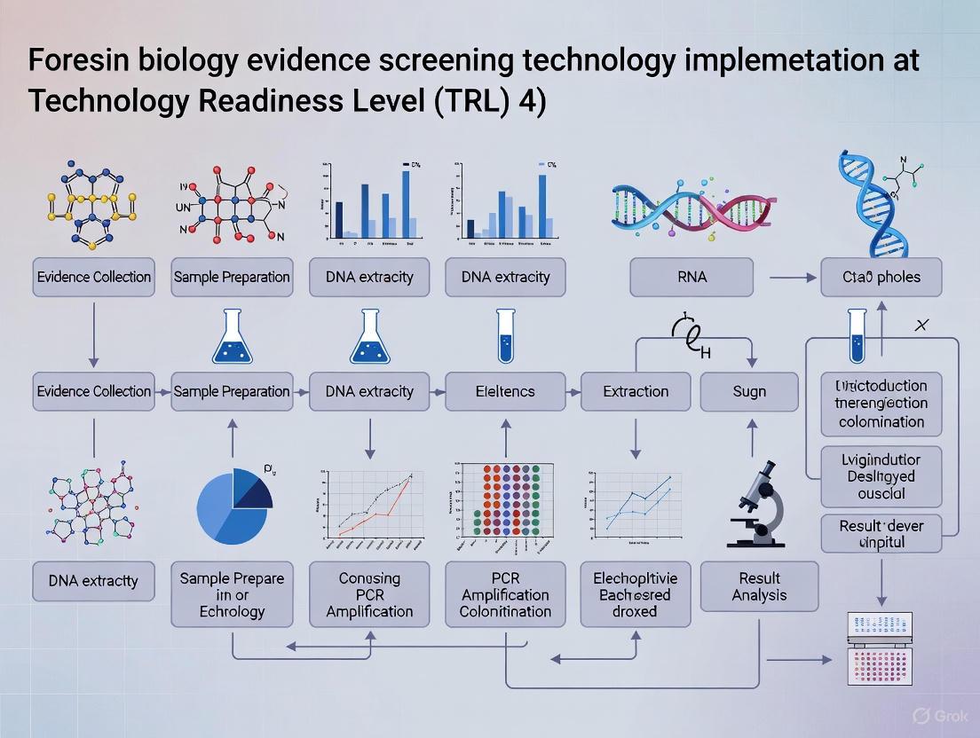
The following diagram illustrates the complete research, development, and implementation pathway for forensic biology evidence screening technologies as guided by the NIJ Strategic Research Plan:
Technology Development Pathway: This workflow illustrates the evidence screening technology development pathway from need identification through impact assessment, showing how NIJ's strategic priorities guide research activities at each stage while maintaining feedback mechanisms for continuous improvement.
The Scientist's Toolkit: Research Reagent Solutions for Evidence Screening
Table 3: Essential Research Reagents and Materials for Evidence Screening Technology Development
| Reagent/Material | Function/Application | Implementation Considerations |
|---|---|---|
| Magnetic Bead-based Extraction Kits | DNA purification from complex biological matrices; Integration with microfluidic systems | Enables automation; Reduces manual handling contamination; Improves yield from low-template samples |
| Microfluidic Chip Platforms | Miniaturized DNA analysis; Portable forensic technology development | Enables rapid, on-site DNA extraction; Reduces reagent consumption; Requires specialized instrumentation |
| Stable Fluorescent Dyes & Tags | Evidence visualization; Body fluid identification and differentiation | Enables non-destructive testing; Must maintain evidence integrity for subsequent DNA analysis |
| Probabilistic Genotyping Software | Complex mixture interpretation; Statistical weight of evidence calculation | Requires extensive validation; Computational resource intensive; Training essential for proper use |
| Reference DNA Standards | Method validation; Quality control; Instrument calibration | Essential for quantitative assays; Must represent diverse population groups; Enables inter-laboratory comparison |
| Rapid DNA Amplification Kits | Field-deployable DNA analysis; Expedited processing workflows | Reduced processing time; May have limitations with complex or degraded samples |
| Surface Sampling Devices | Efficient DNA recovery from various evidence substrates | Material composition affects DNA adsorption/release; Standardization needed across evidence types |
| Stabilization Buffers & Preservation Media | Maintain DNA integrity during storage and transport | Critical for field collections; Temperature stability requirements vary; Affects downstream analysis |
Strategic Integration with Forensic Biology Evidence Screening
The NIJ Strategic Research Plan provides a comprehensive framework for advancing forensic biology evidence screening technologies through coordinated research activities across multiple domains. For researchers and implementers, understanding the interconnected nature of these strategic priorities is essential for developing technologies that are not only scientifically sound but also operationally viable.
The emphasis on workforce development within Strategic Priority IV ensures that the implementation of new evidence screening technologies includes appropriate training, competency assessment, and continuing education programs [1]. This human factor is critical for successful technology transition, as even the most advanced screening platforms require skilled operators to generate reliable, interpretable results. Similarly, Strategic Priority V's focus on coordination across communities of practice facilitates the information sharing and collaborative partnerships necessary for standardized implementation of new screening technologies across diverse laboratory environments [1].
For forensic biology evidence screening technology implementation specifically, the NIJ plan addresses critical needs such as methods to associate cell type with DNA profile, technologies to improve DNA recovery, and approaches to optimize DNA processing workflows [3]. These directed research priorities provide clear guidance for developers and implementers regarding the operational challenges most needing innovative solutions. By aligning evidence screening technology development with these strategically identified needs, researchers can maximize the impact and adoption potential of their work.
Defining Technology Readiness Levels (TRL) in a Forensic Biology Context
Technology Readiness Levels (TRL) are a systematic metric used to assess the maturity level of a particular technology. The scale consists of nine levels, with TRL 1 being the lowest (basic principles observed) and TRL 9 being the highest (actual system proven in operational environment) [4]. This standardized approach provides a common framework for researchers, developers, and funding agencies to communicate about technological development status. Originally developed by NASA, the TRL framework has been widely adopted across multiple sectors including aerospace, energy, and healthcare [5]. In forensic biology, implementing new evidence screening technologies requires careful navigation through each TRL stage to ensure reliability, validity, and eventual admissibility in legal proceedings [6].
The forensic science landscape is undergoing significant transformation, with forensic biology laboratories increasingly processing complex biological evidence while facing heightened scrutiny regarding scientific integrity and evidence reliability [7]. This environment makes the TRL framework particularly valuable for guiding the development and implementation of new technologies in a manner that satisfies both scientific and legal standards. The adoption of emerging technologies in forensic biology—such as next-generation sequencing (NGS), rapid DNA analysis, and artificial intelligence-driven workflows—must be carefully managed through structured readiness assessments to meet the rigorous standards required for courtroom evidence [6] [8].
TRL Framework Adaptation for Forensic Biology
The generalized TRL scale requires contextual adaptation for forensic biology applications to address field-specific requirements, including validation standards, contamination control, and legal admissibility considerations. The table below outlines a tailored TRL framework for forensic biology evidence screening technologies.
Table 1: Technology Readiness Levels Adapted for Forensic Biology Context
| TRL | Definition | Forensic Biology Specific Criteria | Validation Requirements |
|---|---|---|---|
| 1 | Basic principles observed and reported | Literature review of fundamental biological principles; identification of potential forensic markers | Review of scientific knowledge base; assessment of foundational research [9] |
| 2 | Technology concept formulated | Practical application of principles to forensic scenarios; initial hypothesis for evidence screening | Concept generation; development of experimental designs [9] |
| 3 | Analytical and experimental proof-of-concept | Laboratory studies with controlled samples; initial demonstration of forensic applicability | Characterization of preliminary candidates; feasibility demonstration [9] |
| 4 | Component validation in laboratory environment | Basic forensic components integrated; testing with mock biological evidence | Optimization for assay development; finalization of critical design requirements [9] |
| 5 | Component validation in relevant environment | Breadboard system tested with forensically relevant samples; integration of key subsystems | Product development of reagents, components, and subsystems; pilot scale manufacturing preparations [9] |
| 6 | System model demonstration in relevant environment | Prototype testing with authentic case-type samples; evaluation in simulated forensic laboratory | System integration and testing with alpha/beta instruments; pilot lot production [4] [9] |
| 7 | Prototype demonstration in operational environment | Working prototype demonstrated in forensic laboratory setting; comparison with standard methods | Analytical verification with contrived and retrospective samples; preparation for clinical/validation studies [9] |
| 8 | Actual system completed and qualified | Technology validated for specific forensic applications; establishment of standard operating procedures | Clinical studies/evaluation; FDA clearance/approval for diagnostic components; finalization of GMP manufacturing [9] |
| 9 | Actual system proven in operational setting | Routine use in casework; successful challenge in legal proceedings; integration into quality management systems | Actual technology proven through successful deployment in operational setting [4] |
Experimental Protocols for TRL Advancement in Forensic Biology
Protocol for TRL 3-4 Transition: Proof-of-Concept Validation
Objective: To establish analytical proof-of-concept for a novel forensic biology screening technology using controlled samples and laboratory conditions.
Materials and Reagents:
- Reference DNA standards (purified human genomic DNA)
- Mock biological samples (saliva, blood, touch DNA samples on various substrates)
- Extraction and purification kits (magnetic bead-based systems recommended)
- Positive and negative control materials
- All necessary buffers and solutions
Procedure:
- Prepare dilution series of reference DNA standards covering expected forensic range (0.1-50 ng/μL)
- Spike mock samples with known quantities of DNA standards
- Process samples through the proposed screening technology following manufacturer's instructions
- Analyze results for sensitivity, specificity, and reproducibility
- Compare results with current standard methods (e.g., conventional PCR, electrophoresis)
- Perform statistical analysis to determine significant differences (p < 0.05 considered significant)
- Document all procedures, results, and observations in controlled laboratory notebooks
Acceptance Criteria:
- Technology demonstrates ability to detect biological material at forensically relevant concentrations
- False positive rate < 5% in negative controls
- Coefficient of variation < 15% for replicate analyses
Protocol for TRL 5-6 Transition: Simulated Environment Testing
Objective: To validate technology components in a simulated forensic environment using forensically relevant samples and conditions.
Materials and Reagents:
- Authentic-type forensic samples (bloodstains, saliva swabs, touched objects)
- Environmental challenged samples (UV-exposed, humidified, heated)
- Inhibitor-containing samples (soil, dye, humic acid)
- Miniaturized extraction kits for field deployment
- Portable power sources and environmental control equipment
Procedure:
- Collect authentic-type samples using standard forensic collection techniques
- Subject sample subsets to environmental challenges reflecting casework conditions
- Process samples using the proposed technology in a simulated operational environment
- Incorporate intentional contamination controls to assess robustness
- Train multiple operators with varying experience levels to assess usability
- Compare results with laboratory-based standard methods
- Assess ease of use, processing time, and required technical expertise
- Document any procedural challenges or failure modes
Acceptance Criteria:
- Technology performs reliably with environmentally challenged samples
- Multiple operators can achieve consistent results with minimal training
- Technology demonstrates superiority or equivalence to existing methods in at least one key parameter
Protocol for TRL 7-8 Transition: Operational Environment Qualification
Objective: To qualify the complete technology system in an operational forensic laboratory environment following established quality standards.
Materials and Reagents:
- Casework-type samples (blind-coded)
- ISO/IEC 17025 compliant quality control materials
- Documentation systems for chain of custody
- Reference standard methods and equipment
- Proficiency test materials
Procedure:
- Establish technology within accredited forensic laboratory workflow
- Process blind-coded casework-type samples alongside routine casework
- Implement full chain-of-custody documentation and evidence handling procedures
- Conduct comparative analysis with standard methods using statistical equivalence testing
- Perform robustness testing under varying laboratory conditions
- Validate according to SWGDAM guidelines or equivalent international standards
- Assess data integration with laboratory information management systems (LIMS)
- Document all validation data for regulatory submission purposes
Acceptance Criteria:
- Technology meets all relevant forensic validation standards (SWGDAM, ENFSI, etc.)
- Successful integration with laboratory quality management system
- Demonstration of fitness-for-purpose for intended forensic applications
Technology Development Workflow
The following diagram illustrates the complete technology development pathway from basic research to operational implementation in forensic biology.
Technology Development Pathway in Forensic Biology
Forensic Biology Research Reagent Solutions
The successful development and implementation of forensic biology technologies requires specific research reagents and materials tailored to evidentiary applications. The table below details essential solutions for technology development across TRL stages.
Table 2: Key Research Reagent Solutions for Forensic Biology Technology Development
| Reagent/Material | Function | TRL Application Range | Forensic-Specific Considerations |
|---|---|---|---|
| Reference DNA Standards | Quantification calibration and method comparison | TRL 3-9 | Should include degraded DNA and low-copy number samples to mimic forensic evidence [8] |
| Mock Biological Samples | Controlled testing without evidentiary constraints | TRL 3-6 | Should include blood, saliva, semen, and touch DNA on various substrates [8] |
| Inhibitor Panels | Assessment of inhibition resistance | TRL 4-7 | Should include common forensic inhibitors: hematin, humic acid, tannins, dyes [8] |
| Stabilization Buffers | Preservation of biological integrity during testing | TRL 5-9 | Must maintain DNA stability under various storage conditions [8] |
| Rapid Extraction Kits | Efficient DNA recovery from complex substrates | TRL 5-9 | Should be optimized for minimal hands-on time and maximum yield from trace samples [8] |
| Portable Detection Kits | Field-based screening and analysis | TRL 6-9 | Must include environmental controls and user-friendly interface [8] |
| Quality Control Materials | Validation and proficiency testing | TRL 7-9 | Should be traceable to international standards and commutabile with casework samples [7] |
| LIMS Integration Modules | Data management and chain of custody | TRL 7-9 | Must comply with forensic standards for data integrity and security [7] |
Legal and Regulatory Considerations for TRL Progression
Advancement through TRL stages in forensic biology requires careful attention to legal admissibility standards alongside technical validation. Courtroom admissibility standards including the Frye Standard, Daubert Standard, Federal Rule of Evidence 702 in the United States, and the Mohan Criteria in Canada establish rigorous requirements for scientific evidence [6]. These standards demand that forensic technologies demonstrate reliability, known error rates, peer review acceptance, and standardized protocols—factors that must be addressed throughout TRL progression.
The transition from TRL 7 to TRL 8 particularly requires thorough documentation of validation studies, error rate analysis, and proficiency testing to satisfy these legal standards [6]. Technologies intended for forensic implementation must also integrate with established quality management systems, typically based on ISO/IEC 17025 standards for forensic testing laboratories [7]. This integration includes implementing appropriate chain-of-custody procedures, evidence tracking systems, and casework documentation protocols that will withstand legal scrutiny.
The structured framework of Technology Readiness Levels provides an essential roadmap for developing and implementing novel technologies in forensic biology. By systematically addressing technical, operational, and legal requirements at each TRL stage, researchers and developers can effectively advance promising technologies from basic concept to court-admissible applications. The specialized protocols, validation criteria, and reagent solutions outlined in this document offer practical guidance for navigating this complex pathway while maintaining scientific rigor and legal compliance. As forensic biology continues to evolve with emerging technologies such as next-generation sequencing, rapid DNA analysis, and artificial intelligence applications, the TRL framework will remain crucial for ensuring that innovation translates into reliable, validated forensic practice.
Within the framework of Technology Readiness Level (TRL) research for implementing novel forensic biology evidence screening technologies, establishing foundational validity and reliability is a critical precursor to admissibility and widespread adoption. The forensic science community faces increasing scrutiny regarding the scientific underpinnings of its methods, particularly for non-DNA evidence [10]. Courts, guided by standards such as those from the Daubert ruling, require that expert testimony be based on scientifically valid methods with known error rates and adherence to established standards [10]. The National Institute of Justice (NIJ) explicitly identifies "Foundational Validity and Reliability of Forensic Methods" as a strategic priority, emphasizing the need to understand the fundamental scientific basis of forensic disciplines and to quantify measurement uncertainty [1]. This document outlines application notes and experimental protocols designed to address these core needs, providing a structured pathway for researchers to rigorously evaluate new forensic biology screening technologies before they are deployed in the criminal justice system.
Application Notes: Quantitative Frameworks for Evidence Analysis
The evolution from purely qualitative assessments to integrated quantitative and statistical frameworks is central to modern forensic biology. The following application notes summarize key methodologies and data for establishing the validity and reliability of biological evidence screening.
Analytical Techniques for Qualitative and Quantitative Analysis
Forensic analysis often involves a tandem approach, where qualitative analysis identifies the presence or absence of specific substances, and subsequent quantitative analysis determines the concentration or amount of those substances [11]. This is crucial in forensic biology for applications such as DNA quantitation, toxicology, and body fluid identification.
Table 1: Common Analytical Techniques in Forensic Biology
| Technique | Primary Use in Forensic Biology | Qualitative/Qantitative Application | Key Metric |
|---|---|---|---|
| Massively Parallel Sequencing (MPS) | STR/SNP sequencing, mixture deconvolution, body fluid ID [12] | Both | Detects sequence variation in alleles; high discrimination power [12] |
| Quantitative PCR (qPCR) | DNA quantitation, body fluid identification via RNA analysis [12] | Quantitative | Measures DNA concentration; determines presence of body fluid-specific RNA [13] [12] |
| Liquid Chromatography-Mass Spectrometry (LC-MS) | Drug detection, biomarker identification [11] [14] | Both (Confirmatory & Quantitative) | Identifies and quantifies compounds based on mass-to-charge ratio |
| Immunochromatography | Presumptive tests for drugs, specific proteins [14] | Qualitative | Rapid, visual identification of target substances |
| Probabilistic Genotyping | DNA mixture interpretation [12] | Quantitative (Statistical) | Calculates Likelihood Ratios (LR) to weigh evidence [12] |
Performance Metrics for Technology Validation
Evaluating a new forensic technology requires assessing its performance against benchmark standards. The following metrics are essential for establishing validity and reliability.
Table 2: Key Validation Metrics for Forensic Screening Technologies
| Performance Metric | Definition | Target for New Technology | Experimental Protocol Reference |
|---|---|---|---|
| Sensitivity | The minimum amount of target analyte (e.g., DNA, specific RNA) that can be reliably detected. | ≤10 pg DNA for advanced methods [12] | Protocol 3.1 |
| Specificity | The ability to distinguish the target analyte from similar, non-target substances. | >95% for body fluid-specific mRNA markers [12] | Protocol 3.2 |
| Accuracy/Precision | The closeness of agreement between a measured value and a true value (accuracy), and the repeatability of measurements (precision). | CV < 10% for quantitative measurements | Protocol 3.3 |
| Stochastic Threshold | The DNA quantity below which allele drop-out due to random effects becomes significant. | Determined empirically via validation studies [12] | Protocol 3.1 |
| Error Rate | The frequency of false positives and false negatives in controlled tests. | Must be established and reported for the method and lab [10] | Protocol 3.4 |
| Limit of Detection (LOD) | The lowest concentration of an analyte that can be consistently identified. | Defined by statistical models from validation data | Protocol 3.1 |
Experimental Protocols
Protocol for Determining Sensitivity and Stochastic Threshold
Objective: To establish the minimum quantity of DNA that can be reliably amplified and profiled using a specific screening technology (e.g., STR kit, MPS panel), and to determine the stochastic threshold.
- Sample Preparation: Serially dilute a control DNA standard of known concentration (e.g., 1:10 dilutions from 1 ng/µL to 0.5 pg/µL).
- Replication: For each dilution level, prepare a minimum of n=10 replicates to account for stochastic effects.
- Amplification & Analysis: Process all replicates through the entire analytical workflow (extraction, quantitation, amplification, separation/sequencing).
- Data Collection: Record the following for each replicate:
- Whether a full, partial, or no profile was obtained.
- The number of alleles detected versus expected.
- Peak heights (for CE) or read depths (for MPS).
- Presence of allelic drop-in.
- Data Analysis:
- Sensitivity (LOD): Plot the profile success rate (%) against the input DNA quantity. The LOD is typically the lowest quantity where ≥95% of replicates produce a usable profile.
- Stochastic Threshold: Calculate the average peak height/read depth of heterozygous alleles at each dilution. The stochastic threshold is the peak height/read depth value below which heterozygous balance falls outside an acceptable range (e.g., <60%) and allelic drop-out is observed in >5% of replicates.
Protocol for Assessing Specificity and Cross-Reactivity
Objective: To verify that the screening technology detects only the intended target (e.g., a specific body fluid, drug metabolite) and does not cross-react with common interferents.
- Test Panel Creation: Assemble a panel of samples including:
- Positive Controls: Pure, known target substances.
- Common Interferents: Biologically relevant substances that may be present at a crime scene (e.g., other body fluids, soil, cleaning agents, common pharmaceuticals).
- Mixtures: Samples containing the target mixed with interferents in various ratios.
- Negative Controls: Samples known to lack the target (e.g., water).
- Blinded Analysis: Code the samples and analyze them using the screening technology under validation.
- Data Collection & Scoring: Record the result (positive/negative, or quantitative value) for each sample.
- Data Analysis:
- Calculate the false positive rate (number of interferents incorrectly identified as target / total number of interferents).
- Calculate the false negative rate (number of positive controls incorrectly called negative / total number of positive controls).
- For quantitative assays, report the degree of signal suppression or enhancement caused by interferents in mixture samples.
Protocol for Intra- and Inter-Laboratory Reproducibility
Objective: To measure the precision (repeatability and reproducibility) of the screening technology within a single laboratory and across multiple laboratories.
- Sample & Standard Preparation: Prepare a set of homogeneous, stable reference samples or standards with known properties. These should span low, medium, and high concentrations of the target analyte.
- Intra-Laboratory Study (Repeatability):
- Within a single lab, have two or more analysts test each reference sample multiple times (e.g., n=5) over a short period (e.g., one day) using the same equipment and reagents.
- Calculate the standard deviation and coefficient of variation (CV) for the results from each sample level.
- Inter-Laboratory Study (Reproducibility):
- Distribute identical sets of the reference samples to a minimum of three participating laboratories.
- Each lab follows a standardized protocol (the one being validated) to analyze the samples.
- Each sample should be tested a minimum of n=3 times per lab.
- Statistical Analysis:
- Perform an analysis of variance (ANOVA) to partition the total variance into components representing between-lab and within-lab variability.
- Report the overall reproducibility standard deviation and the interlaboratory CV.
Protocol for Black Box Study to Estimate Error Rates
Objective: To empirically measure the false positive and false negative rates of a forensic feature-comparison method under realistic conditions [10].
- Study Design: Create a set of trial specimens that includes:
- True Matches: Pairs of samples known to originate from the same source.
- True Non-Matches: Pairs of samples known to originate from different sources.
- The prevalence of matches and non-matches should not be 50:50 but should reflect a more realistic, challenging ratio.
- Blinding & Presentation: The set of trials is presented to examiners in a "black box" format where they have no contextual information about the cases. The order of trials is randomized.
- Examination & Conclusion: Each examiner compares the pairs and selects a conclusion from a predefined scale (e.g., Identification, Inconclusive, Exclusion).
- Data Analysis:
- False Positive Rate (FPR): (Number of incorrect "Identification" decisions on true non-match pairs / Total number of true non-match pairs).
- False Negative Rate (FNR): (Number of incorrect "Exclusion" decisions on true match pairs / Total number of true match pairs).
- Report rates with confidence intervals to quantify uncertainty.
Visualizing Workflows and Logical Frameworks
Foundational Research Workflow
Evidence Evaluation Logic
The Scientist's Toolkit: Essential Research Reagents & Materials
Table 3: Key Reagents and Materials for Foundational Validation Studies
| Item | Function in Research & Development |
|---|---|
| Standard Reference Material (SRM) | Provides a material with certified properties (e.g., DNA concentration, sequence) for calibrating instruments, assessing accuracy, and ensuring consistency across experiments and laboratories [1]. |
| Control DNA | Used in sensitivity, stochastic, and reproducibility studies to monitor assay performance and distinguish true results from artifacts. Includes male/female, single-source, and mixture controls. |
| Characterized Body Fluid Samples | Saliva, blood, semen, etc., samples collected under IRB approval and used as positive controls and knowns for developing and validating body fluid identification assays [12]. |
| Probabilistic Genotyping Software | Sophisticated software that uses statistical models to interpret complex DNA mixtures, accounting for stutter, drop-out, and drop-in, and providing a Likelihood Ratio to weigh the evidence [12]. |
| MPS Platform & Kits | Enables high-throughput, sequence-based analysis of multiple marker types (STRs, SNPs) from a single sample, increasing discriminatory power and aiding in the analysis of challenging samples [12]. |
| Inhibitor Stocks | Purified substances (e.g., humic acid, hematin, tannin) that are added to samples during validation to test the robustness of the DNA extraction and amplification process to common PCR inhibitors. |
| Synthetic Oligonucleotides | Custom-designed DNA sequences used as positive controls for qPCR assays, as components in multiplex assay design, and for creating synthetic DNA mixtures with precisely known genotypes. |
Microbiome analysis represents a paradigm shift in forensic science, moving beyond traditional human DNA analysis to exploit the unique microbial communities associated with human bodies and environmental samples. The microbiome comprises all microorganisms—bacteria, fungi, viruses, and their genes—inhabiting a specific environment [15]. In forensics, this approach provides investigative leads for individual identification, geolocation inference, and post-mortem interval (PMI) estimation [15]. The field has evolved significantly from culture-dependent techniques to next-generation sequencing (NGS) technologies, enabling comprehensive characterization of microbial communities without the limitations of laboratory cultivation [15] [16].
Key Analytical Methods in Forensic Microbiome Research
Sequencing Technologies
Two primary NGS methods are employed in forensic microbiome analysis, each with distinct advantages and limitations for forensic applications:
Table 1: Comparison of Microbiome Sequencing Methods
| Feature | Amplicon Sequencing | Shotgun Metagenomic Sequencing |
|---|---|---|
| Target | Marker genes (e.g., 16S rRNA, ITS) [15] | All genomic DNA in sample [15] |
| Resolution | Genus to species level [15] | Species to strain level [15] |
| Cost | Lower [15] | Higher [15] |
| Suitable for Low-Biomass | Better performance [15] | Poorer performance [15] |
| Information Output | Taxonomic composition [15] | Taxonomic & functional gene information [15] |
| Common Platforms | Illumina MiSeq, Oxford Nanopore, PacBio [15] | Illumina platforms [15] |
Bioinformatics Pipelines
Data analysis pipelines are critical for converting raw sequencing data into forensically actionable information. The bioinformatics workflow for amplicon sequencing typically involves:
- Sequence Denoising: Using algorithms like DADA2 or Deblur to generate exact amplicon sequence variants (ASVs), which are more precise than traditional operational taxonomic units (OTUs) [15].
- Taxonomic Classification: Assigning taxonomy to ASVs using reference databases such as SILVA, Greengenes, or UNITE [15].
- Statistical Analysis: Multivariate statistical methods to identify discriminatory microbial signatures for forensic applications.
For shotgun metagenomic data, analysis includes:
- Quality Control & Host Depletion: Using tools like KneadData and Trimmomatic to remove low-quality sequences and host DNA contamination [15].
- Taxonomic Profiling: Utilizing tools like MetaPhlAn2 or Kraken 2 for comprehensive taxonomic assignment [15].
- Functional Profiling: Analysis with tools like HUMAnN3 to determine microbial metabolic capabilities, which may provide additional contextual information [15].
Figure 1: Bioinformatics Workflow for Forensic Microbiome Analysis
Experimental Protocols
Sample Collection and Preservation
Proper sample collection is critical for reliable microbiome analysis. Forensic samples may include:
- Skin swabs: Collected from palmar surfaces using sterile swabs moistened with molecular-grade water [16].
- Soil samples: Collected from potential crime scenes at depths of 0-5 cm using sterile corers [16].
- Body fluids: Vaginal fluid, saliva, and fecal material collected with sterile swabs or containers [16].
- Decomposition samples: Swabs from skin, soil beneath remains, or internal organs during autopsy [16].
Preservation Protocol: Immediately after collection, samples should be placed in DNA/RNA stabilization buffer and stored at -80°C to prevent microbial community shifts. Storage duration and conditions should be standardized and documented, as variations can affect bacterial community analysis [16].
DNA Extraction and Library Preparation
DNA Extraction:
- Use mechanical lysis (bead beating) combined with chemical lysis for comprehensive cell disruption.
- Employ commercial kits (e.g., DNeasy PowerSoil Kit) optimized for environmental samples.
- Include extraction controls to monitor contamination.
- Quantify DNA yield using fluorometric methods (e.g., Qubit).
16S rRNA Amplicon Library Preparation:
- Amplify the V4 hypervariable region of the 16S rRNA gene using primers 515F/806R.
- Attach dual-index barcodes and Illumina sequencing adapters.
- Clean amplified libraries using magnetic bead-based purification.
- Quantify libraries and pool in equimolar ratios.
- Validate library quality using capillary electrophoresis.
Shotgun Metagenomic Library Preparation:
- Fragment genomic DNA to ~350 bp using acoustic shearing.
- Perform end repair, A-tailing, and adapter ligation.
- Size-select fragments using magnetic beads.
- Amplify library with index primers.
- Quantify and normalize libraries for sequencing.
Sequencing and Data Analysis
Sequencing Parameters:
- Illumina MiSeq: 2×250 bp for 16S rRNA amplicons
- Illumina NovaSeq: 2×150 bp for shotgun metagenomics
Bioinformatics Analysis Protocol:
- Quality Filtering: Remove low-quality reads (Q-score <20) and trim adapters.
- Denoising: For 16S data, use DADA2 to infer exact amplicon sequence variants (ASVs).
- Taxonomic Assignment: Classify sequences against the SILVA database for 16S data or RefSeq for shotgun data.
- Contamination Removal: Identify and remove potential contaminants using the decontam package (R).
- Statistical Analysis: Perform alpha and beta diversity analyses, differential abundance testing, and machine learning classification for forensic applications.
Forensic Applications and Validation
Individual Identification
The human skin microbiome is highly personalized and stable over time, making it valuable for associating individuals with objects or locations [15] [16]. Key findings include:
- Individuals leave unique microbial fingerprints on surfaces like computer keyboards and phones [15].
- Microbial signatures can persist for weeks to months on touched objects [15].
- Cohabiting individuals develop similar microbial profiles, enabling relationship inference [15].
Post-Mortem Interval (PMI) Estimation
Thanatomicrobiome (microbes inhabiting the body after death) and epinecrotic communities (microbes on the body surface) follow predictable successional patterns that correlate with time since death [16]. Research demonstrates:
- Distinct microbial community shifts occur at specific decomposition stages [16].
- Soil microbiota beneath decomposing remains changes predictably [16].
- Machine learning models using microbial markers can estimate PMI with increasing accuracy [16].
Body Fluid Identification
Microbial signatures can differentiate body fluids when conventional methods are inconclusive:
- Vaginal fluid: Characterized by Lactobacillus crispatus and Lactobacillus jensenii [16].
- Saliva: Identified by Streptococcus salivarius and other oral commensals [16].
- Feces: Distinguished by Bacteroides species and other gut microbiota [16].
Geolocation Inference
Soil microbiomes exhibit biogeographical patterns that can associate samples with specific regions [15] [16]. Applications include:
- Linking suspects to crime scenes through soil microbial signatures [15].
- Determining the origin of illicit materials based on environmental microbiomes [15].
- Regional identification using distinctive microbial taxa [15].
Table 2: Technology Readiness Levels for Forensic Microbiome Applications
| Application | Current TRL | Key Validation Needs | Legal Considerations |
|---|---|---|---|
| Individual Identification | 3-4 (Experimental Proof) | Standardized protocols, error rate analysis, population studies [3] | Meets Daubert standards for reliability and peer review with further validation [6] |
| PMI Estimation | 3 (Analytical Formulation) | Succession model validation across environments, precision estimates [3] | Requires known error rates and general acceptance in forensic pathology [6] |
| Geolocation | 2-3 (Technology Concept) | Comprehensive spatial databases, statistical models for probability [3] | Necessitates demonstration of reliability under varying environmental conditions [6] |
| Body Fluid ID | 4 (Lab Validation) | Multi-center validation studies, mixture interpretation protocols [3] | Must establish specificity and sensitivity thresholds for courtroom admissibility [6] |
Figure 2: Technology Readiness Level Framework for Forensic Implementation
The Scientist's Toolkit: Research Reagent Solutions
Table 3: Essential Research Reagents for Forensic Microbiome Analysis
| Reagent/Kit | Function | Application Notes |
|---|---|---|
| DNeasy PowerSoil Pro Kit | DNA extraction from soil and difficult samples | Optimal for inhibitor removal; includes bead beating for cell lysis [15] |
| Illumina 16S Metagenomic Sequencing Library Preparation Reagents | 16S rRNA amplicon library prep | Includes primers targeting hypervariable regions; compatible with dual indexing [15] |
| ZymoBIOMICS Microbial Community Standard | Positive control for sequencing runs | Validates entire workflow from extraction to analysis; quantifies technical variation [15] |
| Qubit dsDNA HS Assay Kit | Accurate DNA quantification | Fluorometric method preferred over spectrophotometry for low-biomass samples [15] |
| DADA2 (R package) | Amplicon sequence variant inference | More exact than OTU clustering; reduces spurious sequence variants [15] |
| MetaPhlAn2 | Taxonomic profiling from shotgun data | Species-level resolution using clade-specific marker genes [15] |
| SILVA Database | 16S rRNA reference database | Curated alignment and taxonomy; regularly updated [15] |
Current Challenges and Future Directions
Despite its potential, forensic microbiome analysis faces several challenges that must be addressed for routine implementation:
- Standardization: Lack of standardized protocols for sample collection, storage, and analysis [16].
- Validation: Need for established error rates, reproducibility metrics, and population-specific databases [16] [3].
- Bioinformatics: Requirement for user-friendly, validated bioinformatics pipelines accessible to forensic laboratories [15].
- Legal Admissibility: Meeting Daubert standards (testing, peer review, error rates, acceptance) and Mohan criteria (relevance, necessity, reliability) for courtroom evidence [6].
Future development should focus on:
- Reference Databases: Expanding curated microbial databases for forensic applications [3].
- Quantitative Models: Developing statistical frameworks for evaluating evidence weight [3].
- Integration: Combining microbiome data with other forensic markers for stronger associations [3].
- Rapid Detection: Creating field-deployable tools for preliminary microbiome screening [3].
The Critical Role of Reference Collections and Databases for Method Development
In forensic biology, the implementation of new evidence screening technologies relies on robust method development and validation. Central to this process are forensic reference collections and databases, which provide the foundational materials and data necessary to develop, optimize, and validate analytical methods. These resources enable the transition of technologies from basic research to operational use by providing authenticated materials for testing and comparison. The National Institute of Justice (NIJ) emphasizes this need through its Research and Development Technology Working Group, which has identified specific operational requirements where improved databases and reference materials would significantly advance forensic capabilities [3]. This document outlines the critical function of these resources within the technology readiness level (TRL) framework for forensic biology evidence screening.
Forensic reference collections encompass both physical samples and digital data records that serve as benchmarks for analytical method development. These resources provide known materials against which unknown evidentiary samples can be compared, ensuring analytical accuracy and reliability. The following table summarizes key forensic databases and their characteristics relevant to method development.
Table 1: Key Forensic Reference Databases and Collections
| Database/Collection Name | Maintaining Organization | Primary Content | Scale of Collection | Access Method |
|---|---|---|---|---|
| International Forensic Automotive Paint Data Query (PDQ) [17] | Royal Canadian Mounted Police (RCMP) | Chemical and color information of original automotive paints | ~13,000 vehicles; ~50,000 paint layers | Annual CD release to member law enforcement agencies |
| FBI Lab - Forensic Automobile Carpet Database (FACD) [17] | Federal Bureau of Investigations (FBI) | Carpet fibers from vehicles; microscopic characteristics, IR microscopy | ~800 samples | Request-based analysis by FBI for law enforcement |
| European Collection of Automotive Paints (EUCAP) [17] | French Police Lab (Germany BKA handles data analysis) | Paint samples from salvage vehicles | Not Specified | Internet access with secure ID and password |
| FBI Lab - Fiber Library [17] | Federal Bureau of Investigations (FBI) | FTIR spectra of textile fibers; physical fiber samples | 86 spectral records | Analysis request to FBI; library in OMNIC software |
| Barcode of Life Database (BOLD) [18] | University of Guelph, Canada | DNA sequences for species identification | 1.8M+ specimen records; 111,289+ species with barcodes | Free online access with optional login |
These resources address the critical need for authenticated reference materials that underpin reliable method development. As noted by NIJ's research priorities, there remains a pressing need for "additional characterization of existing databases and further development of population data of forensically relevant genetic markers" particularly for "populations that are currently underrepresented in existing databases" [3]. This is especially true in forensic biology, where population genetics statistics are essential for calculating evidential weight.
Quantitative Analysis of Database Utility
The effectiveness of reference databases in method development can be quantitatively assessed through parameters such as sample diversity, analytical precision, and discrimination power. The following table summarizes key quantitative metrics for evaluating database utility in forensic method development.
Table 2: Quantitative Metrics for Database Utility in Method Development
| Performance Parameter | Application in Method Development | Target Threshold | Impact on Technology Implementation |
|---|---|---|---|
| Sample Diversity | Assessing method robustness across different sample types | Comprehensive population coverage | Reduces false negatives in evidence screening |
| Discrimination Power | Evaluating method's ability to distinguish between sources | >0.99 for highly similar materials | Ensures evidentiary value of matches |
| Analytical Precision | Establishing reproducibility of measurements | CV <5-15% depending on analyte | Validates repeatability across instruments and operators |
| Limit of Detection (LOD) | Determining minimum detectable quantity of target | Substance-dependent (e.g., ng/mL for drugs) | Defines application range for trace evidence |
| False Positive Rate | Validating method specificity | <1% for confirmatory methods | Ensures reliability in casework applications |
Quantitative data analysis methods, including descriptive analysis and diagnostic analysis, are essential for interpreting these performance parameters [19]. For instance, statistical analysis of database records helps establish appropriate match thresholds and confidence intervals for new screening technologies. The move toward probabilistic genotyping for DNA mixture interpretation exemplifies how database-driven statistical models are transforming forensic biology [3].
Experimental Protocols for Method Validation
Protocol 1: Method Development Lifecycle for Forensic Screening
The development of analytical methods follows a structured lifecycle to ensure reliability and admissibility in legal contexts. This protocol adapts the pharmaceutical industry's rigorous approach to method development for forensic applications [20].
1. Requirements Identification
- Define the analytical need based on operational gaps (e.g., detecting new synthetic drugs)
- Review existing/compendial procedures for adaptability
- Establish target performance characteristics (specificity, sensitivity, precision)
2. Method Development
- Select appropriate analytical techniques (e.g., LC-MS/MS, GC×GC-MS)
- Optimize instrument parameters through systematic testing
- Establish sample preparation procedures
- Develop preliminary acceptance criteria
3. Validation Planning & Execution
- Prepare a validation protocol defining experiments and acceptance criteria
- Evaluate specificity, limit of detection, limit of quantitation, linearity, accuracy, range, and precision
- Document all procedures and results thoroughly
4. Implementation & Monitoring
- Develop Standard Operating Procedures (SOPs) for routine execution
- Define criteria for revalidation and system suitability tests
- Implement continuous monitoring for fitness-for-purpose
5. Technology Transfer
- Qualify receiving laboratories through comparative testing
- Document transfer under pre-approved protocol
- Ensure procedural knowledge is effectively communicated
This structured approach aligns with international guidelines from ICH, FDA, and EMA, adapted for forensic contexts [20]. The process emphasizes documentation rigor and regulatory compliance, which are equally essential in forensic science where evidence must withstand legal challenges.
Protocol 2: GC×GC-MS for Complex Mixture Analysis in Trace Evidence
Comprehensive two-dimensional gas chromatography-mass spectrometry (GC×GC-MS) provides enhanced separation for complex forensic samples. This protocol details its application for trace evidence analysis, based on published methodologies [21].
Sample Preparation
- Lubricant Analysis: Conduct hexane solvent extraction of suspected lubricant samples (50-100 µL)
- Paint/Polymer Analysis: Employ flash pyrolysis with Pyroprobe 4000: start at 50°C for 2s, ramp to 750°C at 50°C/s, hold for 2s
- Sample Size: Use minimal material (~50 µg) to preserve evidence integrity
Instrument Configuration
- GC System: Agilent 7890B gas chromatograph with split-splitless injector
- MS System: Agilent 5977 quadrupole mass spectrometer
- Columns: Combination of non-polar/moderately polar columns for two-dimensional separation
- Modulator: Differential flow modulation (DFM) for comprehensive 2D separation
- Temperature Program: Optimized gradient from 50°C to 280°C based on analyte properties
Data Acquisition & Analysis
- Mass Spectrometry Conditions: Electron ionization (EI) mode at 70 eV; mass range 35-550 m/z
- Data Processing: Use specialized software to generate 2D chromatographic "fingerprints"
- Pattern Recognition: Compare sample fingerprints to reference database entries
- Component Identification: Leverage increased peak capacity to resolve co-eluting compounds
This protocol demonstrates a significant advancement over traditional GC-MS, particularly for discriminating between sources of complex mixtures like sexual lubricants, automotive paints, and tire rubber [21]. The enhanced separation power addresses a key limitation in trace evidence analysis where coelution can prevent correct identification.
Research Reagent Solutions for Forensic Method Development
The following table details essential materials and reagents required for implementing advanced analytical methods in forensic biology evidence screening.
Table 3: Essential Research Reagents for Forensic Biology Method Development
| Reagent/Material | Function in Method Development | Application Examples | Critical Specifications |
|---|---|---|---|
| Authenticated Reference Standards | Method calibration and quantitative accuracy | Drug quantification, toxicology testing, DNA quantification | Purity >95%, concentration verification, stability data |
| DNA Extraction Kits | Recovery of amplifiable DNA from diverse evidence types | Low-copy number DNA, touch DNA, challenging substrates (e.g., metal) | Yield efficiency, inhibitor removal, compatibility with downstream analysis |
| Chromatography Columns | Separation of complex mixtures | GC×GC-MS for lubricants, paints; LC-MS/MS for drugs | Stationary phase selectivity, column efficiency, temperature stability |
| Mass Spectrometry Calibrants | Instrument calibration and mass accuracy confirmation | Daily MS system tuning, quantitative method validation | Known m/z ratios, chemical stability, compatibility with analysis mode |
| Population DNA Databases | Statistical assessment of evidentiary value | Probabilistic genotyping, mixture interpretation, kinship analysis | Representative population coverage, quality-controlled data, ethical compliance |
| Optical Glass Standards | Microscope calibration for physical evidence analysis | Refractive index measurements of glass fragments | Certified refractive index range (e.g., 1.34 to 2.40), traceable documentation |
These research reagents form the foundational toolkit for developing and implementing new screening technologies. Their quality directly impacts the reliability of analytical results and the eventual admissibility of evidence in legal proceedings. As highlighted by NIJ, there is a particular need for "improved DNA collection devices or methods for recovery and release of human DNA (e.g., from metallic items)" [3], demonstrating how reagent development directly addresses operational challenges.
Reference collections and databases serve as the cornerstone of method development in forensic biology evidence screening. They provide the essential materials for technology validation, performance assessment, and operational implementation. As analytical techniques evolve toward greater sensitivity and specificity, the role of comprehensive, well-characterized reference resources becomes increasingly critical. The ongoing development of these resources, particularly for emerging evidence types and underrepresented populations, will directly determine the success of new technology implementation across the forensic science community. Future advancements should focus on expanding database diversity, improving accessibility, and enhancing integration with statistical interpretation tools to maximize their impact on forensic method development.
Forensic laboratories worldwide are operating in an era of unprecedented technological transformation. They face growing pressure to implement advanced analytical methods while managing increasing case backlogs, complex evidence, and high expectations for expedited results. This application note details the current technological demands, provides validated experimental protocols for key forensic analyses, and visualizes core workflows to support researchers and scientists in the field of forensic biology evidence screening. The content is framed within the context of Technology Readiness Level (TRL) research to facilitate the implementation and maturation of emerging forensic technologies.
Market Dynamics and Quantitative Landscape
The global forensic technology market is experiencing significant growth, driven by technological advancements and increasing demand for reliable criminal identification procedures. The table below summarizes key quantitative data for the DNA forensics market, a critical segment of forensic laboratories' technological portfolio.
Table 1: Global DNA Forensics Market Projections and Segment Analysis
| Metric | Details |
|---|---|
| 2024 Market Value | $3.1 billion [22] |
| 2025 Projected Market Value | $3.3 billion [22] |
| 2030 Projected Market Value | $4.7 billion [22] |
| CAGR (2025-2030) | 7.7% [22] |
| Dominant Product Segment | Kits and Consumables [22] |
| Key Techniques | PCR, STR, Next-Generation Sequencing (NGS) [22] |
| Major Applications | Criminal Testing, Paternity & Familial Testing [22] |
| Regional Market Leader | North America ($1.1B in 2024) [22] |
This growth is fueled by several factors: rising crime rates necessitating accurate tools, increased government funding for forensic science, technological developments in testing, and the expansion of national DNA databases which now exist in over 70 countries [22]. Furthermore, the integration of artificial intelligence (AI) and machine learning into forensic processes is emerging as a key trend, enabling improved analysis and automation [22].
Key Technological Pressures and Advanced Protocols
Pressure: Demand for Rapid Results
Solution: Rapid DNA Integration
A significant pressure is the demand to reduce turnaround times. The Federal Bureau of Investigation (FBI) has approved the integration of Rapid DNA technology into the Combined DNA Index System (CODIS), effective July 1, 2025 [23]. This allows law enforcement to process DNA samples in hours instead of days or weeks and compare them directly to the national database.
Table 2: Research Reagent Solutions for DNA Quantitation via qPCR
| Item | Function |
|---|---|
| Quantitative PCR (qPCR) Kits | Provide optimized primers, probes, enzymes, and buffers for specific quantification of human DNA [24]. |
| Human DNA Quantitation Standard (e.g., NIST SRM 2372) | Serves as a quality control measure to assess the accuracy of quantitation results, ensuring data reliability [24]. |
| DNA Extraction Kits | Isolate and purify DNA from complex biological samples prior to quantitation and downstream analysis. |
| Fluorescent Dyes/Probes | Intercalate with or bind to DNA, emitting a fluorescent signal that is monitored during PCR cycles to measure amplification [24]. |
Protocol 1: Data Analysis for Quantitative PCR (qPCR) Purpose: To accurately determine the quantity and quality of human DNA in a forensic sample prior to downstream STR analysis [24].
Methodology:
- Amplification: The qPCR process is monitored in real-time via a fluorescent signal. The process occurs in three phases:
- Exponential Phase: Theoretical doubling of amplicons per cycle. The baseline fluorescence (background noise) is established, and a fluorescence threshold is set. The Cycle Threshold (CT) is the point at which a sample's fluorescence exceeds this threshold [24].
- Linear Phase: Reagent consumption impedes efficiency; this phase is not used for data analysis [24].
- Plateau Region: Amplification ceases due to critical reagent depletion [24].
- Standard Curve Generation: A regression line is derived by plotting the CT values of known DNA standards against the log of their concentrations [24].
- Sample Quantitation: The concentration of an unknown sample is determined by comparing its CT value against the standard curve. A lower CT indicates a higher initial DNA concentration [24].
The workflow for this protocol is logically sequenced below:
Pressure: Casework Backlog and Resource Allocation
Solution: AI-Powered Predictive Modeling
Forensic labs face substantial backlogs. AI and machine learning can transform case management through predictive modeling [25].
Protocol 2: AI for Case Prioritization and Resource Allocation Purpose: To leverage historical case data to predict processing times, prioritize evidence, and optimize resource allocation [25].
Methodology:
- Data Aggregation: Compile historical case data including evidence type, complexity, processing time, and analyst workload.
- Model Training: Train a machine learning model on this data to identify patterns and correlations between case characteristics and required resources.
- Prediction and Prioritization:
- Case Duration Estimation: Input characteristics of new cases to predict processing time, aiding in staffing and equipment planning [25].
- Evidence Prioritization: The model automatically scans and ranks incoming evidence based on its predicted investigative value, helping to prioritize high-yield samples [25].
- Human Verification: A critical guardrail. All AI-generated recommendations must be verified by a qualified forensic analyst to prevent misclassification that could have serious consequences [25]. An audit trail documenting all AI inputs and conclusions is essential [25].
The decision process for integrating AI into the workflow is outlined as follows:
Pressure: Integration of Complex Data Types
Solution: Forensic Data Analysis (FDA) and Intelligence Synthesis
Labs must handle diverse data from DNA, latent prints, and trace evidence. Forensic Data Analysis (FDA) provides a structured process to examine this data for patterns of fraudulent or non-standard activities [26].
Protocol 3: The Forensic Data Analysis (FDA) Process Purpose: To identify risk areas, detect non-standard activities, and synthesize intelligence from multiple forensic data streams [26].
Methodology: The FDA process is iterative and consists of four stages:
- Acquisition: Identify and gather relevant data from multiple sources (e.g., databases, logs) into a centralized structure like a data warehouse [26].
- Examination: Use exploratory data analysis and data visualization to look at data set characteristics and identify patterns of activity [26].
- Analysis: Create queries, process results, and review emerging patterns. This stage involves creating and testing hypotheses through iterative simulation; if no evidence is found, a new hypothesis is developed [26].
- Reporting: Present findings via written reports, graphical dashboards, or other business intelligence techniques [26].
The cyclical nature of this analytical process is shown in the following workflow:
Discussion and Forward-Facing Considerations
The current landscape requires forensic labs to be agile in adopting new technologies like Rapid DNA and AI, while maintaining rigorous scientific standards. The implementation of these technologies aligns with higher TRLs, facilitating their transition from research to operational use. Key challenges that must be managed include:
- Accuracy and Validation: For Rapid DNA, ensuring proper sample collection and processing to prevent contamination is critical for maintaining evidence credibility [23].
- AI Oversight and Trust: AI systems require careful human oversight and proven reliability before deployment. The "black box" nature of some models necessitates robust audit trails and a framework for responsible AI use in forensics [25].
- Standardization: A lack of universal standards for data forensics and AI applications presents an administrative challenge, highlighting the need for community-wide consensus on best practices [26] [25].
Forensic labs are navigating a complex environment shaped by the pressure to do more, faster, and with greater analytical depth. The protocols for qPCR, AI-driven resource management, and forensic data analysis detailed in this application note provide a roadmap for leveraging current technologies to meet these demands. Successful implementation within a TRL framework requires a balanced approach that embraces innovation while adhering to the foundational principles of validation, standardization, and expert oversight.
Cutting-Edge Tools: From Spectroscopy to Genomics in Evidence Screening
Estimating the time since deposition (TSD) of a bloodstain is often considered a "holy grail" in forensic science, as it can provide critical information for reconstructing the timeline of a crime [27]. While numerous analytical techniques have been explored, attenuated total reflectance Fourier transform infrared (ATR FT-IR) spectroscopy has emerged as a particularly powerful method due to its non-destructive nature, rapid analysis time, and high sensitivity to the biochemical changes occurring in blood as it ages [28] [29]. This Application Note details the experimental protocols and performance data for implementing ATR FT-IR spectroscopy for bloodstain age determination, framed within the context of advancing the Technology Readiness Level (TRL) of this methodology for forensic practice.
Experimental Protocols
Sample Preparation Protocol
The following protocol ensures the generation of reliable and reproducible bloodstain samples for age estimation studies.
- Blood Collection: Obtain fresh whole blood samples via venipuncture from healthy, consenting donors. The use of anticoagulants (e.g., EDTA) should be consistent and documented, as their absence may be preferable to mimic real-world conditions [28]. Institutional ethical approval and donor informed consent are mandatory.
- Substrate Deposition: Deposit blood droplets (typical volume 5-50 µL) onto relevant substrates. For a comprehensive model, include both non-rigid (e.g., white cotton fabric, cellulose paper) and rigid (e.g., glass) surfaces, as surface properties significantly impact spectral data and model performance [30].
- Aging Conditions: Age stains under controlled conditions that simulate real crime scene environments. Key parameters to control and document include:
- Temperature and Humidity: Use environmental chambers for stability.
- Light Exposure: Differentiate between indoor (dim/dark) and outdoor (full spectrum light) conditions [28].
- Time Series: Collect data across a relevant time frame. Studies have successfully monitored changes from hours up to 212 days [28] [30]. A suggested initial time course is 1, 3, 7, 12, 19, 30, 50, 85, and 107 days.
Spectral Acquisition Parameters
Standardized instrument settings are crucial for cross-laboratory reproducibility.
- Instrumentation: Use an FT-IR spectrometer equipped with an ATR accessory featuring a diamond crystal.
- Spectral Range: Collect data in the mid-infrared region, typically 900–1800 cm⁻¹ (the "biofingerprint" region) [28]. Some methods focus on key peaks within 1800–1300 cm⁻¹ [31].
- Resolution and Scans: Set a resolution of 4 cm⁻¹ and co-add 32 scans per spectrum to ensure a high signal-to-noise ratio [28].
- Replication: Acquire multiple technical replicates (e.g., 3-5 spectra from different spots on the same stain) and average them to create a single representative spectrum for each sample.
Data Preprocessing and Chemometric Analysis
Extracting meaningful age-related information from complex spectral data requires a robust chemometric workflow.
- Preprocessing: Apply the following steps to minimize non-chemical spectral variations:
- Baseline Correction: Removes instrumental offsets and scattering effects.
- Standard Normal Variate (SNV) or Multiplicative Scatter Correction (MSC): Further corrects for light scattering, particularly important for stains on porous substrates [28] [32].
- Smoothing: Use techniques like Savitzky-Golay smoothing to reduce high-frequency noise [32] [33].
- Multivariate Analysis:
- Regression for Age Prediction: Use Partial Least Squares Regression (PLSR) to model the relationship between spectral data (X-matrix) and the known TSD (Y-matrix) [28] [30] [33]. The model's complexity (number of latent variables) should be optimized via cross-validation to prevent overfitting.
- Machine Learning for Age Prediction: Advanced techniques like Support Vector Machines for Regression (SVMr) and Artificial Neural Networks (ANNs) trained with the Levenberg-Marquardt algorithm have shown superior performance, achieving coefficients of determination (R²) above 0.92 in prediction [34] [31].
- Validation: Always validate models using an external test set of samples not included in the model building. Internal validation via 10-fold cross-validation is also recommended [28].
Figure 1: ATR-FTIR workflow for bloodstain age estimation, showing key steps from sample preparation to final TSD prediction.
Performance Data and Model Validation
The following tables summarize the predictive performance of ATR FT-IR spectroscopy for bloodstain age estimation as reported in recent literature.
Table 1: Performance of ATR-FTIR for bloodstain age estimation under different conditions.
| Model Type / Condition | Time Range | R² | RMSEP | RPD | Key Techniques | Source |
|---|---|---|---|---|---|---|
| Indoor Model | 7-85 days | 0.94 | 5.83 days | 4.08 | PLSR | [28] |
| Outdoor Model | 7-85 days | 0.96 | 4.77 days | 5.14 | PLSR | [28] |
| Non-Rigid Surfaces (Global Model) | Up to 212 days | >0.90 | - | >3.00 | PLSR | [30] |
| Neural Network (Key Peaks) | 7 days | 0.92 | - | - | ANN (Levenberg-Marquardt) | [31] |
| Optimized PLSR | 7 days | 0.97 | 0.33 days | 6.11 | CARS Variable Selection | [33] |
Table 2: Comparison of ATR-FTIR with other spectroscopic techniques for TSD estimation.
| Technique | Reported Time Range | Key Performance Metrics | Notable Advantages/Limitations |
|---|---|---|---|
| ATR-FTIR | Up to 212 days [30] | R² > 0.90, RPD > 3 [28] [30] | Advantages: High chemical specificity, non-destructive, minimal sample prep. Limitations: Surface-dependent results [30]. |
| NIR Spectroscopy | 16 days [32] | RMSEP ≈ 55 hours [32] | Advantages: Robust, less sensitive to preprocessing. Limitations: Overlapping bands can complicate interpretation [29] [32]. |
| UV-Vis Spectroscopy | 16 days [32] | RMSEP ≈ 40 hours [32] | Advantages: Simple, fast. Limitations: Less specific, primarily probes hemoglobin color changes [29] [32]. |
| Colorimetric Analysis | Up to 60 days [34] | Correlation (r) = 0.83 with TSD [34] | Advantages: Very low-cost, simple instrumentation. Limitations: Substrate color and ambient light can interfere [34]. |
The Scientist's Toolkit: Essential Research Reagents and Materials
Table 3: Key reagents, materials, and equipment for ATR-FTIR bloodstain dating research.
| Item | Specifications / Examples | Function / Application Note |
|---|---|---|
| ATR-FTIR Spectrometer | Diamond ATR crystal; Spectral range covering 4000-600 cm⁻¹. | Core analytical instrument. The diamond crystal provides durability and requires minimal sample preparation. |
| Substrates | Glass slides, white cotton fabric, filter paper, chromatographic silica gel [30] [33] [31]. | Simulates various surfaces encountered at crime scenes. Non-rigid, porous substrates often yield better models [30]. |
| Chemometrics Software | PLS Toolbox (Eigenvector), Unscrambler (CAMO), in-house code in MATLAB or Python. | Essential for data preprocessing, building regression/classification models, and validation. |
| Data Preprocessing Algorithms | Standard Normal Variate (SNV), Multiplicative Scatter Correction (MSC), Savitzky-Golay Smoothing/Derivatives. | Corrects for physical light scattering effects and enhances chemical signal in spectra [28] [32]. |
| Variable Selection Algorithms | Competitive Adaptive Reweighted Sampling (CARS) [33]. | Identifies the most informative spectral wavelengths, improving model accuracy and robustness. |
Critical Considerations for Implementation
Advancing the TRL of ATR FT-IR for bloodstain dating requires addressing several real-world challenges.
- Environmental and Substrate Effects: Models are highly sensitive to the storage environment and substrate type. A "global model" that incorporates data from multiple non-rigid surfaces and environmental conditions is more versatile and forensically relevant than models built under ideal, controlled laboratory conditions [28] [30].
- Model Specificity and Validation: A model trained on blood from one set of donors must be rigorously validated on blood from completely different individuals to ensure generalizability [28]. Overfitting is a significant risk that must be managed through robust validation protocols.
- Pathway to Operational Use: The ultimate goal is a method that is reproducible, precise, and reliable enough for casework. Future research must focus on standardizing protocols, creating extensive reference spectral libraries, and establishing guidelines for interpreting results and communicating uncertainty in a forensic context [27] [29].
Figure 2: Data analysis and model validation pathway, highlighting critical steps from raw data to a validated predictive model for TSD.
ATR FT-IR spectroscopy, when coupled with advanced chemometric and machine learning techniques, represents a mature and highly promising approach for estimating the age of bloodstains. The method provides a non-destructive, rapid, and chemically specific analysis, with validated models now capable of accurate prediction over periods from a few days to several months. Ongoing research focused on standardizing methodologies, improving model robustness across diverse environmental conditions and substrates, and establishing definitive protocols for forensic validation is critical for the final implementation of this powerful technique into routine forensic practice.
The paradigm of forensic evidence analysis is shifting from centralized laboratories to the crime scene itself, driven by advancements in portable spectroscopic technologies. Handheld X-ray Fluorescence (XRF) and Laser-Induced Breakdown Spectroscopy (LIBS) spectrometers have emerged as powerful tools for non-destructive, in-situ elemental analysis. These instruments provide forensic scientists and law enforcement personnel with the capability to obtain rapid, preliminary chemical characterization of evidence without the need for sample transport or complex preparation, thereby preserving specimen integrity and maintaining chain of custody [35] [36] [37].
The implementation of these technologies aligns with the broader framework of Technology Readiness Level (TRL) research in forensic biology evidence screening. While laboratory-based techniques like Inductively Coupled Plasma Mass Spectrometry (ICP-MS) offer superior sensitivity and precision, they require destructive sampling, extensive preparation, and cannot provide immediate investigative leads. Handheld XRF and LIBS fill a critical gap in the initial screening phase of forensic investigations, enabling real-time decision-making at the crime scene while identifying materials that warrant more comprehensive laboratory analysis [38] [37].
Technology Fundamentals and Comparative Analysis
Operational Principles
Handheld X-Ray Fluorescence (XRF) operates by directing focused X-rays onto a sample, which causes the atoms to emit characteristic secondary (fluorescent) X-rays. The energy of these emitted X-rays is element-specific, allowing for qualitative identification, while the intensity provides quantitative concentration data. Portable XRF (pXRF) instruments can perform rapid, non-destructive elemental characterization of materials ranging from magnesium (Mg) to uranium (U) with detection limits typically at parts per million (ppm) levels for many elements [35] [36] [39].
Laser-Induced Breakdown Spectroscopy (LIBS) utilizes a highly focused pulsed laser to ablate a microscopic amount of material (typically nanograms to picograms), creating a transient plasma. As the plasma cools, the excited atoms and ions emit element-specific wavelengths of light, which are dispersed and detected to provide a full elemental spectrum. LIBS is considered minimally destructive due to the tiny sample quantity consumed during analysis and offers exceptional capabilities for light element detection (lithium to phosphorus) that are challenging for conventional XRF [37] [40] [41].
Comparative Technology Assessment
Table 1: Comparative Analysis of Handheld XRF and LIBS Technologies
| Parameter | Handheld XRF | Handheld LIBS |
|---|---|---|
| Elemental Range | Typically Mg (12) to U (92) [39] | Light elements (Li, Be, B, C) to heavy elements [37] [40] |
| Detection Limits | Low ppm for many elements [42] [39] | Varies; picogram absolute masses demonstrated [37] [41] |
| Analysis Speed | Seconds to minutes [40] | 1-3 seconds per analysis [40] |
| Depth Resolution | Bulk analysis (microns to mm penetration) [35] | Surface analysis (~nm-µm) with depth profiling capability [37] [41] |
| Sample Throughput | High | Very High |
| Destructiveness | Non-destructive [35] [36] | Micro-destructive (ng-pg material consumption) [37] [40] |
| Safety Considerations | Radiation hazard requiring regulatory compliance [40] | Laser safety (protective eyewear) [40] |
| Key Strengths | Quantitative analysis, heavy elements, proven field use [35] [39] | Light elements, spatial mapping, depth profiling, minimal regulation [37] [40] |
| Technology Readiness Level (Forensics) | 8-9 (Established application) [35] [36] [39] | 6-7 (Demonstration in relevant environments) [38] [37] [41] |
Forensic Application Notes
Gunshot Residue (GSR) and Firearm Investigations
LIBS has demonstrated exceptional capability for in-situ GSR analysis due to its high sensitivity to antimony (Sb), barium (Ba), and lead (Pb) - key primer components. Recent proof-of-concept studies utilizing mobile LIBS technology have successfully identified GSR particles on shooters' hands and bullet entry holes with 95% detection rate across various substrates including drywall, glass, and automotive materials [38]. The technology enables rapid single-particle analysis and sensitive multi-elemental detection of trace residues, providing crucial information for shooting reconstruction. LIBS can also characterize the transfer of metal shavings from bullets or cartridge cases to a shooter's hands, with detection rates varying from 33% to 100% depending on bullet type and substrate interactions [38].
Handheld XRF serves as a complementary technique for bullet and casing analysis, providing rapid alloy composition data for projectile lead and copper/zinc jackets. This enables differentiation of ammunition manufacturers through controlled and uncontrolled trace element signatures [39]. XRF can identify characteristic GSR elements (Pb, Ba, Sb) at low ppm levels on various surfaces, though with less spatial resolution than LIBS for particulate analysis [39].
Cigarette Ash and Tobacco Brand Differentiation
Recent research demonstrates the forensic discrimination of tobacco brands through elemental analysis of cigarette ash using handheld XRF. A 2024 study analyzed the 10 most smoked brands in Portugal using an Oxford Instruments X-MET7500 HHXRF spectrometer, identifying distinctive elemental profiles for brand differentiation [42].
Table 2: Elemental Markers in Cigarette Ash Analysis via HHXRF
| Element Category | Specific Elements | Forensic Significance |
|---|---|---|
| Major Components | Al, Ca, Cl, Cu, Fe, K, Mn, P, Rb, S, Si, Sr, Ti, Zn [42] | Primary discriminators between brands |
| Measurement Approach | Five replicate measurements per sample (275 total analyses) [42] | Ensures statistical reliability |
| Statistical Analysis | One-way ANOVA with Tukey's post-hoc test; hierarchical cluster classification [42] | Confirms significant inter-brand differences |
| Key Advantage | Non-destructive analysis preserves sample for DNA testing [42] | Maintains evidence integrity for additional forensic analysis |
This application provides investigators with a valuable associative evidence tool when cigarette butts are recovered from crime scenes, potentially linking suspects or witnesses through tobacco brand identification where DNA evidence may be insufficient or unavailable [42].
Paint, Polymer, and Coating Analysis
The depth profiling capability of LIBS makes it particularly valuable for the analysis of multi-layer coatings such as automotive paints. Recent sensor developments have demonstrated the ability to resolve all four characteristic layers of automotive paint systems: electrocoat primer, primer surfacer, basecoat, and clear coat [37] [41]. This stratification information provides valuable forensic signatures for vehicle identification in hit-and-run investigations. LIBS achieves superior depth resolution compared to micro-XRF, enabling precise layer-by-layer characterization while detecting both organic and inorganic components [37].
Handheld XRF provides complementary bulk elemental composition of paint layers, particularly useful for identifying specific pigment formulations containing titanium (Ti), barium (Ba), or chromium (Cr). This approach enables rapid classification of paint samples without the consumptive sampling required for traditional laboratory methods [39].
Geological and Soil Evidence
Portable XRF has established itself as a rapid screening tool for forensic geoscience, enabling the comparative analysis of soils and geological materials associated with crime scenes, vehicles, or suspects. The technique provides in-situ chemical characterization with sufficient precision to identify areas of interest for further investigation [35] [36]. Field-based pXRF analysis offers time-efficiency and cost-effectiveness compared to traditional laboratory techniques, though laboratory-based pXRF measurements yield greater accuracy when evidentiary requirements demand higher precision [35] [36].
Applications include soil characterization for provenance determination, heavy metal contamination assessment in environmental crimes, and the identification of geological trace evidence transferred between locations. The non-destructive nature of pXRF preserves samples for subsequent analyses, maintaining evidence integrity throughout the forensic workflow [35] [36].
Controlled Substances and Hazardous Materials
Handheld XRF has proven valuable for the identification of clandestine drug manufacturing through detection of precursor elements. Specifically, the technology can identify phosphorus (P) and iodine (I) at concentrations as low as 500 ppm and 20 ppm respectively, characteristic of red phosphorus methamphetamine production methods [39]. This enables rapid screening of suspicious residues at suspected "kitchen clan labs" without the need for laboratory submission.
Both XRF and LIBS technologies show capability for explosives and hazardous materials identification. XRF can identify and quantify components in black powder (KClO₃, KClO₄, KNO₃) and flash powders [39], while LIBS has demonstrated capacity to detect energetic materials through characteristic elemental signatures and spectral pattern recognition, including standoff detection capabilities for safe analysis [37].
Experimental Protocols
Standardized Evidence Screening Protocol
Handheld XRF Analysis of Cigarette Ash
Objective: To discriminate between tobacco brands through elemental analysis of cigarette ash using handheld XRF spectrometry [42].
Materials and Equipment:
- Oxford Instruments X-MET7500 HHXRF spectrometer or equivalent
- Pre-cleaned plastic cylinder sample containers
- Certified reference materials for calibration
- Commercially available cigarette brands (minimum 10 replicates per brand)
Sample Preparation Protocol:
- Smoking Simulation: Smoke cigarettes using a mechanical smoking machine or standardized human smoking protocol until approximately 5mm remains above the filter.
- Ash Collection: Gently tap ash into pre-cleaned plastic cylinder containers. Avoid contamination from external sources.
- Sample Presentation: Fill containers uniformly to ensure consistent analysis geometry and minimize air gaps.
- Replication: Prepare five replicate samples for each brand to account for inherent variability.
Instrumental Parameters:
- Analysis Mode: Soil/Geochemistry mode or custom method
- Measurement Time: 30-60 seconds per spot
- Voltage/Current: 40-50 kV, auto-current optimization
- Beam Filter: Main filter for intermediate element range
- Atmosphere: Air (no purge required)
- Quality Control: Analyze certified reference materials every 10 samples to verify calibration
Data Collection:
- Elemental Menu: Program instrument to quantify Al, Ca, Cl, Cu, Fe, K, Mn, P, Rb, S, Si, Sr, Ti, Zn
- Replicate Measurements: Collect five spectra from different regions of each ash sample
- Data Export: Export elemental concentrations in ppm format for statistical analysis
Statistical Analysis:
- Data Screening: Calculate average concentrations and standard deviations for each element by brand
- Normality Testing: Apply Kolmogorov-Smirnov test to verify data distribution normality
- Variance Analysis: Perform one-way ANOVA to identify significant inter-brand concentration differences
- Post-hoc Testing: Apply Tukey's HSD test to identify homogeneous brand subsets
- Classification: Employ hierarchical cluster analysis to visualize brand discrimination
Mobile LIBS for Gunshot Residue Analysis
Objective: To detect and characterize gunshot residue particles on shooter's hands and impacted surfaces using mobile LIBS technology [38] [37].
Materials and Equipment:
- Mobile LIBS instrument with pulsed laser (≥10 mJ/pulse)
- Sampling kits including tape lifts or swabs
- Aluminum sampling stubs for particle collection
- Standard reference materials containing Pb, Ba, Sb
- Personal protective equipment (laser safety glasses)
Sample Collection Protocol:
- Hand Sampling: Collect GSR particles from shooter's hands using adhesive tape lifts or alcohol-moistened swabs
- Surface Sampling: Collect particles from bullet entry holes and surrounding surfaces using similar methods
- Control Samples: Collect control samples from non-exposed surfaces using identical materials
- Sample Preservation: Secure samples in clean containers to prevent contamination during transport
Instrumental Parameters (Prototype System):
- Laser Source: Nd:YAG, 1064 nm, 10-50 mJ/pulse
- Spot Size: 10-100 μm diameter
- Repetition Rate: 1-10 Hz
- Detection Wavelength Range: 200-800 nm
- Detector Type: Intensified CCD with time-gated detection
- Atmosphere: Ambient air or argon purge for enhanced sensitivity
- Spectral Resolution: ≤0.1 nm
Data Collection Workflow:
- Instrument Calibration: Verify performance using certified reference materials
- Spectral Acquisition: Collect 10-20 spectra from different locations on each sample
- GSR Signature Identification: Monitor for characteristic Pb, Ba, and Sb emission lines
- Multi-element Detection: Record additional elements (Al, Ca, Fe, Si, Sn, Zn, Cu, Ti, Sr) associated with modern ammunition
- Data Storage: Save complete spectra with metadata for subsequent pattern recognition
Data Interpretation:
- Spectral Processing: Apply background subtraction and peak identification algorithms
- Elemental Mapping: Create spatial distribution maps of GSR particles when applicable
- Statistical Classification: Utilize chemometric pattern recognition to distinguish GSR from environmental particles
- Control Comparison: Contrast sample spectra with control spectra to identify significant differences
Essential Research Reagent Solutions and Materials
Table 3: Essential Materials for Handheld XRF and LIBS Forensic Applications
| Category | Specific Items | Application Purpose |
|---|---|---|
| Instrumentation | Oxford Instruments X-MET7500 HHXRF [42] | High-performance handheld XRF analysis |
| Custom LIBS Sensor (ENEA/Fraunhofer) [37] [41] | Trace evidence analysis with handheld/tabletop operation | |
| DELTA Handheld XRF Analyzer (Evident) [39] | Commercial XRF for field investigations | |
| Calibration Standards | Certified Reference Materials (CRM) [42] [35] | Instrument calibration and quality assurance |
| 21-element standard plotted on silica wafer [37] | LIBS sensitivity verification (pg-level detection) | |
| Sample Collection | Plastic cylinder containers [42] | XRF ash sample holding and presentation |
| Adhesive tape lifts and swabs [38] [37] | GSR and trace particle collection | |
| Aluminum sampling stubs [38] | Particle retention for LIBS analysis | |
| Safety Equipment | Laser safety eyewear [40] | LIBS laser radiation protection |
| XRF radiation warning signage [40] | Safe operational zone demarcation | |
| Data Analysis | IBM SPSS Statistics software [42] | Statistical analysis of elemental data |
| Chemometric pattern recognition algorithms [37] | Spectral classification and identification |
Technology Implementation Considerations
Analytical Validation Framework
The implementation of handheld XRF and LIBS technologies in forensic workflows requires systematic validation aligned with legal admissibility standards including the Daubert Standard (U.S.) and Mohan Criteria (Canada). Key validation parameters must address reliability, error rates, and general scientific acceptance [6]. For courtroom applications, analytical methods must demonstrate:
- Repeatability and Reproducibility: Intra- and inter-laboratory validation studies
- Error Rate Quantification: Known rates of false positive/negative identification
- Standardization: Established protocols and acceptance criteria
- Peer Review: Publication in recognized scientific literature [6]
Operational Limitations and Mitigation Strategies
Both technologies present implementation challenges that require consideration:
Matrix Effects: Both XRF and LIBS signals are influenced by sample matrix composition, potentially affecting accuracy. Mitigation: Use of matrix-matched calibration standards and empirical correction methods.
Spatial Resolution: Conventional XRF analyzes larger areas (mm-scale), potentially diluting trace signals. Mitigation: Micro-XRF attachments or LIBS for small particle analysis.
Light Element Limitations: XRF struggles with elements lighter than magnesium. Mitigation: Complementary LIBS analysis for light elements.
Regulatory Compliance: XRF instruments require radiation safety protocols and regulatory compliance. Mitigation: Comprehensive training programs and administrative controls [40].
Handheld XRF and LIBS technologies represent complementary approaches to elemental analysis in forensic science, each with distinct advantages for specific evidence types. XRF excels in non-destructive screening of bulk materials with quantitative capability, while LIBS provides superior spatial resolution and light element sensitivity for trace evidence. The ongoing development of more portable, sensitive, and user-friendly instruments continues to expand their implementation potential across diverse forensic applications.
The integration of these technologies into forensic workflows requires careful consideration of their evidentiary limitations and complementary roles within the analytical hierarchy. When implemented with appropriate validation and quality assurance, handheld XRF and LIBS provide powerful tools for rapid, on-site screening that can guide investigative directions while preserving precious evidence for subsequent confirmatory analysis using traditional laboratory methods.
Next-Generation Sequencing (NGS) is catalyzing a paradigm shift in forensic genetics, offering solutions to long-standing limitations of traditional Short Tandem Repeat (STR) profiling via Capillary Electrophoresis (CE). While CE-based STR analysis has been the gold standard for decades, it faces notable constraints in multiplexing capability, analysis of degraded DNA, mixture deconvolution, and resolution of complex kinship relationships [43]. NGS technologies address these challenges by providing massively parallel sequencing, which enables simultaneous analysis of hundreds to thousands of genetic markers with single-base resolution, revealing sequence-level variation that is invisible to CE methods [44] [45]. This application note details how NGS overcomes specific STR profiling limitations, with quantitative performance comparisons and detailed experimental protocols for forensic implementation.
Technical Comparison: NGS vs. CE-STR
Key Advantages of NGS Technology
Table 1: Performance Comparison of CE-STR vs. NGS Systems
| Parameter | CE-STR Profiling | NGS-Based Approaches |
|---|---|---|
| Multiplexing Capacity | Limited (typically 20-30 STRs) due to spectral overlap [43] | High (55 X-STRs [44] or 10,230 SNPs [46]) |
| Marker Information | Length polymorphism only [43] | Sequence-level polymorphism + length polymorphism [44] |
| Degraded DNA Performance | Limited due to large amplicon sizes (100-450 bp) [43] | Superior with most amplicons <150 bp [46] |
| Mixture Deconvolution | Minor contributor detection typically up to 1:19 ratio [44] | Enhanced; minor alleles detectable at 1:19 (male-male) and 1:9 (female-male) ratios [44] |
| Mutation Rate | Relatively high (10⁻⁶ to 10⁻² per generation) [43] | Lower for SNPs; enables extended kinship analysis [46] |
| Kinship Discrimination | Limited beyond 1st-degree relatives [43] | Effective for 2nd-degree (80.47-93.20% accuracy [44]) to 5th-degree relationships [46] |
Economic and Implementation Context
The U.S. next-generation sequencing market is projected to grow from USD 2.85 billion in 2025 to USD 12.52 billion by 2035, reflecting a compound annual growth rate (CAGR) of 15.95% [47]. This growth is driven by rapidly expanding genomics applications and declining sequencing costs, making NGS increasingly accessible for forensic laboratories [47] [48].
Application-Specific NGS Solutions
Complex Kinship Analysis
NGS dramatically extends kinship analysis capabilities beyond the limitations of traditional STRs. Where conventional STR typing is typically limited to first-degree relationships and struggles with second-degree kinship, NGS panels can discriminate second-degree relationships with 80.47% to 93.20% accuracy and extend to fifth-degree relationships in optimal conditions [44] [46].
Protocol 1: Kinship Analysis Using NGS SNP Panels
- DNA Extraction: Use silica-based magnetic bead extraction for high-quality DNA from various sample types.
- Library Preparation: Employ the ForenSeq Kintelligence kit (Qiagen/Verogen) following manufacturer specifications.
- Target Amplification: Amplify 10,230 SNP markers using PCR conditions: 98°C for 2 min, 25 cycles of [98°C for 30 sec, 60°C for 30 sec, 68°C for 45 sec], final extension at 68°C for 5 min [46].
- Sequencing: Load libraries onto MiSeq FGx Sequencing System using MiSeq Reagent Kit v3 (600-cycle).
- Data Analysis: Process data with ForenSeq Universal Analysis Software (UAS) using identity-by-descent (IBD) algorithms with relationship predictions based on shared centimorgans (cM) [46].
Analysis of Degraded DNA and Human Remains
NGS demonstrates superior performance with degraded DNA samples common in forensic casework and historical remains. The shorter amplicon sizes (<150 bp for most SNPs vs. 100-450 bp for STRs) enable more complete profile recovery from compromised samples [46].
Table 2: Performance Comparison on Aged Skeletal Remains
| Method | Samples Successfully Typed | Genetic Information Obtained | Kinship Leads Generated |
|---|---|---|---|
| CE-STR (PowerPlex ESX17/Y23) | 6/20 samples (30%) | Partial to complete STR profiles | Limited to direct matching |
| NGS-SNP (ForenSeq Kintelligence) | 18/20 samples (90%) | 7,000-10,000 SNPs on average | 5/16 generated possible 5th-degree kinship associations [46] |
Protocol 2: Working with Degraded DNA Samples
- DNA Quantification: Use quantitative PCR (qPCR) methods specifically designed for degraded DNA assessment.
- Library Preparation Optimation: For highly degraded samples, increase PCR cycles to 28-30 and extend elongation time.
- Hybridization Capture: For SNP panels, use biotinylated probes and streptavidin-coated magnetic beads for target enrichment.
- Quality Control: Assess library fragment size distribution using Bioanalyzer or TapeStation systems.
- Sequencing and Analysis: Sequence with minimum 0.5ng input DNA; utilize specialized bioinformatic pipelines for damage pattern analysis [46].
Mixture Deconvolution
NGS provides enhanced capability for resolving DNA mixtures, a significant challenge in forensic casework. The single-base resolution of NGS allows for more precise allele calling and improved detection of minor contributors in mixtures [44].
Protocol 3: Mixture Analysis Using NGS
- Sample Preparation: Create mixture ratios from known contributors (1:1 to 1:19).
- Library Preparation: Use 55-plex X-STR NGS panel with multiplexed PCR capture approach.
- Sequencing: Sequence on Illumina platforms with minimum 0.5ng total DNA input.
- Data Analysis:
- For STRs: Utilize sequence-based allele calling to distinguish isometric alleles (same length, different sequence).
- For SNPs: Employ quantitative thresholding based on read depth ratios.
- Interpretation: Establish minimum allele frequency thresholds of 1-5% for minor contributor detection [44].
Research Reagent Solutions
Table 3: Essential Research Reagents for Forensic NGS
| Reagent/Kit | Manufacturer | Function | Key Applications |
|---|---|---|---|
| ForenSeq Kintelligence Kit | Qiagen/Verogen | Amplification of 10,230 SNPs | Kinship, bioancestry, phenotype analysis [46] |
| 55-Plex X-STR NGS Panel | Custom | Multiplex PCR capture of 55 X-STRs | Complex kinship, male mixture analysis [44] |
| MiSeq FGx Sequencing System | Illumina/Verogen | Forensic-grade sequencing platform | All forensic NGS applications [46] |
| GlobalFiler PCR Amplification Kit | Thermo Fisher Scientific | CE-STR reference standard | Method comparison studies [43] |
| PowerPlex Fusion 6C System | Promega | CE-STR reference standard | Method comparison studies [43] |
Workflow and Experimental Design
NGS Implementation Pathway
The following diagram illustrates the logical pathway for implementing NGS technology in a forensic workflow, particularly for overcoming specific STR limitations:
NGS Solutions for STR Limitations
Degraded DNA Analysis Workflow
The specialized workflow for analyzing degraded DNA samples demonstrates the practical advantage of NGS in challenging forensic contexts:
Degraded DNA Analysis Workflow
Implementation Considerations
Validation Requirements
For forensic laboratories implementing NGS technology, comprehensive validation is essential. The 55-plex X-STR NGS panel was validated according to SWGDAM guidelines, assessing:
- Repeatability and Reproducibility: Inter-run and intra-run concordance studies
- Sensitivity: Optimal performance with minimum 0.5ng DNA input [44]
- Inhibitor Resistance: Robust performance with humic acid (up to 7 ng/μL) and hematin (up to 30 μM) [44]
- Mixture Studies: Determination of optimal mixture ratios and detection thresholds [44]
- Species Specificity: Confirmation of human-specific amplification [44]
Bioinformatics Infrastructure
Successful NGS implementation requires substantial bioinformatics capabilities, including:
- High-performance computing resources for data processing
- Secure data storage solutions for massive datasets (terabytes per project) [48]
- Specialized software for variant calling and interpretation (e.g., ForenSeq UAS) [46]
- Bioinformatics expertise for pipeline development and maintenance
Ethical and Privacy Considerations
NGS implementation must address important ethical challenges:
- Informed Consent: Complexity increases with the volume and potential implications of genetic data [49]
- Data Privacy: Genomic data requires robust protection against re-identification risks [49]
- Data Sharing: Balance between scientific collaboration and individual privacy protection [49]
Next-Generation Sequencing represents a transformative technology for forensic genetics, directly addressing core limitations of traditional STR profiling. Through massively parallel sequencing, NGS enables enhanced multiplexing, superior performance with degraded DNA, improved mixture deconvolution, and extended kinship analysis capabilities. As the technology continues to evolve with decreasing costs and improving accessibility, NGS is positioned to become an indispensable tool for forensic laboratories handling complex casework where conventional STR analysis reaches its operational limits. Implementation requires careful validation, bioinformatics infrastructure development, and consideration of ethical implications, but offers unprecedented analytical power for forensic genetic analysis.
The Rise of Forensic Genetic Genealogy (FGG) and Dense SNP Testing
Forensic Genetic Genealogy (FGG) represents a paradigm shift in forensic science, leveraging dense Single Nucleotide Polymorphism (SNP) testing and genealogical research to generate investigative leads in criminal cases and unidentified human remains investigations [50]. Unlike traditional forensic methods that compare DNA profiles directly against criminal databases, FGG utilizes consumer genealogy databases containing millions of genetic profiles from individuals seeking ancestral information [51]. This approach has revolutionized forensic investigations by enabling identification through distant familial relationships, successfully solving decades-old cold cases that had previously exhausted all conventional investigative leads [51] [50].
The integration of massively parallel sequencing (MPS) technologies has been the primary catalyst for adopting FGG in forensic practice [50]. While traditional forensic methods rely on Short Tandem Repeat (STR) typing via capillary electrophoresis, FGG employs dense SNP testing that provides a vastly richer dataset of hundreds of thousands to millions of genetic markers [50] [52]. This technological advancement has transformed forensic genetics from a purely identification tool into an investigative method capable of generating leads de novo,
Comparative Analysis of Genetic Markers in Forensic Science
Table 1: Comparison of STR and Dense SNP Technologies in Forensic Applications
| Parameter | STR Typing (CE) | Dense SNP Testing |
|---|---|---|
| Marker Type | Short Tandem Repeats (15-30 loci) | Single Nucleotide Polymorphisms (100,000-1,000,000+) |
| Primary Application | Direct comparison against offender databases | Familial searching through genealogy databases |
| Discriminatory Power | High (probabilities of identity 10-26 to 10-31) [52] | Extremely high (enables distant kinship inference) |
| Degraded DNA Performance | Limited (especially for standard-length STRs) | Superior (works with smaller DNA fragments) [50] |
| Mixture Deconvolution | Limited to typically 2 contributors | Enhanced capability for complex mixtures |
| Kinship Resolution | Primarily 1st degree relationships | 2nd, 3rd degree, and distant relatives [50] |
| Additional Information | Identity only | Biogeographical ancestry, phenotypic traits [50] |
| Cost per Sample | Lower | Higher, but decreasing [50] |
| Database Infrastructure | CODIS/NDNAD (government-controlled) | Consumer databases (GEDmatch, FamilyTreeDNA) |
Technical Advantages of Dense SNP Testing
Dense SNP testing provides several critical advantages over traditional STR analysis that make it particularly suitable for forensic genetic genealogy. The stability of SNPs throughout the genome, combined with their distribution in high density, enables robust kinship analysis well beyond first-degree relationships [50]. Unlike STRs, which have a relatively high mutation rate (between 10-6 and 10-2 per generation) [52], SNPs are more stable genetically, providing more reliable matching across generations.
Furthermore, SNPs can be detected in significantly smaller DNA fragments than STRs, making them particularly advantageous for analyzing degraded forensic samples that would otherwise yield incomplete or no STR data [50]. This capability has been enhanced through methods adapted from ancient DNA (aDNA) research, allowing for recovery of genetic information from highly compromised evidence [50].
The information content derived from SNP testing extends beyond mere identification, enabling forensic DNA phenotyping for physical characteristics such as eye color, hair color, skin pigmentation, and biogeographical ancestry inference [50] [52]. This ancillary information provides crucial investigative context when no suspect information is available.
Technology Readiness Level (TRL) Assessment
Current TRL Status and Validation Requirements
Forensic Genetic Genealogy utilizing dense SNP testing currently operates at TRL 7-8, indicating that the technology has been proven to work in operational environments and is transitioning to routine implementation [6]. The technology has demonstrated repeated success in solving cold cases and identifying human remains, with one of the largest U.S. providers alone showing a significant cumulative rise in case solve announcements in recent years [50].
However, for complete integration into forensic laboratory workflows, FGG must meet rigorous legal and analytical standards [6]. In the United States, admissibility standards including the Frye Standard, Daubert Standard, and Federal Rule of Evidence 702 require that scientific evidence be generally accepted in the relevant scientific community, peer-reviewed, tested with known error rates, and properly administered [6]. Similarly, Canada's Mohan criteria emphasize relevance, necessity, absence of exclusionary rules, and proper expert qualification [6].
Table 2: Technology Readiness Level (TRL) Assessment for FGG Implementation
| TRL Level | Stage Description | FGG Implementation Status |
|---|---|---|
| TRL 1-3 | Basic principles observed and proof-of-concept established | Completed (early research and foundational cases) |
| TRL 4-6 | Technology validation in laboratory and relevant environments | Completed (multiple validation studies and case applications) |
| TRL 7 | System prototype demonstration in operational environment | In progress (successful case resolutions across multiple jurisdictions) |
| TRL 8 | System complete and qualified through test and demonstration | In progress (establishing standardized protocols and error rate analysis) |
| TRL 9 | Actual system proven through successful mission operations | Future (routine implementation in forensic laboratories) |
Remaining Barriers to Full Implementation
Despite its demonstrated effectiveness, several significant barriers impede the full integration of FGG into routine forensic practice. Privacy concerns represent a major ethical challenge, as consumers who submit DNA to genealogy databases typically do so for ancestral purposes without explicit consent for law enforcement use [51] [53]. The "third-party doctrine" in U.S. law, which suggests no reasonable expectation of privacy for information shared with third parties, is complicated by the deeply personal nature of genetic information [53].
Database diversity limitations present another substantial hurdle, as current genealogy databases predominantly contain profiles from individuals of European descent [53]. This bias reduces the efficacy of FGG for cases involving individuals from underrepresented populations, potentially creating disparities in justice outcomes.
Technical challenges include working with degraded or contaminated samples, though advancements in ancient DNA techniques are continuously improving this capability [50] [53]. Additionally, the legal framework surrounding FGG evidence admissibility continues to evolve, with courts determining appropriate standards for methodology validation, error rates, and expert testimony [6] [53].
Experimental Protocols and Workflows
FGG Analytical Workflow
Detailed Protocol: From Sample to SNP Profile
Sample Preparation and DNA Extraction
Principle: Forensic samples often contain degraded or limited DNA, requiring specialized extraction methods to maximize yield while minimizing contamination. Protocols adapted from ancient DNA research are particularly valuable for compromised samples [50].
Procedure:
- Sample Collection: Collect biological material using sterile swabs or tools. For highly degraded samples, collect multiple subsamples when possible.
- DNA Extraction: Use silica-based magnetic bead extraction methods optimized for low-yield samples. Incorporate carrier RNA if sample DNA concentration is expected to be very low.
- Inhibition Removal: Apply additional purification steps if PCR inhibitors are suspected (common in soil-contaminated samples).
- Quantification: Utilize quantitative PCR (qPCR) methods specifically designed for degraded DNA assessment, focusing on short target sequences.
Library Preparation and Sequencing
Principle: Whole genome sequencing via massively parallel sequencing platforms provides comprehensive SNP data required for distant kinship matching [50] [52].
Procedure:
- Library Preparation: Use commercial library preparation kits optimized for degraded DNA. Include unique dual indexes to enable sample multiplexing while preventing cross-contamination.
- Target Enrichment (Optional): For extremely low-yield samples, employ targeted enrichment approaches focusing on forensic SNP panels.
- Quality Control: Assess library quality and quantity using capillary electrophoresis or qPCR methods.
- Sequencing: Perform whole genome sequencing on Illumina or similar platforms to achieve minimum 1-2x coverage across the genome. Higher coverage (10-30x) is preferred when sample quality permits.
Genealogical Research Protocol
Principle: Genetic matches from genealogy databases require traditional genealogical research to build family trees and identify potential candidates [51].
Procedure:
- Match Analysis: Identify genetic matches sharing 20-500 cM of DNA with the unknown sample. Closer matches (100-500 cM) typically enable more direct identification.
- Cluster Analysis: Organize matches into genetic networks corresponding to different ancestral lines.
- Document Research: Utilize public records (birth, marriage, death certificates), census data, and newspaper archives to build family trees for each genetic cluster.
- Tree Integration: Identify intersection points between family trees where the unknown individual could logically fit based on age, location, and genetic relationships.
- Candidate Prioritization: Generate a list of potential candidates ranked by genetic and genealogical consistency.
Essential Research Reagent Solutions
Table 3: Key Research Reagents and Materials for FGG Implementation
| Reagent/Material | Function | Example Products |
|---|---|---|
| DNA Extraction Kits | Isolation of high-quality DNA from challenging forensic samples | Qiagen Investigator Kit, Promega DNA IQ System |
| Library Prep Kits | Preparation of sequencing libraries from forensic DNA samples | Illumina DNA Prep, Twist NGS Library Preparation Kit |
| Whole Genome Amplification Kits | Amplification of low-input DNA samples | REPLI-g Single Cell Kit, GenomiPhi DNA Amplification Kit |
| SNP Microarrays | High-density SNP genotyping | Illumina Global Screening Array, Thermo Fisher Axiom Precision Medicine Array |
| Quality Control Assays | Assessment of DNA quantity and quality | Qubit dsDNA HS Assay, TapeStation Genomic DNA ScreenTape |
| Data Analysis Software | Bioinformatics analysis of sequencing data | BWA, GATK, Plink, ERSA (Estimated Relationship from SNPs) |
| Genealogy Research Tools | Family tree building and genetic match analysis | GEDmatch, Genetic Affairs, Family Tree Builder |
Implementation Considerations for Forensic Laboratories
Computational and Data Management Requirements
The implementation of FGG in forensic laboratories demands significant computational infrastructure and bioinformatics expertise. Sequencing data from a single whole genome can require hundreds of gigabytes of storage, with processing demands exceeding typical forensic laboratory capabilities [50]. A recommended approach involves establishing dedicated bioinformatics teams or partnerships with academic institutions possessing the necessary computational resources.
Data security protocols must meet highest standards due to the sensitive nature of genetic information. Encryption of data both in transit and at rest, coupled with strict access controls, is essential for maintaining privacy and chain of custody requirements [7]. Forensic laboratories should implement specialized Laboratory Information Management Systems (LIMS) capable of handling both traditional STR data and genomic sequencing data while maintaining ISO/IEC 17025 compliance [7].
Quality Assurance and Validation Framework
Implementation of FGG requires comprehensive validation studies addressing specific forensic requirements [6]. These include:
- Sensitivity Studies: Determining minimum input DNA requirements for reliable SNP profiling across different sample types and degradation states.
- Mixture Studies: Establishing limits for detecting minor contributors in DNA mixtures and developing interpretation guidelines.
- Error Rate Analysis: Quantifying false positive and false negative rates for kinship matching across different relationship degrees.
- Population Studies: Validating kinship inference algorithms across diverse population groups to ensure equitable application.
Standard operating procedures must be developed covering the entire workflow, from sample reception through genealogical research and confirmatory testing. These procedures should explicitly address the transition from investigative leads to legally admissible evidence, including documentation requirements for genealogical research methods and findings [6] [7].
Forensic Genetic Genealogy represents a transformative advancement in forensic science, effectively overcoming the limitations of traditional STR profiling for the most challenging cases [50]. The technology's current TRL of 7-8 reflects its proven capability in operational environments while acknowledging the need for continued standardization and validation before full routine implementation [6].
The future evolution of FGG will likely focus on increased automation of genealogical research processes, with emerging tools utilizing graph-based models of genealogical records and DNA match data to enable AI-assisted family tree construction [50]. This automation will not only improve efficiency but also enhance objectivity and transparency by reducing reliance on subjective expert interpretation [50].
As the field progresses, regulatory frameworks must evolve to balance the immense public safety benefits of FGG with legitimate privacy concerns [53]. Clear guidelines regarding appropriate use cases, data handling protocols, and oversight mechanisms will be essential for maintaining public trust while delivering justice for victims and their families [51] [50] [53]. With proper implementation, FGG promises to revolutionize forensic practice, providing answers in cases that were previously considered unsolvable.
Application Notes
The integration of automation and artificial intelligence (AI) is fundamentally transforming forensic biology, enhancing the objectivity, speed, and informational yield of evidence screening and analysis. These technologies are critical for addressing growing caseloads and the complexity of modern evidence. The application of AI in forensic science spans multiple domains, from traditional DNA profiling to novel digital tools, collectively raising the Technology Readiness Level (TRL) of forensic biology practices.
AI in Forensic Genetic Analysis
The application of AI in forensic genetics has predominantly focused on two key areas: the analysis of Short Tandem Repeat (STR) profiles and haplogroup classification for ancestry inference [54]. AI and machine learning (ML) models minimize the risk of misinterpretation in complex DNA mixtures, a long-standing challenge in the field [54]. Beyond traditional STRs, dense single nucleotide polymorphism (SNP) testing represents a force multiplier. SNP testing, powered by massively parallel sequencing (MPS), accesses hundreds of thousands of genetic markers, enabling analysis of degraded samples and kinship inferences well beyond first-degree relationships [55].
Table 1: AI and Machine Learning Applications in Forensic Biology
| Application Area | AI/Technology Used | Key Function | Reported Performance/Impact |
|---|---|---|---|
| Forensic Triage & Classification | Gradient Boosting Machine (GBM), AutoML [56] | Predicts patient disposition/hospital admission from emergency department triage data. | AUC ROC of 0.8256 [56] |
| STR & Mixture Analysis | Machine Learning Models [54] | Interprets complex DNA mixture profiles to minimize misinterpretation. | Reduces mis-triage rates by 0.3-8.9% in analogous medical contexts [57] |
| Post-Mortem Analysis | Convolutional Neural Networks (CNNs) [58] | Detects cerebral hemorrhage and head injuries from post-mortem CT scans. | 70% to 94% accuracy [58] |
| Ancestry & Haplogroup Analysis | Machine Learning Algorithms [54] | Classifies DNA samples into haplogroups for biogeographical ancestry inference. | Enables high-resolution ancestry estimation [55] |
| Wound Pattern Analysis | Deep Learning Systems [58] | Classifies gunshot wound patterns from imagery. | 87.99% to 98% accuracy [58] |
| Forensic Genetic Genealogy | Dense SNP testing with automated genealogy tools [55] | Builds familial connections across multiple generations to identify unknown individuals. | Solves cold cases; identifies human remains [55] |
| Cause of Death Analysis | Multi-agent AI system (FEAT) with LLM [59] | Automates cause-of-death analysis by synthesizing autopsy, toxicology, and scene data. | Outperformed state-of-the-art AI systems; high expert concordance [59] |
Automation and Rapid Analysis Technologies
Automation is crucial for scaling forensic analyses. Rapid DNA technology automates the process of generating DNA profiles from reference samples in hours instead of days, enabling integration with national databases like CODIS [23]. Next-Generation Sequencing (NGS) is another transformative technology, with workflows becoming increasingly automated. NGS allows for the analysis of over 150 genetic markers from a single sample—a significant increase over the approximately 24 markers used with traditional capillary electrophoresis—even from low-quantity or degraded samples [60]. The implementation of Laboratory Information Management Systems (LIMS) and digital workflows for latent prints and questioned documents has also been key to improving evidence tracking, digitizing case documentation, and decreasing turnaround times [61].
Experimental Protocols
Protocol: AutoML Model for Predictive Triage
This protocol outlines the methodology for developing a machine learning model to predict outcomes, such as hospital admission, based on initial triage data [56]. It serves as a template for creating similar classification systems in forensic biology, for example, to prioritize evidence samples for analysis.
2.1.1. Data Sourcing and Preprocessing
- Data Source: Utilize a relevant database. For medical triage, the MIMIC-IV-ED database was used [56]. For forensic applications, a database of forensic case data with known outcomes would be required.
- Data Extraction: Employ SQL or similar querying language to extract necessary variables (e.g., vital signs, basic metrics, demographic data) from relevant tables [56].
- Data Preprocessing: Implement a context-aware preprocessing methodology to handle irregular, incomplete, and anomalous data patterns common in real-world data [56]. This includes handling missing values, normalizing numerical features, and encoding categorical variables.
2.1.2. Model Training with AutoML
- Platform Selection: Use an AutoML platform, such as H2O.ai, to streamline model selection and hyperparameter tuning [56].
- Model Training: Train the model on the preprocessed dataset. The process involves the AutoML platform automatically testing multiple algorithms (e.g., Gradient Boosting Machines, Random Forest, GLM) to identify the best-performing model [56].
- Model Selection: The final model is selected based on performance metrics. For example, a Gradient Boosting Machine (GBM) model may be chosen for its high AUC ROC (Area Under the Receiver Operating Characteristic Curve) [56].
2.1.3. Model Interpretation with Explainable AI (XAI)
- Implementation: Incorporate Explainable AI (XAI) techniques to ensure the transparency of the predictive processes [56].
- Analysis: Use XAI to identify and visualize key variables that significantly influence the model's predictions (e.g., acuity level, waiting hours) [56]. This step is critical for fostering trust and facilitating ethical AI use in forensic settings.
AutoML Predictive Modeling Workflow
Protocol: Next-Generation Sequencing for Forensic DNA Analysis
This protocol describes the steps for implementing NGS in a forensic laboratory for enhanced DNA marker analysis, based on training provided to state crime labs [60].
2.2.1. Sample Preparation and Library Construction
- DNA Extraction: Extract DNA from evidence samples using standardized forensic methods. For challenging, low-quantity, or degraded samples, apply techniques adapted from ancient DNA research to improve yield [55].
- Library Preparation: Use robotic workstations (e.g., from Opentrons) to automate the preparation of DNA sequencing libraries. This involves fragmenting DNA, attaching adapters, and amplifying the library [60].
- Target Enrichment: Employ commercial kits (e.g., from Nimagen) to enrich the library for specific forensic markers, such as STRs, SNPs, and identity-informative SNPs [60].
2.2.2. Sequencing and Data Analysis
- Sequencing: Load the prepared libraries onto a sequencer (e.g., from Qiagen) for Massively Parallel Sequencing (MPS) [60].
- Bioinformatic Processing: Use specialized software (e.g., from NicheVision) to analyze the raw sequence data. The workflow includes:
- Demultiplexing: Assigning sequences to individual samples.
- Alignment: Mapping sequences to the human reference genome.
- Variant Calling: Identifying STR alleles and SNPs from the aligned data [60].
- Interpretation and Reporting: Generate a final report detailing the genetic markers found. The increased number of markers provided by NGS offers higher resolution for human identification and ancestry inference [60].
NGS Forensic DNA Analysis Workflow
The Scientist's Toolkit: Essential Research Reagents & Materials
The following table details key reagents, technologies, and software platforms essential for implementing advanced AI and automated methods in forensic biology research.
Table 2: Essential Research Reagents and Solutions
| Item/Tool | Type | Function in Forensic AI & Automation |
|---|---|---|
| H2O.ai AutoML Platform | Software | An open-source AutoML platform that automates the process of training and tuning a large number of machine learning models, simplifying predictive model development [56]. |
| Massively Parallel Sequencer | Instrumentation | Enables Next-Generation Sequencing (NGS), allowing for the simultaneous analysis of hundreds to thousands of genetic loci from multiple samples, providing vastly more data than traditional methods [55] [60]. |
| Forensic NGS Kits | Reagent Kit | Commercial kits designed for forensic applications that contain the necessary reagents for library preparation and target enrichment of specific STR and SNP markers [60]. |
| Robotic NGS Workstation | Automation | Automates the liquid handling steps required for preparing DNA sequencing libraries, increasing throughput, reproducibility, and efficiency while reducing human error [60]. |
| Dedicated Forensic SNP Analysis Software | Software | Specialized bioinformatic tools for processing NGS data, performing tasks such as STR and SNP genotyping, mixture deconvolution, and ancestry/phenotype prediction [60]. |
| Rapid DNA Instrument | Instrumentation | Automated system that performs DNA extraction, amplification, and analysis in approximately 90 minutes, generating profiles that can be uploaded to databases like CODIS [23] [61]. |
| Laboratory Information Management System (LIMS) | Software | Manages evidence and sample tracking, workflow assignments, and data reporting, digitizing the forensic laboratory for improved efficiency and accountability [61]. |
| Thermal Ribbon Analysis Platform (TRAP) | Software/Instrumentation | An automated system developed in-house to significantly improve the efficiency of analyzing financial documents and counterfeit identification instruments [61]. |
The field of forensic biology is undergoing a transformative shift, straddling two parallel technological revolutions. On one end, high-throughput laboratory sequencing provides unparalleled depth and accuracy for complex evidence analysis. On the other, rapid DNA technologies deliver actionable intelligence in field settings where time is critical. This application note details the implementation workflows for both technological extremes, providing forensic researchers and scientists with structured protocols, performance metrics, and integration frameworks essential for technology readiness level (TRL) advancement in evidence screening.
The evolution from traditional DNA analysis methods to these advanced platforms addresses fundamental challenges in forensic science: the need for greater processing efficiency, higher sample throughput, and faster turnaround times without compromising analytical rigor. High-throughput workflows enable public health and forensic laboratories to process thousands of samples while meeting stringent quality metrics, as demonstrated by implementations that process over 5,000 genomes annually with median turnaround times of 7 days [62]. Simultaneously, rapid DNA systems generate forensic DNA results in approximately 90 minutes, providing immediate investigative leads while suspects are still in custody or at active crime scenes [63].
Table 1: Technology Comparison for Forensic DNA Analysis
| Technology Type | Throughput Capacity | Typical Turnaround Time | Key Applications | Primary Implementation Setting |
|---|---|---|---|---|
| High-Throughput Laboratory Sequencing | 96-384 samples per batch | 4-10 days | Whole genome sequencing, outbreak investigation, antimicrobial resistance monitoring | Centralized Public Health and Reference Laboratories |
| Rapid DNA Analysis | 5 samples per run (RapidHIT system) | ~90 minutes | Suspect identification, crime scene leads, booking station processing | Decentralized Field Environments, Police Stations |
High-Throughput Laboratory Genomics Workflow
Nucleic Acid Extraction and Normalization
The foundation of any reliable genomic workflow begins with standardized nucleic acid extraction. For high-throughput bacterial whole genome sequencing—highly relevant for public health and forensic microbiology—the Wadsworth Center Bacteriology Laboratory implemented an automated extraction protocol that processes 96 samples per run with specific modifications for Gram-positive and Gram-negative organisms [62].
Protocol: High-Throughput DNA Extraction Using QIAcube HT
- Sample Preparation: Sort samples by Gram stain result, project, and identifier in a 96-well S-block. Implement distinct pre-lysis treatments:
- Gram-negative bacteria: 180 µL buffer ATL + 20 µL Proteinase K (50 µg/µL)
- Gram-positive bacteria: 180 µL enzymatic lysis buffer (ELB) + 1.5 µL lysozyme (100 mg/mL), incubate 30 minutes, then add 20 µL Proteinase K for additional 30 minutes
- Heat Treatment: Incubate S-block at 56°C at 300 rpm for 60 minutes total (with Proteinase K addition to Gram-positive samples at 30-minute mark)
- RNase Treatment: Add 4 µL RNase A (100 mg/mL) to each well, incubate 4 minutes at room temperature
- Automated Extraction: Load S-block onto QIAcube HT using "Gram-Bacterialpellets QCHT" program with modified dual vacuum steps and visual confirmation between steps
- Elution: Elute DNA in 70 µL 10 mM Tris-HCl pH 8.0
- Quantification: Use Quant-iT HS dsDNA Assay Kit according to manufacturer's instructions [62]
For laboratories requiring ultra-high throughput plasmid sequencing, automated systems like the Opentrons Flex can process 96 samples in under 3 hours with strong shaking lysis (3000 rpm, 90 seconds), achieving consistency (CV = 12.7%) and high-quality data (Q30 > 90%) [64].
Library Preparation and Sequencing
The transition from extracted DNA to sequence-ready libraries represents the most variable component of high-throughput workflows. The Rochester Genomics Center documents several kit-based approaches tailored to application needs [65]:
Table 2: High-Throughput Library Preparation Systems
| Library Preparation Method | DNA Input | Target Capture | Primary Applications |
|---|---|---|---|
| TruSeq DNA PCR-Free | 1-2 µg | DNA | Whole genome sequencing, SNP/InDel identification, high GC-rich regions |
| Illumina DNA Prep (Nextera Flex) | 100-500 ng | DNA | Whole genome sequencing, SNP/InDel identification, gene fusions |
| NEBNext Ultra II FS | 100 pg - 500 ng | DNA | Reliable fragmentation regardless of DNA input amount or GC content |
| Automated Smart-seq3 | Single-cell input | Full-length cDNA | Single-cell transcriptomics with high gene detection sensitivity |
For bacterial whole genome sequencing, the Wadsworth Center implemented a cost-effective quarter-volume library preparation method using Illumina DNA Prep with reaction volumes reduced to 25% of standard protocol, maintaining data quality while significantly reducing per-sample costs [62]. This approach demonstrates how protocol optimization can enhance throughput within budgetary constraints—a critical consideration for public health laboratories.
After library preparation, sequencing platforms are selected based on application requirements. PacBio's HiFi microbial workflow provides highly accurate long reads that enable complete genome assemblies, while Illumina short-read platforms offer high throughput for variant detection and outbreak tracing [66] [67].
High-Throughput Laboratory Genomics Workflow
Quality Control and Data Analysis
Rigorous quality control checkpoints throughout the workflow ensure data integrity. The Wadsworth Center implemented a three-tiered quality system:
- Pre-sequencing QC: DNA quantification via fluorometric assays and quality assessment through spectral ratios (A260/A280 ≥ 1.8)
- Library QC: Quantification via real-time PCR and fragment size analysis via electrophoresis
- Post-sequencing QC: Base calling, adapter trimming, demultiplexing, and alignment metrics [62] [67]
Bioinformatic processing follows a structured pipeline: raw data cleanup (base calling, adapter trimming), sequence analysis (alignment, variant calling), and biological interpretation (pathway analysis, biomarker identification) [67]. For microbial applications, automated assembly pipelines within SMRT Link software enable automated demultiplexing, assembly, circularization, and polishing of both chromosomes and plasmids, achieving consensus accuracies >99.99% [66].
Rapid DNA Field Kits: Technology and Implementation
Rapid DNA Systems and Protocols
Rapid DNA technologies represent a paradigm shift in forensic operations, moving analysis from centralized laboratories directly to field settings. These integrated systems automate the entire DNA analysis process—extraction, amplification, separation, and detection—in a single instrument [63].
Protocol: RapidHIT ID System Operation for Reference Samples
- Sample Collection: Insert buccal (cheek) swab or liquid blood sample directly into the sample cartridge
- Cartridge Loading: Place sample cartridge and buffer cartridge into the RapidHIT ID System
- Automated Processing: System automatically performs:
- DNA extraction: Solid-phase extraction without manual intervention
- PCR amplification: Amplification of STR loci in approximately 90 minutes total processing time
- Capillary electrophoresis: Separation and detection of amplified fragments
- Data Analysis: Automated genotype interpretation and comparison against DNA databases
- Reporting: Generate hit/no-hit confirmation report for investigative leads [63]
The Expressmarker 16 system demonstrates a developmental validation approach for rapid DNA-STR kits, incorporating 15 gene loci (including 13 CODIS loci) with amplification time reduced to approximately 1 hour. Validation studies demonstrated full profiles obtained with as little as 0.1 ng DNA input, with high concordance to conventional STR kits [68].
Performance Validation and Limitations
Field deployment of rapid DNA technology requires careful attention to operational limitations and validation requirements. Critical performance characteristics include:
- Sensitivity: Optimal performance with high-quality, single-donor samples; reduced sensitivity with low-quantity or degraded DNA
- Mixture Detection: Limited capability for complex mixture interpretation compared to laboratory systems
- Sample Types: Primarily validated for reference samples (buccal swabs, blood); more variable performance with forensic evidence samples [69]
A comprehensive field study comparing rapid DNA analysis to laboratory methods found that while investigative timelines were significantly reduced, rapid DNA techniques demonstrated lower sensitivity than regular DNA analysis equipment. The technology was primarily suitable for visible blood traces with high DNA quantity from a single donor, with limited effectiveness for saliva traces from items like cigarette butts due to inhibition challenges [69].
Rapid DNA Field Analysis Workflow
Implementation Framework: Technology Readiness Assessment
Successful implementation of forensic genomics technologies requires systematic evaluation across multiple dimensions. The following framework supports TRL assessment for both high-throughput laboratory and rapid field technologies:
Table 3: Technology Readiness Assessment for Forensic Genomics
| Assessment Dimension | High-Throughput Laboratory Genomics | Rapid DNA Field Systems |
|---|---|---|
| Analytical Sensitivity | 0.1 ng DNA for full profiles [62] | 0.1 ng DNA for full profiles [68] |
| Sample Throughput | 96-384 samples per batch [66] [65] | 5 samples per run (RapidHIT) [69] |
| Turnaround Time | 4-10 days (median 7 days) [62] | ~90 minutes [63] |
| STR Loci Analyzed | Genome-wide | 15-24 loci [69] [68] |
| Data Quality Metrics | >99.99% consensus accuracy [66] | >98% concordance with standard kits [68] |
| Implementation Cost | High instrumentation cost, lower per-sample cost | Lower instrumentation cost, higher per-sample cost |
| Personnel Requirements | Highly trained technical staff | Minimal training required |
| Regulatory Status | Laboratory-developed protocols | FBI NDIS-approved for reference samples |
Integration Strategies and Operational Considerations
The complementary strengths of high-throughput laboratory genomics and rapid DNA systems enable tiered implementation strategies:
- Operational Prioritization: Deploy rapid DNA for time-sensitive investigations requiring immediate leads; utilize high-throughput sequencing for complex evidentiary analysis and population-level studies
- Sample Segregation: Implement triage protocols to direct high-quality, single-source samples to rapid systems and complex/mixed samples to laboratory workflows
- Data Integration: Establish informatics pipelines that allow data sharing between systems while maintaining chain of custody and quality standards
For forensic laboratories, the integration of automated extraction platforms like the QIAcube HT with streamlined library preparation methods (e.g., quarter-volume reactions) enables processing of thousands of samples annually within budgetary constraints [62]. This approach maintained testing capacity even when 90% of laboratory staff were reassigned during the COVID-19 pandemic, demonstrating operational resilience.
The Scientist's Toolkit: Essential Research Reagents and Platforms
Table 4: Research Reagent Solutions for Forensic Genomics
| Product/Platform | Manufacturer/Provider | Primary Function | Application Context |
|---|---|---|---|
| QIAcube HT | Qiagen | Automated DNA extraction platform | High-throughput nucleic acid purification from diverse sample types |
| Illumina DNA Prep | Illumina | Library preparation for NGS | Whole genome sequencing, variant detection |
| RapidHIT ID System | Thermo Fisher Scientific | Fully integrated rapid DNA analysis | STR profiling from reference samples in field settings |
| Expressmarker 16 | AGCU ScienTech | Rapid DNA-STR kit | Forensic genotyping with 15 loci including CODIS |
| SMRTbell Prep Kit 3.0 | PacBio | Long-read sequencing library prep | HiFi sequencing for complete genome assembly |
| Smart-seq3 | Multiple providers | Full-length scRNA-seq protocol | Single-cell transcriptomics with high sensitivity |
| Quant-iT HS dsDNA Assay | Thermo Fisher Scientific | Fluorometric DNA quantification | Accurate DNA concentration measurement for library prep |
The implementation landscape for forensic genomics spans a technological continuum from high-throughput laboratory workflows to rapid field-deployable kits, each with distinct advantages and operational considerations. High-throughput systems provide comprehensive genetic information with robust quality metrics, while rapid DNA technologies deliver expedited results for tactical investigative support. Successful implementation requires careful matching of technology capabilities to operational needs, rigorous validation protocols, and strategic resource allocation. As these technologies continue to evolve, forensic laboratories must maintain flexible integration frameworks that leverage the complementary strengths of both approaches to advance justice through scientific innovation.
Navigating Real-World Hurdles: Backlogs, Bias, and Implementation Barriers
The implementation of forensic biology evidence screening technologies at high Technology Readiness Levels (TRLs) is critically hampered by systemic case backlogs and pervasive resource constraints. These challenges directly impact the criminal justice system by causing significant delays in investigations, prolonging the detention of innocent individuals, and allowing recidivist offenders to remain at large [70]. Current data from operational laboratories reveals turnaround times for forensic DNA analysis stretching from months to over a year in severely affected jurisdictions [71]. This application note synthesizes quantitative data on these constraints and provides detailed experimental protocols for implementing Rapid DNA technology and triage methodologies specifically designed to enhance throughput within overburdened forensic systems. The integration of these technologies, supported by strategic federal funding mechanisms such as the DNA Capacity Enhancement for Backlog Reduction (CEBR) program, presents a viable pathway toward sustainable forensic operations [72]. The recommendations and protocols herein are structured to provide researchers and laboratory managers with actionable strategies for improving operational efficiency while maintaining the highest standards of analytical quality.
Quantitative Analysis of Forensic Backlogs & Resource Constraints
The following tables consolidate empirical data on case backlogs and resource limitations across various forensic laboratories, highlighting the critical need for improved evidence screening technologies and resource allocation strategies.
Table 1: Documented Forensic Case Backlogs and Turnaround Times in U.S. Jurisdictions
| Jurisdiction | Case Type / Discipline | Backlog Volume | Current Avg. Turnaround Time | Target / Optimal Turnaround Time |
|---|---|---|---|---|
| Colorado | Sexual Assault Kits (SAKs) | 1,200+ kits [71] | 570 days (~1.5 years) [71] | 90 days [71] |
| Colorado | Toxicology (Blood Alcohol & Drugs) | Not Specified | 99 days (~3 months) [71] | 70 days (by end of 2026) [71] |
| Connecticut | All Disciplines (DNA, Firearms, etc.) | None Reported | 20 days [71] | Maintain < 30 days [71] |
| Connecticut | DNA & Sexual Assault Evidence | None Reported | 27 days [71] | N/A |
| Oregon | Sexual Assault Kits (SAKs) | 474 kits (as of June 2025) [71] | Halts DNA for property crimes until SAK backlog cleared [71] | Clear backlog by end of 2025 [71] |
| Multiple (U.S.) | Forensic DNA Case Entries | Dynamic, lab-dependent [70] | > 30 days is considered backlogged per NIJ standard [70] | ≤ 30 days [70] |
Table 2: Key Federal Funding Programs for Backlog Reduction & Laboratory Capacity
| Program Name | Administering Agency | Primary Focus & Use of Funds | FY 2025/2026 Funding Status (Proposed) |
|---|---|---|---|
| DNA Capacity Enhancement for Backlog Reduction (CEBR) [72] | Bureau of Justice Assistance (BJA) | Personnel, overtime, equipment, supplies to process, analyze, and interpret DNA evidence [72]. | FY2025 grants open. FY2026 proposal: $120M (below $151M cap) [71]. |
| Debbie Smith DNA Backlog Grant Program [71] | Not Specified | Process backlogged evidence, including SAKs; support CODIS expansion [71]. | FY2026 proposal: $120M (same as FY2024/25) [71]. |
| Paul Coverdell Forensic Science Improvement Grants [71] | Not Specified | Replace equipment, train personnel, reduce backlogs across all forensic disciplines [71]. | FY2026 proposal: Cut by 71% ($35M to $10M) [71]. |
Experimental Protocols for Backlog Reduction & Technology Implementation
Protocol: Implementation and Validation of Rapid DNA Technology
Objective: To integrate and validate Rapid DNA instrumentation for on-site or in-lab analysis of reference samples and single-source crime scene evidence to reduce turnaround times and alleviate laboratory backlogs [23].
Background: Rapid DNA technology utilizes automated systems to produce DNA profiles from buccal swabs or evidentiary samples in under two hours, a significant acceleration compared to traditional laboratory processing that can take days or weeks [23]. The FBI has approved the integration of validated Rapid DNA profiles into the Combined DNA Index System (CODIS), effective July 2025, vastly expanding its utility for generating immediate investigative leads [23].
Materials & Equipment:
- Commercial Rapid DNA Instrumentation (e.g., ANDE, RapidHIT)
- Single-use Rapid DNA Test Cartridges
- Buccal Swab Collection Kits or Single-Source Evidence Swabs
- Laptop Computer with Instrument Control Software
- External Data Communication System (for CODIS upload)
Methodology:
- Sample Collection & Loading: Collect a reference buccal sample using a sterile swab provided in the kit. For crime scene evidence, this protocol is restricted to single-source stains or swabs. Load the sample swab and required reagents into the designated chamber of the pre-packaged, single-use test cartridge according to the manufacturer's instructions.
- Cartridge Insertion & Run Initiation: Insert the sealed cartridge into the designated bay of the Rapid DNA instrument. Using the control software, initiate the automated run sequence. The system will automatically perform:
- Lysis of cells and DNA extraction.
- PCR amplification of the core CODIS STR loci.
- Capillary electrophoresis and fluorescence detection of amplified alleles.
- Genotype calling and profile generation.
- Data Analysis & Quality Check: Upon run completion (typically 60-120 minutes), the software will generate an electronic electropherogram and a DNA profile. A qualified analyst must review the raw data and the called profile to confirm:
- The profile meets the required analytical thresholds for peak height and balance.
- No evidence of contamination, pull-up, or other analytical artifacts is present.
- The profile is suitable for comparison and database entry.
- Database Upload (If Applicable): For validated systems and sample types approved for CODIS entry, the resulting DNA profile can be uploaded to the local DNA Index System (LDIS) for a search against the state (SDIS) and national (NDIS) levels of CODIS [23].
Validation Parameters:
- Precision & Reproducibility: Process a minimum of 50 replicates of a control DNA sample to determine concordance with standard laboratory methods and intra-/inter-instrument reproducibility.
- Sensitivity & Stochastic Threshold: Establish the minimum input DNA quantity required to generate a full, reliable profile. Determine the stochastic threshold for the system.
- Inhibitor Tolerance: Test the system's performance with common forensic inhibitors (e.g., humic acid, hematin, tannin) at various concentrations.
- Mock Casework Samples: Validate the system using a panel of mock evidence samples, including blood and saliva on various substrates.
Protocol: Evidence Triage for Forensic DNA Analysis
Objective: To establish a standardized, risk-based methodology for prioritizing forensic DNA casework that maximizes the probative value of evidence analyzed while managing laboratory resources and reducing systemic backlogs [70].
Background: Triage is a strategic approach that involves prioritizing tests based on the underlying investigative requests and the potential probative value of the evidence [70]. This is essential when demand for forensic testing outpaces laboratory capacity, forcing difficult decisions about case priority [71].
Materials & Equipment:
- Standardized Evidence Triage Form (Digital or Paper)
- Multidisciplinary Triage Committee (Investigators, Prosecutors, Forensic Analysts)
- Secure Evidence Storage and Tracking System
Methodology:
- Case Intake and Initial Assessment: Upon receipt of evidence, log all items and associated case information (e.g., crime type, suspects, victims) into the laboratory information management system (LIMS).
- Multidisciplinary Triage Review: Convene the triage committee to review the case details against established priority criteria. The review should consider:
- Immediate Threat to Public Safety: Cases involving active, violent serial offenders or missing persons where time is critical.
- Severity of Crime: Violent personal crimes (homicide, sexual assault) are typically prioritized over property crimes [71].
- Investigative Need: The potential for DNA analysis to provide a definitive investigative lead, such as identifying a suspect in an otherwise "cold" case.
- Court Dates: Upcoming trial dates that necessitate expedited analysis.
- Sample Suitability: The quantity and quality of the biological material, prioritizing samples most likely to yield a single-source, interpretable profile.
- Priority Tier Assignment: Based on the review, assign the case to one of the following tiers:
- Tier 1 (Critical): Process immediately (e.g., active threat, urgent court date).
- Tier 2 (High): Process within 30 days (e.g., violent crimes like sexual assault and homicide without an immediate trial date).
- Tier 3 (Medium): Process within 90 days (e.g., non-violent felonies, property crimes with high-value loss).
- Tier 4 (Low / Intelligence): Process as capacity allows or use for intelligence-led data generation.
- Workflow Integration & Re-assessment: Integrate the prioritized case into the laboratory's workflow. The triage committee should meet regularly (e.g., weekly) to review the priority queue and adjust assignments based on new information or changes in laboratory capacity.
Visual Workflows for Backlog Management Strategies
Figure 1. Evidence Triage and Prioritization Workflow. This diagram outlines the decision-making process for prioritizing forensic casework based on public safety risk, crime severity, and investigative need, enabling efficient resource allocation [70] [71].
Figure 2. Rapid DNA Analysis and CODIS Integration Workflow. This chart illustrates the streamlined, automated process for generating DNA profiles from reference samples using Rapid DNA technology, culminating in potential CODIS entry for immediate suspect identification [23].
The Scientist's Toolkit: Research Reagent & Technology Solutions
Table 3: Essential Research Reagents and Technologies for Forensic Biology Evidence Screening
| Item / Technology | Function & Application in Evidence Screening | Key Considerations for Implementation |
|---|---|---|
| Rapid DNA Instrumentation & Cartridges [23] | Automated, integrated system for rapid DNA extraction, amplification, separation, and analysis. Used for fast processing of reference samples and single-source evidence to generate immediate leads. | Restricted to specific sample types. Requires rigorous internal validation before implementation and CODIS upload. Reduces burden on central lab. |
| STR Amplification Kits | Fluorescently-labeled primer sets for PCR amplification of Short Tandem Repeat (STR) loci. The core chemistry for generating DNA profiles compatible with the CODIS database. | Select kits based on required loci, sensitivity, and inhibitor tolerance. A foundational, validated consumable for all forensic DNA labs. |
| Automated Liquid Handlers | Robotics for performing precise, high-volume liquid transfers. Automates repetitive steps like PCR setup and extraction, increasing throughput, reducing human error, and freeing analyst time. | Significant capital investment. Requires validation of automated protocols. Key for labs processing high volumes of database or casework samples. |
| Specialized DNA Extraction Kits | Chemical reagents and purification systems (magnetic bead, silica-based) for isolating DNA from complex forensic substrates while removing PCR inhibitors. | Critical for recovering DNA from challenging evidence (e.g., touch DNA, degraded bones, inhibited samples). Choice of kit depends on sample type. |
| Laboratory Information Management System (LIMS) | Software for tracking evidence, managing casework workflow, storing analytical data, and reporting results. Essential for maintaining chain of custody and managing triage priorities. | Enables efficient workflow management and data integrity. Can be configured to support triage protocols and track key performance indicators like turnaround time. |
| Federal Grant Funding (CEBR) [72] | A critical "resource" for acquiring the above technologies, funding personnel, and supporting overtime. Directly targets backlog reduction and capacity building. | Requires application process. Funds can be used for personnel, equipment, and supplies. Essential for sustaining and modernizing laboratory operations. |
The effective implementation of new forensic biology evidence screening technologies is contingent upon a robust and stable laboratory workforce. As public health and forensic laboratories adopt advanced analytical techniques—ranging from comprehensive two-dimensional gas chromatography (GC×GC) to forensic genetic genealogy (FGG)—they face significant challenges in recruiting, training, and retaining qualified scientific staff [73] [6]. This application note examines evidence-based workforce development strategies within the context of technology implementation, focusing on practical protocols for maintaining institutional knowledge and operational excellence during technological transitions. The persistent atrophy of the public health workforce due to mass retirements, under-funding, and limited advancement opportunities underscores the critical need for structured workforce planning [74]. By integrating strategic retention initiatives with technology readiness level (TRL) advancement, laboratories can create sustainable environments that support both cutting-edge forensic capabilities and career development pathways for scientific professionals.
Current Workforce Challenges and Quantitative Landscape
Table 1: Public Health Laboratory Workforce Retention Metrics and Influencing Factors [73]
| Metric Category | Specific Measure | Finding/Value |
|---|---|---|
| Retention Intentions | Planning to leave within 4 years | 38.6% (2024 survey) |
| Planning to leave within 1 year | Slight decrease from previous surveys | |
| Planning to retire in 2 years | 16% | |
| Recruitment Drivers | Top factor for new hires | Job Security |
| Second most important factor | Work/Life Balance | |
| Third most important factor | Benefits | |
| Retention Drivers | Most important retention factor | Work/Life Balance |
| Second most important retention factor | Job Security | |
| Third most important retention factor | Safe/Secure Work Environment |
The public laboratory workforce faces a complex set of challenges that directly impact the successful implementation and sustainability of new forensic technologies. Recent survey data from the Association of Public Health Laboratories (APHL) reveals that 38.6% of laboratory staff intend to leave their positions within four years, with 16% of those planning to retire entirely from the public health laboratory workforce [73]. This impending exodus represents a significant risk to institutional knowledge, particularly concerning the specialized expertise required for operating and maintaining advanced evidence screening technologies.
Funding instability exacerbates these staffing challenges. Recent terminations of COVID-19 funding supporting public health infrastructure have created what laboratory directors describe as a "funding seesaw" that impedes long-term workforce planning [73]. This financial uncertainty affects laboratories' ability to compete with private sector salaries and invest in the continuous training necessary for emerging technologies. Additionally, the lack of advancement opportunities in flat organizational structures and the physical isolation of some laboratory locations further complicate retention efforts [73] [74].
Workforce Development Experimental Protocols
Protocol: Career Ladder Implementation for Forensic Technology Specialists
Objective: Establish structured advancement pathways for laboratory staff specializing in emerging forensic technologies to increase retention and facilitate expertise development.
Materials:
- Organizational charts and current position descriptions
- Technology implementation roadmap
- Training needs assessment tools
- Stakeholder interview questionnaires
Procedure:
- Conduct Technology-Focused Gap Analysis
- Map current staff competencies against future technology requirements
- Identify specific skill gaps for implementing targeted technologies (e.g., GC×GC, forensic genetic genealogy)
- Document proficiency levels for each technology stack component
Develop Tiered Position Descriptions
- Create a progression series (e.g., Forensic Biology Specialist I-III) with clear competency milestones
- Define specific technology proficiencies required for each level
- Establish non-competitive promotion pathways for staff meeting documented requirements [73]
Implement Cross-Training Framework
- Develop rotation schedules through different technology platforms
- Create mentored operation protocols for new equipment
- Document knowledge transfer through standardized checklists
Establish Evaluation Metrics
- Track promotion timelines and retention rates by technology specialty
- Monitor technology-specific proficiency development
- Assess impact on technology implementation timelines
Expected Outcomes: Implementation of this protocol should result in increased retention of technology specialists, reduced time to proficiency for new analytical platforms, and clearer advancement pathways for staff working with specialized evidence screening technologies.
Protocol: Technology Integration Mentorship Program
Objective: Create a structured knowledge transfer system that pairs experienced staff with early-career scientists during technology implementation projects.
Materials:
- Mentor-mentee pairing assessment tools
- Technology-specific training curricula
- Knowledge transfer documentation templates
- Progress evaluation forms
Procedure:
- Mentor Identification and Training
- Identify subject matter experts for specific technology platforms
- Provide mentorship training focused on technology transfer
- Establish clear expectations for mentorship time commitments and outcomes
Structured Technology Training Sequence
- Develop phase-based training modules aligned with technology implementation milestones
- Incorporate both theoretical and practical competency components
- Schedule regular knowledge assessment checkpoints
Documentation and Evaluation
- Maintain training logs for each technology platform
- Conduct regular mentor-mentee progress reviews
- Adjust training intensity based on proficiency development metrics
Expected Outcomes: This protocol aims to accelerate technology adoption, preserve institutional knowledge, and enhance staff engagement through structured relationship building and clearly defined technology proficiency development pathways.
Strategic Retention Initiatives Diagram
Research Reagent Solutions: Workforce Development Toolkit
Table 2: Essential Resources for Laboratory Workforce Development Programs
| Tool/Resource | Function | Implementation Example |
|---|---|---|
| Workforce Surveys | Tracks employment trends, job satisfaction, and factors influencing retention decisions. | APHL's workforce profile survey identifies that work/life balance and job security are top retention factors [73]. |
| Career Ladder Frameworks | Provides formal paths for advancement with education, certification, and performance requirements. | Arizona Bureau of State Laboratory Services enables non-competitive promotion for staff satisfying career track requirements [73]. |
| Fellowship & Internship Programs | Develops next-generation laboratorians through temporary assignments and mentoring. | APHL-CDC Public Health Laboratory Fellowship and Internship Programs jump-start scientific careers in public service [73]. |
| Retention Scorecard | Metrics-driven tool to assess and improve staff retention through data analysis. | APHL's Retention Scorecard helps laboratory leaders have informed career growth conversations with scientists [73]. |
| Cross-Training Matrix | Documents and evenly distributes high-strain or undesirable tasks across team members. | Coverage matrices prevent task burnout and build redundancy for critical methods [75]. |
| Authorship Policies | Ensures fair credit distribution in multi-author work through transparent contribution thresholds. | Publicly posted policies integrated into project intake forms document contributions from the start [75]. |
Technology Implementation and Workforce Integration
The integration of advanced evidence screening technologies requires parallel development of workforce capabilities. As forensic laboratories adopt techniques such as comprehensive two-dimensional gas chromatography (GC×GC) and forensic genetic genealogy (FGG), they must simultaneously address the legal readiness requirements for courtroom admissibility, including the Daubert Standard and Federal Rule of Evidence 702 [6]. These legal frameworks necessitate that laboratory staff not only achieve technical proficiency but also understand the validation requirements, error rate analysis, and standardization protocols demanded for expert testimony.
Table 3: Workforce Competencies for Advanced Forensic Technologies
| Technology Platform | Required Staff Competencies | Workforce Development Approach |
|---|---|---|
| Comprehensive 2D Gas Chromatography (GC×GC) | Method development, data interpretation, courtroom testimony preparation | Progression from assisted to independent operation, mock testimony exercises, validation protocol training |
| Forensic Genetic Genealogy (FGG) | Bioinformatics, kinship analysis, ethical considerations, database management | Cross-training with genomics specialists, ethics training, phased responsibility increase |
| Rapid DNA Technology | Quality control, contamination prevention, results interpretation | Intensive initial training with competency assessment, quarterly proficiency testing |
| Spectroscopic Techniques | Instrument calibration, multivariate analysis, chemometrics | Vendor-facilitated training, inter-laboratory comparison studies, reference material analysis |
The implementation of Rapid DNA technology into CODIS by 2025 exemplifies the workforce planning necessary for successful technology adoption [23]. This development requires not only technical training on the new platforms but also education on the legal standards for evidence admissibility, data integrity protocols, and testimony requirements. Laboratories must approach these technological transitions as integrated systems requiring both equipment acquisition and human capital development.
Successful implementation of forensic biology evidence screening technologies depends on strategic workforce development initiatives that address the full employee lifecycle from recruitment through retention. By integrating structured career ladders, technology-focused mentorship programs, and equitable recognition systems, public laboratories can create environments that support both technological innovation and professional growth. The protocols and frameworks presented in this application note provide actionable approaches for building sustainable workforce capabilities that align with advancing technology readiness levels. As forensic technologies continue to evolve, laboratories that prioritize parallel investment in human capital and equipment acquisition will achieve more successful implementation outcomes and greater long-term operational stability.
Mitigating Cognitive and Institutional Bias in Forensic Analysis
Forensic science is undergoing a significant transformation, moving from a discipline where results were admitted with minimal scrutiny to one demanding greater scientific rigor and recognition of human factors [76]. Cognitive bias, the unconscious influence of extraneous information and mental shortcuts on decision-making, presents a substantial threat to objective forensic analysis. Institutional biases can embed these errors into laboratory workflows and protocols, creating systemic vulnerabilities. In forensic biology evidence screening, where outcomes directly impact judicial proceedings, implementing effective strategies to mitigate these biases is an ethical and scientific imperative. This document provides application notes and detailed protocols for integrating bias mitigation into the technological implementation pathway for forensic biology methods.
The pioneering work of cognitive neuroscientist Itiel Dror provides a essential framework for understanding these challenges. Dror's research demonstrates that even ostensibly objective forensic data is susceptible to bias driven by contextual, motivational, and organizational factors [77]. This is particularly critical in forensic mental health evaluations but also extends to all interpretive disciplines. Dror identified six expert fallacies that increase vulnerability to bias, including the belief that bias only affects unethical or incompetent practitioners, and proposed a pyramidal model showing how biases infiltrate expert decisions [77]. Mitigating these unconscious influences requires more than self-awareness; it demands structured, external strategies integrated into laboratory workflows and evidence screening technologies [77].
Theoretical Foundations: Understanding Bias Pathways
Dual Process Theory and Expert Fallacies
Human cognition operates through two primary systems, as described by Kahneman [77]. System 1 thinking is fast, intuitive, and requires low cognitive effort, while System 2 thinking is slow, analytical, and deliberate. Forensic experts routinely employing System 1 for pattern recognition may inadvertently apply these shortcuts to complex interpretive tasks, leading to systematic errors. This cognitive vulnerability is exacerbated by several commonly held misconceptions among forensic experts.
Table 1: Six Expert Fallacies and Their Implications for Forensic Biology
| Fallacy Name | Core Misconception | Relevance to Evidence Screening |
|---|---|---|
| Unethical Practitioner Fallacy | Only unethical peers are biased [77] | Creates false confidence in one's own objectivity |
| Incompetence Fallacy | Bias results only from technical incompetence [77] | Overlooks how bias affects even technically sound analyses |
| Expert Immunity Fallacy | Expertise itself provides protection from bias [77] | Encourages cognitive shortcuts based on experience patterns |
| Technological Protection Fallacy | Technology and algorithms eliminate bias [77] | Overlooks how human input and interpretation remain vulnerable |
| Bias Blind Spot | Perception that others are vulnerable but not oneself [77] | Prevents self-recognition of biased decision-making |
| Simple Solution Fallacy | Basic measures like blinding are sufficient [77] | Underestimates the multifaceted nature of bias mitigation |
Institutional and Systemic Bias Dimensions
Bias operates not only at the individual cognitive level but also through institutional practices and laboratory workflows. The multiple comparisons problem illustrates how institutional protocols can systematically increase error rates without malicious intent [78]. When conducting numerous comparisons (e.g., database searches, wire cut surface alignments), the probability of false discoveries increases substantially, a concern highly relevant to DNA database utilization and mixture interpretation.
Table 2: Impact of Multiple Comparisons on False Discovery Rates
| Single-Comparison False Discovery Rate (FDR) | Family-Wise FDR after 10 Comparisons | Family-Wise FDR after 100 Comparisons |
|---|---|---|
| 0.45% [78] | 4.5% [78] | 36.6% [78] |
| 0.70% [78] | 6.8% [78] | 50.7% [78] |
| 2.00% [78] | 18.3% [78] | 86.7% [78] |
| 7.24% [78] | 52.8% [78] | 99.9% [78] |
Core Mitigation Framework: Linear Sequential Unmasking-Expanded (LSU-E)
Principles and Workflow
Linear Sequential Unmasking-Expanded (LSU-E) provides a structured approach to information management that mitigates cognitive bias by controlling the sequence and timing of exposure to potentially biasing information [79]. This method ensures that analysts form initial impressions based solely on the evidence itself before encountering contextual information that could influence interpretation.
Implementation Protocol for Forensic Biology Screening
Protocol Title: Implementation of LSU-E for Forensic DNA Evidence Screening
Objective: To minimize cognitive bias during the analysis and interpretation of forensic biology evidence, specifically DNA profiles and mixtures.
Materials Required:
- Case management software with information segregation capability
- Standardized laboratory worksheets
- Blind verification assignment system
- Information Management Toolkit [79]
Procedure:
Pre-Analysis Information Assessment (Conducted by Case Manager)
- Document all available case information including investigative context, reference samples, and witness statements.
- Classify information as either:
- Task-Relevant: Essential for technical execution (e.g., specimen type, collection method)
- Potentially Biasing: Not essential for analysis but could influence interpretation (e.g., suspect confession, other forensic results)
- Record this assessment using the standardized worksheet.
Blinded Technical Analysis
- Assign the case to an analyst providing only task-relevant information.
- The analyst performs all technical procedures including:
- DNA extraction and quantification
- PCR amplification
- Capillary electrophoresis
- Initial profile interpretation without reference samples
- Document all analytical results and preliminary conclusions before proceeding.
Sequential Information Revelation
- Provide reference sample data to the analyst after initial documentation.
- The analyst performs comparisons and documents findings.
- Expose potentially biasing information only after all comparisons are documented, if required for final interpretation.
Blind Verification
- Assign the case for verification to a second analyst using the same sequential information approach.
- The verifier should work independently without knowledge of the first analyst's conclusions.
Documentation and Transparency
- Maintain records of the information management process.
- Use the Information Management Toolkit to document decision points [79].
Validation Metrics:
- Document instances where initial conclusions were modified after contextual information exposure
- Track inter-analyst concordance rates
- Monitor implementation fidelity through random case audits
Technological Solutions and Quantitative Approaches
Probabilistic Genotyping and Statistical Frameworks
Technological solutions provide essential safeguards against cognitive bias through standardized statistical approaches. In DNA mixture interpretation, probabilistic genotyping software implements quantitative models that compute Likelihood Ratios (LRs) to evaluate evidence under competing propositions [80]. Different software implementations (e.g., STRmix, EuroForMix, LRmix Studio) employ distinct mathematical approaches, producing varying LR values for the same evidence [80].
Table 3: Comparison of Probabilistic Genotyping Software Approaches
| Software | Model Type | Data Utilized | Typical Output Characteristics |
|---|---|---|---|
| LRmix Studio | Qualitative [80] | Allele presence/absence [80] | Generally more conservative LRs [80] |
| STRmix | Quantitative [80] | Allele peaks and heights [80] | Generally higher LRs [80] |
| EuroForMix | Quantitative [80] | Allele peaks and heights [80] | Intermediate LR values [80] |
Advanced Quantitative Matching Techniques
Emerging technologies in fracture matching and toolmark analysis demonstrate the evolution toward objective, quantitative forensic comparisons. One novel framework employs spectral analysis of fracture surface topography combined with multivariate statistical learning to classify matches and non-matches with minimal subjective input [81]. This approach analyzes the unique, non-self-affine properties of fracture surfaces at microscopic scales (typically >50-70μm) where surface roughness characteristics become distinctive [81].
Institutional Implementation Strategy
Laboratory Integration Protocol
Successful implementation of bias mitigation requires systematic institutional commitment. The Department of Forensic Sciences in Costa Rica demonstrated a successful pilot program incorporating various research-based tools including LSU-E, Blind Verifications, and case managers [76]. This program systematically addressed key barriers to implementation and provides a model for resource allocation [76].
Protocol Title: Institutional Implementation of Cognitive Bias Mitigation
Objective: To integrate structured bias mitigation strategies into laboratory quality management systems.
Implementation Framework:
Pilot Program Initiation
- Select a specific section (e.g., Questioned Documents, DNA) for initial implementation [76]
- Form an implementation team with management representation
- Establish baseline metrics for current decision-making processes
Stakeholder Engagement and Training
- Conduct workshops on cognitive bias mechanisms and fallacies
- Address resistance by emphasizing this enhances rather than questions expertise
- Train on specific protocols like LSU-E and blind verification
Resource Allocation and Tool Deployment
- Implement case management systems for information segregation
- Provide the Information Management Toolkit to all analysts [79]
- Adjust workload metrics to account for additional procedural steps
Monitoring and Continuous Improvement
- Track implementation fidelity through case record reviews
- Monitor key performance indicators and compare to baseline
- Establish feedback mechanisms for protocol refinement
The Scientist's Toolkit: Essential Research Reagents and Solutions
Table 4: Key Research Reagents and Solutions for Bias-Aware Forensic Implementation
| Tool/Reagent | Function/Purpose | Implementation Role |
|---|---|---|
| Information Management Toolkit [79] | Guides evaluation of case materials | Training tool and practical solution for analysts [79] |
| Probabilistic Genotyping Software [80] | Computes likelihood ratios for DNA evidence | Provides quantitative, transparent statistical framework [80] |
| Case Management System | Controls information flow to analysts | Enables sequential unmasking and blind verification |
| Standardized Operating Procedures | Documents bias-aware protocols | Institutionalizes mitigation strategies into quality system |
| 3D Topographical Imaging System [81] | Captures microscopic fracture surfaces | Enables quantitative matching using surface topography [81] |
| Validation Framework | Measures error rates and reliability | Establishes empirical basis for method performance [81] |
Mitigating cognitive and institutional bias in forensic analysis requires a multifaceted approach combining theoretical understanding, structured protocols, technological solutions, and institutional commitment. The integration of Linear Sequential Unmasking-Expanded (LSU-E) with quantitative analytical frameworks and probabilistic approaches provides a robust foundation for enhancing the reliability and objectivity of forensic biology evidence screening. Implementation success depends on systematic adoption across the technology readiness level (TRL) pathway, with careful attention to validation, documentation, and cultural transformation within forensic institutions. As forensic science continues its evolution toward greater scientific rigor, these bias mitigation strategies represent essential components of a modern, transparent, and reliable forensic system.
Application Notes: The Value Proposition of Advanced Sequencing in Forensic Investigations
The implementation of sophisticated sequencing technologies in forensic science requires a rigorous cost-benefit analysis to justify resource allocation. The core mission of forensic laboratories is to maximize the value of evidence, a metric predominantly measured through the timeliness of service when price and quality are relatively fixed for consumers [82]. The transformative potential of Next-Generation Sequencing (NGS) must therefore be evaluated against its costs, with the goal of achieving an optimal return on investment that ultimately enhances public safety and investigative outcomes [82] [83].
Quantitative Benefits: Case Study of a Cold Case Initiative
A definitive case study demonstrating the cost-benefit analysis of applying additional resources to forensic biology is Project Resolution, an initiative by the Acadiana Criminalistics Laboratory (ACL) in Louisiana [82]. The project involved the analysis of 605 no-suspect "cold" sexual assault cases dating back to 1985 using outsourced DNA testing.
Table 1: Cost-Benefit Analysis of Project Resolution
| Metric | Value | Investigative Benefit |
|---|---|---|
| Initial Investment | $186,000 | State-funded special project for outsourcing DNA analysis [82] |
| Cases with Male DNA Profile | 285 out of 605 (47% yield) | Production of foreign male DNA profiles eligible for CODIS entry [82] |
| Initial CODIS Hits | 134 matches to 119 offenders | Immediate investigative leads generated, 14.1% linked to out-of-state offenders [82] |
| Long-Term CODIS Hits (10-year update) | 164 matches | Hit rate increased to 58% due to DNA database expansion, demonstrating enduring value [82] |
The data shows a significant net investigative benefit. The initial investment of $186,000 yielded a continuously appreciating asset; the 58% CODIS hit rate a decade later underscores how the value of forensic data grows as DNA databases expand, solving crimes far into the future [82]. This demonstrates a powerful return on investment, where the cost per solved case decreases over time.
Advantages of Next-Generation Sequencing (NGS) in Forensic Microbiology
NGS technologies present numerous advantages over traditional methods, which directly contribute to their investigative value and justify their implementation costs [83].
Table 2: NGS Advantages and Investigative Value
| Advantage of NGS | Description | Investigative Benefit |
|---|---|---|
| High-Throughput Capability | Ability to sequence multiple DNA fragments simultaneously, generating massive data in a single run [83] | Drastically increases laboratory throughput, potentially reducing backlogs and improving timeliness [82] [83] |
| Broad Pathogen Identification | Detects a wide spectrum of microorganisms from complex samples [83] | Crucial for investigating biocrimes, bioterrorism, and outbreaks by determining the source of harmful pathogens [83] |
| Analysis of Trace Concentrations | High sensitivity allows for detection of microorganisms at very low levels [83] | Enhances the ability to obtain results from low-template or degraded samples, increasing the value of evidence [83] |
| Detailed Genomic Characterization | Provides comprehensive data for precise characterization of microbial agents [83] | Enables higher-resolution comparisons and more robust source attribution, strengthening evidence for court [83] |
Experimental Protocols
This section provides detailed methodologies for implementing advanced sequencing and analysis in a forensic context.
Protocol: A Decision-Support Framework for Technology Implementation
A Rational Decision Theory (RDT) approach can guide decisions on using new, resource-intensive technologies by systematically weighing speed against sensitivity [84]. The following protocol is adapted for evaluating sequencing technologies.
1. Principle: To deconstruct the complex decision of implementing a new technology (e.g., rapid but less sensitive NGS vs. slower, more sensitive lab methods) into manageable segments, minimizing intuitive but potentially biased decision-making [84].
2. Materials:
- Case information (crime type, time sensitivity)
- Sample information (expected DNA/analyte quality and quantity)
- Technology performance data (validation studies for sensitivity/success rates)
- Decision Support System (DSS) framework
3. Procedure:
- Step 1: Determine Technology Success Rate (TSR). Based on validation studies, assign a probability that the specific technology will yield a usable result from the sample type in question. For example, a Rapid DNA device might have a lower TSR for touch DNA compared to a high-template sample [84].
- Step 2: Establish a Decision Threshold (T). This threshold is the minimum acceptable success rate for using the technology and is based on the context of the case. It is calculated by weighing the cost of being late (C~late~) against the cost of being wrong (C~wrong~).
T = C~wrong~ / (C~wrong~ + C~late~)C~wrong~includes the risk of consuming the sample and losing potential evidence.C~late~includes the risk that a delayed result allows a perpetrator to re-offend or evade capture [84].
- Step 3: Make the Rational Decision.
- If
TSR > T, the rational decision is to use the new technology. - If
TSR < T, the rational decision is to use the traditional, more sensitive/slower laboratory method [84].
- If
Protocol: mRNA Profiling for Body Fluid and Tissue Identification
Identifying the source of a DNA profile is crucial for activity-level evaluations, helping to address questions about how a person's DNA was deposited on evidence [85].
1. Principle: Detect tissue-specific messenger RNA (mRNA) biomarkers to identify the body fluid or tissue present in a forensic sample, providing context that DNA profiling alone cannot [85].
2. Materials:
- RNA-free consumables (tips, tubes)
- RNase-free work environment (decontaminated with RNaseZap or equivalent)
- Extraction kit for co-purification of DNA and RNA (e.g., AllPrep DNA/RNA Mini Kit)
- Reverse Transcription kit for cDNA synthesis
- Quantitative PCR (qPCR) instrument
- Tissue-specific mRNA assays (e.g., for blood, semen, saliva, vaginal material, skin)
- Positive and negative controls
3. Procedure:
- Step 1: Sample Collection. Collect a sub-sample of the stain using sterile technique to avoid contamination.
- Step 2: Nucleic Acid Co-extraction. Extract total nucleic acids from the sample, following manufacturer protocols. This allows for both DNA and RNA analysis from a single sample.
- Step 3: Reverse Transcription. Convert the extracted RNA into complementary DNA (cDNA) using a reverse transcriptase enzyme.
- Step 4: Quantitative PCR (qPCR). Amplify the cDNA using tissue-specific mRNA assays.
- Run all samples, controls, and a no-template control (NTC) in parallel.
- The qPCR cycle threshold (Ct) value indicates the presence and relative abundance of the target mRNA.
- Step 5: Interpretation.
- A low Ct value for a tissue-specific marker indicates a positive identification of that body fluid.
- The results are interpreted in the context of the case using a Bayesian framework to weigh the probability of the findings under the prosecution and defense scenarios [85].
The Scientist's Toolkit: Key Research Reagent Solutions
The following table details essential materials and their functions in forensic sequencing and biology evidence screening.
Table 3: Essential Reagents for Forensic Sequencing and Biology
| Research Reagent / Solution | Function in Forensic Analysis |
|---|---|
| NGS Library Prep Kits | Prepare DNA or RNA libraries for high-throughput sequencing on NGS platforms; crucial for metagenomic studies in forensic microbiology [83]. |
| Tissue-Specific mRNA Assays | qPCR assays containing primers and probes for biomarkers specific to body fluids/tissues (e.g., hemoglobin beta for blood); enable activity-level evaluations [85]. |
| Nucleic Acid Co-extraction Kits | Simultaneously purify DNA and RNA from a single sample substrate; preserves limited evidence and allows for both STR profiling and cell type identification [85]. |
| Chemiluminescent Sprays (e.g., Bluestar) | Used at crime scenes to locate latent or washed bloodstains by catalyzing a luminescent reaction with hemoglobin; non-destructive to subsequent DNA analysis [85]. |
| Kinetic Analysis Software (e.g., KinTest) | Utilizes STR frequency data to calculate Likelihood Ratios for familial relationships; assists in solving cases through familial DNA searching [82]. |
Optimizing for Degraded and Low-Input DNA Samples with Ancient DNA Methods
The recovery and analysis of degraded, low-input DNA is a critical challenge in fields ranging from paleogenomics to forensic science. Success in these endeavors depends on specialized methods designed to maximize the yield and authenticity of endogenous DNA from samples where it is present in low copy numbers and is highly fragmented. This document details optimized protocols and application notes for working with such challenging samples, framing them within the context of implementing advanced forensic biology evidence screening technologies. The methodologies presented here, adapted from ancient DNA (aDNA) research, provide a robust framework for obtaining reliable genetic data from compromised forensic evidence.
Key Research Reagent Solutions
The following table catalogues essential reagents and their functions for optimizing DNA recovery from degraded and low-input samples.
Table 1: Essential Research Reagents for Degraded DNA Work
| Reagent/Material | Primary Function | Application Note |
|---|---|---|
| Silica-based Purification | Binds and concentrates short, fragmented DNA molecules from solution [86]. | Crucial for recovering the ultrashort DNA fragments typical of degraded samples [86]. |
| Power Beads Solution (Qiagen) | Disrupts cells and removes co-extracted humic acid inhibitors from complex samples [86]. | Particularly effective for samples from challenging substrates, improving downstream enzymatic reactions [86]. |
| Proteinase K | Digests and inactivates nucleases that would otherwise degrade DNA [87]. | Essential during the lysis step to protect DNA integrity from enzymatic breakdown [87]. |
| EDTA (Ethylenediaminetetraacetic acid) | Chelates metal ions required for nuclease activity, acting as a demineralizing agent and DNA stabilizer [87]. | Note: Requires balanced use as it is also a known PCR inhibitor [87]. |
| CTAB (Cetyltrimethylammonium bromide) | Precipitates polysaccharides and other plant metabolites that can interfere with DNA purification [86]. | Especially useful for processing plant-derived materials or other tissues high in complex carbohydrates [86]. |
| DTT (Dithiothreitol) | A reducing agent that breaks disulfide bonds to aid in the lysis of tough tissues [86]. | Improves DNA yield from recalcitrant sample types like seeds or hardened tissues. |
| Twist Ancient DNA Enrichment Reagent | A commercial in-solution hybridization bait set that enriches for over 1.2 million target SNPs from sequencing libraries [88]. | Enables cost-effective, population-genetics-grade analysis from samples with low endogenous DNA content (0.1-44%) [88]. |
Optimized DNA Extraction Protocol
This protocol, the Silica-Power Beads DNA Extraction (S-PDE), is adapted from sediment aDNA extraction and optimized for macrofossils, demonstrating superior performance in recovering processable aDNA from waterlogged grape seeds compared to traditional phenol-chloroform or CTAB methods [86].
Workflow Diagram
Detailed Methodology
Sample Preparation and Decontamination
- Remove superficial contaminants with sterile water and tools under a microscope.
- Subject the sample to a 20-minute UV treatment for surface decontamination.
- Critical: Perform all steps in a dedicated aDNA laboratory with stringent contamination controls, including the use of blank extraction controls [86].
Mechanical Disruption
- Fragment the sample into a fine powder using a drill (e.g., Dremel Fortiflex 9100) fitted with a 1.3 mm diameter drill bit.
- Operate at a reduced speed of approximately 100 RPM to minimize heat-induced DNA damage. Use tweezers to secure the sample during drilling [86].
Chemical Lysis and Demineralization
- Incubate the powdered sample in a digestion buffer containing Proteinase K, EDTA, and SDS to digest proteins and demineralize the matrix.
- Incorporate Power Beads Solution to enhance cell disruption and bind environmental inhibitors like humic acids [86].
- Optimization Note: Temperature control during digestion (typically 55°C–72°C) and pH optimization of buffers are critical for preserving DNA integrity while maximizing yield [87].
DNA Binding and Purification
- Bind the released DNA to silica, either in a column or in solution. This step is highly effective at capturing short, fragmented DNA [86].
- Wash the silica-bound DNA with an appropriate buffer to remove salts, proteins, and residual PCR inhibitors.
Elution
- Elute the purified DNA in a low-ionic-strength buffer, such as Tris-EDTA (TE), to stabilize the fragments for downstream applications.
Target Enrichment and Sequencing for Low-Input Samples
For samples with low endogenous DNA content, target enrichment after extraction and library building is essential for cost-effective analysis. The "Twist Ancient DNA" reagent is a benchmarked solution for enriching over 1.2 million genome-wide SNPs [88].
Enrichment Strategy Decision Workflow
Performance Metrics and Experimental Data
The following table summarizes quantitative data from a benchmark study comparing deep shotgun sequencing to one (TW1) and two (TW2) rounds of enrichment with the Twist Ancient DNA reagent across libraries with varying endogenous DNA content [88].
Table 2: Comparison of Sequencing and Enrichment Method Efficacy
| Method | Best For Endogenous DNA Content | Avg. Target SNPs Captured | Key Advantage | Key Limitation/Caveat |
|---|---|---|---|---|
| Deep Shotgun Sequencing | >27% (Cost-effective) | Baseline | No allelic bias from baits; provides full genomic data [88]. | Prohibitively expensive for low-endogenous content samples to achieve sufficient target coverage [88]. |
| One-Round Enrichment (TW1) | >38% | High | Cost-effective; robust enrichment with minimal complexity loss for high-quality libraries [88]. | Introduces mild allelic bias, though less than other commercial baits [88]. |
| Two-Round Enrichment (TW2) | <38% | Highest | Maximizes SNP yield and endogenous proportion for low-quality, inhibitor-rich libraries [88]. | Can be detrimental to SNP yield for high-endo% libraries by reducing library complexity; amplifies allelic bias [88]. |
| Pooled Library Enrichment | <27% (For cost-saving) | Comparable to single-library | Reliable and cost-effective; allows processing of up to 4 libraries per reaction without major efficacy loss [88]. | Requires careful experimental design to avoid index hopping or cross-contamination [88]. |
Experimental Protocol Note: The enrichment protocol follows the manufacturer's (Twist Bioscience) instructions. Libraries are amplified via PCR before enrichment. For low-endogenous content libraries (<38%), two rounds of enrichment are recommended. For pooling, a maximum of four libraries can be combined into a single enrichment reaction to reduce costs without significantly compromising performance [88].
In forensic biology evidence screening, the integrity of analytical results is paramount. Structural independence in laboratory management refers to the implementation of systems, processes, and organizational structures designed to proactively prevent bias, minimize human error, and ensure the integrity and admissibility of scientific evidence. This is particularly critical during the technology implementation phase across various Technology Readiness Levels (TRLs), where protocols are established and validated. The principles outlined in this document provide a framework for laboratories to maintain objectivity, thereby upholding the scientific rigor required in forensic casework and supporting the advancement of reliable screening technologies.
Foundational Principles for Unbiased Operations
Establishing a foundation of unbiased operations requires adherence to core management principles that foster an environment of scientific objectivity and procedural consistency.
Standardized Process Implementation: Define and implement Standardized Operating Procedures (SOPs) for all critical laboratory activities, from sample reception to data analysis. SOPs ensure consistent, accurate testing and analysis across all personnel and shifts, serving as essential training guides for new staff and reducing reliance on ad-hoc, variable methods. This standardization is a primary defense against procedural drift and operator-induced bias [89].
Automated Risk Management: Proactively identify potential sources of bias and procedural failure. Automate risk assessments and set up alerts for Out-of-Specification (OOS) results, allowing laboratory teams to react swiftly to anomalies before they compromise larger datasets or evidence integrity. Automated risk management also ensures that audit trails are thorough and current, minimizing the risk of non-compliance during external audits [89].
Data Integrity and Accuracy: Data integrity is the cornerstone of unbiased reporting. Automate data capture directly from instruments wherever possible to reduce transcription errors. Mandate that all personnel use standardized data formats and predefined fields to ensure information is recorded uniformly. Storing all data in a centralized, accessible location streamlines audits and facilitates fast, accurate data retrieval, which is critical for forensic discovery and testimony [89].
Effective Personnel Management: Structural independence is not solely about technology; it also involves people. Laboratory managers must clearly define roles, delegate tasks based on competency, and create a motivated and efficient laboratory setting. Tools integrated within a Laboratory Information Management System (LIMS) can streamline task delegation, monitor progress, and ensure that personnel are managed in a way that supports objective output [89].
Table 1: Core Principles of Structurally Independent Lab Management
| Principle | Key Actions | Impact on Unbiased Output |
|---|---|---|
| Standardized Processes | Develop and enforce SOPs; use visual assessment databases. | Ensures consistency and repeatability, reducing subjective errors. |
| Automated Risk Management | Implement automated OOS alerts; schedule proactive audits. | Enables early detection of deviations, preventing systematic bias. |
| Data Integrity | Automate data capture; use centralized data storage. | Eliminates transcription errors and ensures a reliable audit trail. |
| Personnel Management | Define clear roles; use LIMS for task delegation and monitoring. | Creates accountability and reduces variability from individual practices. |
Protocols for Implementation
Protocol for Establishing Sample Traceability and Chain of Custody
1. Purpose: To provide an unambiguous and tamper-evident record of a sample's location, status, and handling from receipt through to final disposition, which is non-negotiable in forensic biology.
2. Materials and Reagents:
- LIMS with Barcode Generation: For creating unique, scannable sample identifiers.
- Barcode Labels: Durable, adhesive labels resistant to common laboratory solvents and temperatures.
- Barcode Scanners: Interfaces with the LIMS for rapid data entry.
- Secure Storage: Access-controlled refrigerators, freezers, and ambient storage with defined locations.
3. Methodology:
- Step 1: Sample Login and Unique Identification: Upon receipt, assign a unique identifier to the sample within the LIMS. The system should generate a barcode that is immediately attached to the sample container. Record all sample metadata (e.g., source, date/time received, condition).
- Step 2: Chain of Custody Logging: Every transfer of the sample—between analysts, storage units, or instruments—must be scanned and recorded in the LIMS. The system should capture the identity of the individual handling the sample, the date, time, and purpose of the transfer.
- Step 3: Real-Time Location and Status Tracking: Utilize the LIMS dashboard to monitor the current status and location of all samples in the workflow. Any deviation from the expected path should trigger an automated alert to management.
- Step 4: Final Disposition Recording: Upon completion of analysis, record the final disposition of the sample (e.g., returned to storage, destroyed) in the LIMS, completing the audit trail.
4. Diagram: Sample Traceability Workflow: The following diagram illustrates the closed-loop process for maintaining sample integrity.
Protocol for Integrated Instrument Management and Data Capture
1. Purpose: To ensure laboratory instruments are properly maintained and that data is captured directly from the source, preventing manual transcription errors and ensuring data authenticity.
2. Materials and Reagents:
- Calibrated Instruments: Titrators, chromatography systems, sequencers, etc.
- LIMS with Integration Capabilities: A system compatible with a wide range of laboratory instruments (e.g., via OMNIS or other middleware).
- Calibration Standards: Certified reference materials for instrument calibration.
3. Methodology:
- Step 1: Instrument Interfacing: Link the LIMS directly with laboratory instruments. This enables the electronic capture of raw data and results without manual intervention.
- Step 2: Maintenance and Calibration Scheduling: Use the LIMS to track and schedule regular preventive maintenance and calibration for all critical equipment. The system should automatically generate work orders and alert personnel of upcoming due dates.
- Step 3: Automated Data Acquisition: Configure methods to initiate instrument runs and import resultant data directly into the LIMS. The system should associate the data automatically with the correct sample record.
- Step 4: Data Processing and Calculation: Where applicable, program the LIMS to perform final calculations on the imported raw data, ensuring standardized processing and enhanced accuracy in laboratory operations [89].
4. Diagram: Instrument Data Integrity Pathway: This pathway shows the automated flow from instrument to final result, bypassing manual transcription.
Data Management and Analysis for Objective Outcomes
Robust data management practices are critical for ensuring that analytical results are reliable, auditable, and free from manipulation.
Performance Monitoring with Real-Time Dashboards: Utilize real-time dashboards to monitor key laboratory processes and quality control metrics. This allows laboratory teams and managers to observe productivity, spot inefficiencies, and take corrective action before issues escalate. For example, a comprehensive dashboard can show completed tasks, sample metrics, and overall analysis results with control charts, providing immediate visibility into the lab's operational state [89].
Structured Data for Analysis: Data must be structured appropriately for reliable analysis. In a well-structured data set, each row should represent a single, unique observation (e.g., a specific sample's test result), and each column should represent a specific variable (e.g., sample ID, analyte, concentration). Understanding this granularity is crucial for performing accurate statistical analysis and for using advanced tools like level of detail expressions. This structure prevents aggregation errors and ensures traceability back to the original source [90].
Table 2: Quantitative Performance Metrics for Forensic Screening Labs
| Performance Indicator | Target Value | Measurement Frequency | Rationale |
|---|---|---|---|
| Sample Processing Turnaround Time | < 48 hours | Daily | Ensures timely analysis and reporting for casework. |
| Rate of OOS Results | < 1.0% | Weekly | Monitors process stability and analytical method performance. |
| Instrument Uptime | > 98% | Monthly | Indicates reliability of equipment and preventive maintenance efficacy. |
| Data Entry Error Rate | < 0.1% | Per Batch | Validates the effectiveness of automated data capture protocols. |
The Scientist's Toolkit: Research Reagent Solutions
The following table details essential materials and reagents critical for maintaining consistency and integrity in forensic biology screening workflows.
Table 3: Essential Research Reagent Solutions for Forensic Screening
| Item | Function | Critical Quality Controls |
|---|---|---|
| Sample Lysis Buffers | For the breakdown of cellular components and release of DNA/RNA for downstream analysis. | Must be certified nuclease-free; tested for lot-to-lot consistency in extraction efficiency. |
| PCR Master Mixes | Contains enzymes, dNTPs, and buffers necessary for the amplification of specific DNA targets. | Must be validated for use with human-specific primers; tested for the absence of contaminating DNA. |
| Hybridization Capture Probes | Used to selectively enrich target sequences from complex mixtures for next-generation sequencing. | Specificity and sensitivity must be validated for forensic panels; batch consistency is critical. |
| Positive & Negative Controls | To monitor the performance of the analytical process and detect contamination. | Positive controls must be of known, quantified concentration; negatives must be confirmed analyte-free. |
Achieving and maintaining structural independence is not a one-time effort but an evolving process that requires a commitment to continuous improvement. Laboratory management should institutionalize regular audits and reviews, supported by formal feedback loops from all levels of the organization, to identify areas needing optimization. Implementing a LIMS allows labs to track performance metrics, maintain safety standards, and quickly adapt to new regulations or methodological advancements [89]. By embedding these principles and protocols into the core of laboratory operations, forensic biology laboratories can ensure their evidence screening technologies are implemented with the highest degree of scientific rigor, ultimately producing unbiased, reliable, and defensible results.
Ensuring Scientific Rigor: Standards, Validation, and Impact Assessment
Adhering to Updated FBI Quality Assurance Standards (QAS) for 2025
The Federal Bureau of Investigation (FBI) has approved significant revisions to the Quality Assurance Standards (QAS) for both Forensic DNA Testing Laboratories and DNA Databasing Laboratories, with an effective date of July 1, 2025 [91]. These changes represent a critical inflection point for forensic laboratories, moving beyond traditional serology and toxicology to encompass complex molecular and cyber investigations [7]. The modern forensic laboratory must now navigate an intricate landscape where biological DNA analysis intersects with complex digital evidence, demanding rigorous standardization, specialized handling protocols, and robust security management [7].
The most transformative aspect of the 2025 QAS is the formal integration of Rapid DNA technology into mainstream forensic workflows, particularly for processing forensic samples and qualifying arrestees at booking stations [91]. This integration necessitates systematic re-evaluation of current practices to maintain scientific integrity and achieve reliable outcomes while adapting quality management systems to accommodate these technological advancements [7]. For researchers and forensic professionals, understanding these updated standards is essential for maintaining accreditation and ensuring the admissibility of scientific evidence in judicial proceedings.
Key Changes in the 2025 QAS
The 2025 QAS introduces substantive revisions across multiple domains of forensic practice, with particular emphasis on personnel qualifications, validation requirements, audit procedures, and the dedicated framework for Rapid DNA technologies.
Revised Standards for Personnel, Validation, and Audits
Standard 5 (Personnel): Modifications clarify educational requirements for DNA analysts, specifying that prospective employees must provide substantive documentation (e.g., syllabi, instructor letters) verifying course content if their coursework titles do not explicitly match the required subjects [92]. The QAS mandates four specific courses: biochemistry, genetics, molecular biology, and statistics or population genetics [92].
Standard 8 (Validation): Enhanced validation protocols address both emerging technologies and traditional methods, requiring more comprehensive documentation and testing parameters to ensure analytical reliability across diverse evidence types [93].
Standard 15 (Audits): Revised audit procedures increase the frequency and rigor of internal and external assessments, with specific requirements for documenting non-conformities and implementing corrective actions [93].
New Rapid DNA Standards
The introduction of Standards 18 and 19 creates a consolidated framework for Rapid DNA implementation [93]. These new standards provide specific guidance for:
- Using Rapid DNA on forensic samples, commencing with the FBI's implementation plan for this application [91]
- Operating Rapid DNA systems at booking stations for qualifying arrestees, as outlined in the Standards for the Operation of Rapid DNA Booking Systems by Laboratory Agencies and National Rapid DNA Booking Operational Procedures Manual [91]
Table 1: Summary of Key 2025 QAS Changes
| Standard Category | Key Changes | Implementation Considerations |
|---|---|---|
| Personnel (Std. 5) | Clarified educational requirements; documentation pathways for non-matching course titles [92] | Laboratories must update hiring protocols; establish verification processes for educational compliance |
| Validation (Std. 8) | Enhanced validation parameters for new technologies; revised documentation requirements [93] | Develop comprehensive validation plans addressing sensitivity, specificity, and mixture interpretation |
| Audits (Std. 15) | Modified audit frequency and procedures; enhanced non-conformance tracking [93] | Update quality manuals; implement robust corrective action systems |
| Rapid DNA (Stds. 18-19) | New standards consolidating Rapid DNA requirements; provisions for forensic samples and booking applications [91] [93] | Establish validation protocols; operator training programs; contamination control measures |
Implementation Framework for Rapid DNA Technologies
Integration with National DNA Databases
A pivotal development in the 2025 QAS is the formal approval for integrating Rapid DNA profiles into the Combined DNA Index System (CODIS) [23]. This transformation allows law enforcement agencies to compare crime scene DNA with existing national databases in hours rather than weeks, significantly accelerating criminal investigations and potentially clearing suspects sooner [23]. The technological implementation involves:
- Secure Data Transfer Protocols: Establishing encrypted channels for transmitting DNA profiles from Rapid DNA instruments to laboratory information management systems (LIMS) while maintaining a seamless digital chain-of-custody [7]
- Profile Quality Thresholds: Implementing rigorous quality control measures to ensure Rapid DNA-generated profiles meet established forensic standards for database upload and searching [23]
- Backlog Reduction Strategy: Utilizing Rapid DNA capacity to process routine samples, thereby freeing traditional laboratory resources for complex casework requiring advanced techniques [23]
Validation Requirements for Rapid DNA Systems
The 2025 QAS mandates comprehensive validation of Rapid DNA systems against ISO/IEC 17025 criteria, requiring forensic laboratories to conduct extensive performance verification before implementing these technologies in casework [7]. Key validation parameters include:
- Sensitivity Studies: Establishing minimum input DNA quantities for reliable profile generation across diverse sample types and conditions
- Species Specificity: Verifying system performance with human DNA and assessing potential cross-reactivity with non-human biological material
- Mixture Interpretation Capabilities: Determining the system's limitations for resolving DNA contributions from multiple individuals
- Environmental Stress Testing: Evaluating performance with degraded, inhibited, or environmentally compromised samples typical in forensic contexts
- Failure Mode Analysis: Documenting potential points of failure and establishing protocols for detecting and addressing analytical errors
Operational Considerations and Contamination Control
The decentralized nature of Rapid DNA testing introduces distinct operational challenges, particularly regarding contamination risk management [7]. The 2025 QAS addresses these concerns through:
- Operator Competency Requirements: Establishing training and proficiency assessment programs for non-traditional forensic personnel (e.g., law enforcement officers) who may operate Rapid DNA instruments [7]
- Environmental Monitoring: Implementing regular cleaning, decontamination, and calibration verification protocols for instruments deployed outside traditional laboratory settings [7]
- Sample Collection Standards: Developing standardized procedures for proper sample collection, preservation, and documentation to minimize pre-analytical errors
Experimental Protocols for Technology Validation
Rapid DNA System Validation Protocol
Purpose: To verify that Rapid DNA systems consistently generate reliable, reproducible DNA profiles suitable for forensic casework and database entry under the 2025 QAS Standard 8 requirements [7] [93].
Materials and Equipment:
- Rapid DNA instrument and corresponding consumables
- Quantified human DNA standards (varying concentrations)
- Mock casework samples (blood, saliva, touch DNA)
- Environmental stress samples (heat-treated, UV-exposed, inhibited)
- Species specificity samples (non-human DNA)
- Data analysis software and LIMS integration tools
Methodology:
- Instrument Preparation: Calibrate Rapid DNA instrument according to manufacturer specifications; verify reagent quality and storage conditions
- Sensitivity Analysis: Process DNA standards ranging from 0.1ng to 10ng in triplicate; determine optimal input range and stochastic threshold
- Reproducibility Testing: Analyze reference samples across multiple instruments, operators, and days; calculate profile concordance rates
- Specificity Assessment: Challenge system with non-human DNA sources (common animal species); verify human specificity
- Mock Casework Evaluation: Process simulated forensic samples including single-source, mixtures, and compromised specimens; compare results with traditional STR methods
- Data Integration Testing: Transfer profiles to LIMS and database systems; verify data integrity and search functionality
Validation Criteria: ≥95% profile completeness across optimal input range; ≥99% allele concordance with reference methods; complete species specificity; successful database search and matching
Next-Generation Sequencing (NGS) Validation Protocol
Purpose: To establish performance characteristics of NGS systems for forensic DNA analysis, enabling analysis of over 150 genetic markers from low-quantity or degraded samples [60].
Materials and Equipment:
- NGS platform and compatible chemistry
- DNA extraction and library preparation kits
- Degraded and low-copy number DNA samples
- Reference samples with known genotypes
- Bioinformatics pipeline for data analysis
- Statistical software for genotype interpretation
Methodology:
- Library Preparation Optimization: Test various input DNA amounts and fragmentation conditions; optimize amplification cycles
- Marker Performance Characterization: Evaluate 150+ genetic markers for sensitivity, reproducibility, and mixture detection
- Data Analysis Pipeline Validation: Establish threshold settings for allele calling; verify bioinformatic parameters
- Comparative Analysis: Process reference samples with both NGS and traditional CE methods; assess concordance and additional information gained
- Sensitivity Thresholds: Determine optimal input DNA ranges and establish minimum quality metrics for reliable results
Validation Criteria: Consistent genotype calls across replicates; >99% concordance with reference methods; enhanced mixture deconvolution capability; reliable results from ≤100pg input DNA
Research Reagent Solutions for Forensic Technology Implementation
Table 2: Essential Research Reagents for Forensic Technology Implementation
| Reagent Category | Specific Examples | Function in Forensic Workflows |
|---|---|---|
| DNA Extraction Kits | Qiagen forensic DNA extraction kits [60] | Isolate high-quality DNA from diverse forensic samples while removing inhibitors that could compromise downstream analysis |
| Rapid DNA Consumables | Integrated cartridges for Rapid DNA systems [7] | Provide all necessary reagents in stable, ready-to-use format for automated DNA extraction, amplification, and separation |
| NGS Library Preparation | Nimagen forensic NGS kits [60] | Facilitate target enrichment and library construction for sequencing-based analysis of multiple genetic markers |
| Amplification Master Mixes | STR amplification kits with enhanced polymerases | Enable robust PCR from challenging samples including degraded DNA and low-copy number specimens |
| Quantification Standards | Human-specific quantitative PCR assays | Precisely measure human DNA content while assessing sample quality and potential inhibitors |
| Data Analysis Software | NicheVision forensic analysis tools [60] | Interpret complex DNA profiles including mixtures, degraded samples, and NGS data using probabilistic genotyping |
Technology Readiness Assessment Framework
Implementation of new technologies under the 2025 QAS requires systematic assessment across multiple readiness levels. The following workflow diagram illustrates the pathway from technology evaluation to operational deployment:
The 2025 FBI QAS revisions establish a transformed landscape for forensic biology laboratories, particularly through the integration of Rapid DNA technologies and refined requirements for personnel, validation, and quality auditing. Successful implementation requires a systematic approach beginning with comprehensive technology validation against ISO/IEC 17025 criteria, extending through operational protocol development, and culminating in rigorous competency assessment for personnel [7]. The confluence of biological and digital evidence management demands robust quality systems that address both physical sample integrity and digital data security throughout the analytical process [7].
For the forensic research community, these updated standards create both challenges and opportunities. The explicit framework for Rapid DNA and emerging technologies like Next-Generation Sequencing provides a clear pathway for translating innovative methods from research to practice [60]. By aligning technology development and validation activities with the 2025 QAS requirements throughout the technology readiness levels, researchers can accelerate the adoption of advanced forensic capabilities while maintaining the scientific integrity and legal defensibility that represent the cornerstone of forensic science.
Validation frameworks are essential for establishing the scientific reliability and legal admissibility of new forensic technologies. Within forensic biology evidence screening, two complementary approaches dominate method validation: black box studies and white box testing. Black box studies evaluate the accuracy of a method's outputs without considering its internal mechanisms, focusing purely on performance metrics like error rates [94]. In contrast, white box testing involves thorough examination of the internal logic, data structures, and processing pathways of a system to identify potential flaws at a granular level [95]. For forensic methodologies transitioning through Technology Readiness Levels (TRL), implementing both validation frameworks provides the comprehensive scientific foundation required for courtroom acceptance under standards such as Daubert [10].
The Daubert standard, established by the U.S. Supreme Court, outlines five factors for evaluating scientific testimony: whether the method can be and has been tested, whether it has been subjected to peer review, its known or potential error rate, the existence of standards controlling its operation, and its widespread acceptance within the relevant scientific community [94]. Black box studies directly address the critical factor of determining error rates, while white box validation provides evidence regarding the testing of underlying theories and maintenance of operational standards [10].
Black Box Validation Framework
Conceptual Foundation
Black box validation treats the system or method being evaluated as an opaque entity where inputs are entered and outputs emerge, without considering the specific internal processes transforming inputs to outputs [94]. This approach simultaneously tests both the methodology and its practitioners, measuring the accuracy of conclusions without examining how those conclusions were reached [94]. The theoretical foundation for black box testing originates from Mario Bunge's 1963 "General Black Box Theory," which has been applied across diverse scientific fields including software engineering, physics, and psychology [94].
In forensic science, black box studies are particularly valuable for pattern comparison disciplines such as latent fingerprints, firearms and toolmarks, and forensic biology screening methods. These disciplines often involve subjective decision-making processes that are difficult to standardize, making the empirical measurement of output accuracy through black box testing essential for establishing scientific validity [94].
Experimental Protocol for Black Box Studies
Implementing a robust black box validation study requires careful experimental design to minimize biases and produce statistically meaningful results. The following protocol outlines key considerations:
Double-Blind Design: Neither participants nor researchers should have access to information that could introduce bias. Participants should not know the ground truth of samples they receive, and researchers should be unaware of examiners' identities and organizational affiliations when analyzing results [94].
Open Set Randomization: Present examiners with a realistic mixture of samples where not every test item has a corresponding match. This prevents participants from using process of elimination to determine matches and more closely simulates real-case conditions. Randomization should vary the proportion of known matches and non-matches across participants [94].
Sample Size Calculation: Ensure sufficient scale to produce statistically valid results. The FBI/Noblis latent print study engaged 169 examiners who evaluated approximately 100 print pairs each, for a total of 17,121 individual decisions [94].
Difficulty Stratification: Intentionally include samples spanning a broad range of quality and complexity, including challenging comparisons that represent worst-case scenarios. This ensures that measured error rates represent realistic upper bounds for errors encountered in actual casework [94].
Error Rate Documentation: Record both false positive (incorrect match) and false negative (incorrect exclusion) rates. The FBI latent print study reported a 0.1% false positive rate and 7.5% false negative rate, demonstrating that the discipline was tilted toward avoiding false incriminations [94].
Table 1: Key Performance Metrics from Forensic Black Box Studies
| Metric | Definition | Measurement Approach | Exemplary Value |
|---|---|---|---|
| False Positive Rate | Incorrect association between non-matching samples | Number of incorrect matches divided by total non-matching pairs | 0.1% (latent prints) [94] |
| False Negative Rate | Incorrect exclusion of truly matching samples | Number of incorrect exclusions divided by total matching pairs | 7.5% (latent prints) [94] |
| Inconclusive Rate | Proper declination to make definitive determination | Number of inconclusive decisions divided by total comparisons | Varies by discipline and sample difficulty |
| Sensitivity | Ability to correctly identify matching pairs | True positives divided by sum of true positives and false negatives | 92.5% (derived from 7.5% false negative rate) [94] |
| Specificity | Ability to correctly exclude non-matching pairs | True negatives divided by sum of true negatives and false positives | 99.9% (derived from 0.1% false positive rate) [94] |
Figure 1: Black Box Validation Experimental Workflow
White Box Validation Framework
Conceptual Foundation
White box validation involves thorough examination of a method's internal logic, structures, and data transformation processes [95]. Unlike black box testing that focuses solely on inputs and outputs, white box approaches require complete knowledge of the system's internal mechanisms, including algorithms, code structures, and data processing pathways [95]. In forensic science, this translates to understanding not just whether a method produces correct results, but how it achieves those results through its underlying scientific principles and analytical processes.
The white box approach is particularly valuable for debugging complex analytical systems, verifying that quantitative models perform as intended, and ensuring that all processing steps adhere to established scientific principles [96]. In software product line engineering, for example, white-box validation has been successfully implemented through techniques that integrate Statistical Model Checking (SMC) with Process Mining (PM) to provide insights into the internal dynamics of complex systems with infinite state-spaces [97].
Experimental Protocol for White Box Validation
Implementing white box validation for forensic biology methods requires systematic investigation of internal processing components:
Data Profiling: Conduct deep analysis of internal data structures and value distributions within datasets. Examine column value distributions to detect hidden patterns, anomalies, and inconsistencies that might affect analytical results [95].
Schema Validation: Verify the integrity of database schemas and structures that support forensic analyses. Validate relationships between data entities and confirm adherence to predefined structures and formats [95].
Transformation Logic Verification: Trace data through all processing stages to identify potential transformation errors. For quantitative methods, this involves meticulous validation of mathematical operations and business rules applied to data [95].
Process Mining: Apply process mining techniques to execution logs to reconstruct and visualize internal processes. This approach has been successfully used to analyze software product lines with rich constraints and dynamic configurations [97].
Statistical Model Checking (SMC): Integrate SMC with process mining to handle systems with complex, potentially infinite state-spaces. Generate samples of system dynamics to estimate properties such as event probabilities, then use process mining to create compact graphical representations of observed dynamics [97].
Table 2: White Box Testing Techniques for Forensic Biology Methods
| Technique | Application in Forensic Biology | Implementation Considerations |
|---|---|---|
| Data Profiling | Analysis of signal distributions in DNA sequencing data; quality metrics for electrophoregrams | Requires access to raw analytical data; specialized software for pattern detection |
| Schema Validation | Verification of database structures storing genetic profiles; validation of CODIS compatibility | Must address regulatory requirements for forensic databases; integration with existing systems |
| Transformation Logic Verification | Tracing raw signal data through analysis pipelines to final genotype determinations | Complex in probabilistic genotyping systems; requires documentation of all transformation rules |
| Process Mining | Reconstruction of analytical pathways from instrument logs; identification of process deviations | Dependent on comprehensive logging; may require instrument modification for enhanced logging |
| Statistical Model Checking | Verification of probabilistic genotyping systems; validation of likelihood ratio calculations | Computationally intensive; requires expertise in statistical modeling and simulation |
Figure 2: White Box Validation Methodology
Integrated Validation Approach for TRL Advancement
Complementary Framework Implementation
For forensic biology evidence screening technologies progressing through Technology Readiness Levels, an integrated validation approach combining both black box and white box elements provides the most comprehensive scientific foundation. The white-box validation of quantitative product lines by statistical model checking and process mining offers a exemplary framework that can be adapted to forensic biology contexts [96]. This methodology addresses systems with rich constraints and quantitative aspects, similar to the complex analytical systems used in modern forensic biology.
The integrated approach applies process mining techniques to statistical model checking simulations to enhance analytical utility [97]. When SMC results are unexpected, modelers traditionally must determine whether these stem from actual system characteristics or model bugs in a black-box manner. The integrated methodology improves on this limitation by using process mining to provide a white-box perspective on observed system dynamics [97].
Experimental Protocol for Integrated Validation
Hybrid Study Design: Develop validation protocols that incorporate both output-based accuracy assessment (black box) and internal process verification (white box). Allocate resources based on system complexity and testing objectives [95].
Sequential Implementation: Begin with black box testing to establish baseline performance metrics, then proceed to white box analysis to investigate any anomalies discovered during initial testing.
Cross-Validation: Use white box findings to explain black box results, particularly when unexpected error rates or performance limitations are observed.
Iterative Refinement: Apply insights from white box analysis to refine methodologies, then repeat black box testing to verify improvements in performance metrics.
Table 3: Selection Criteria for Validation Approaches
| Factor | White Box Preferred | Black Box Preferred |
|---|---|---|
| System Complexity | Complex systems with intricate data processing | Systems where internal mechanisms are proprietary |
| Testing Objectives | Validation of internal structures and transformation logic | End-to-end validation and user acceptance testing |
| Resource Constraints | Sufficient resources for in-depth internal examination | Limited time or resources requiring quicker implementation |
| Stage of Development | Early TRL stages requiring debugging and optimization | Later TRL stages requiring performance validation |
| Regulatory Requirements | Requirements for complete process transparency | Requirements for demonstrated reliability and error rates |
The Scientist's Toolkit: Essential Research Reagents and Materials
Table 4: Essential Research Materials for Validation Studies
| Item | Function in Validation | Application Notes |
|---|---|---|
| Reference DNA Standards | Provide ground truth samples with known characteristics | Essential for both black box (performance testing) and white box (process verification) studies |
| Process Mining Software | Reconstruct and visualize analytical processes from system logs | Critical for white box validation of complex analytical pipelines; tools like ProM framework |
| Statistical Analysis Packages | Calculate error rates, confidence intervals, and performance metrics | R, Python with scikit-learn for comprehensive statistical evaluation of black box results |
| Data Profiling Tools | Analyze internal data structures and value distributions | OpenRefine, Talend, or custom scripts for white box examination of data quality and transformations |
| Blinded Test Sets | Enable unbiased performance assessment in black box studies | Must include samples spanning difficulty spectrum; requires careful curation and documentation |
| Version Control Systems | Track changes to analytical algorithms and methodologies | Git, SVN for maintaining audit trails during white box examination of system evolution |
Implementing robust validation frameworks combining both black box and white box approaches provides the scientific foundation necessary for advancing forensic biology screening technologies through Technology Readiness Levels. Black box studies deliver critical performance metrics including error rates that satisfy Daubert criteria, while white box validation offers granular understanding of internal processes that supports method refinement and optimization. The integrated approach, exemplified by methodologies combining statistical model checking with process mining, offers a comprehensive pathway for establishing the scientific validity and reliability required for courtroom acceptance of new forensic technologies.
For decades, Short Tandem Repeat (STR) typing analyzed via Capillary Electrophoresis (CE) has served as the gold standard in forensic DNA analysis [52]. This method, which separates PCR amplicons by size, is the backbone of national DNA databases worldwide, such as the Combined DNA Index System (CODIS) [46] [52]. However, CE-based STR typing faces significant limitations when analyzing challenging samples, including those that are degraded, contain inhibitors, or involve complex mixtures [52] [98]. The emergence of Next-Generation Sequencing (NGS), also known as Massively Parallel Sequencing (MPS), presents a paradigm shift, offering not just length-based but sequence-based discrimination of STR alleles [99]. This application note provides a comparative analysis of these two technologies, focusing on their performance characteristics and practical implementation within forensic workflows.
Performance Comparison: NGS vs. CE
The following tables summarize the key technical and performance characteristics of NGS and CE-based STR typing, synthesizing data from multiple validation studies.
Table 1: Technical and Analytical Comparison
| Characteristic | CE-Based STR Typing | NGS-Based STR Typing |
|---|---|---|
| Primary Output | Fragment length (allele size) | Nucleotide sequence |
| Multiplexing Capacity | Low (~20-30 loci) [46] | High (30-200+ loci, including STRs & SNPs) [46] [99] |
| Typable Markers | STRs, limited SE33 in some kits [99] | STRs, SNPs, microhaplotypes [52] [99] |
| Allele Resolution | Distinguishes alleles by length | Distinguishes iso-alleles (same length, different sequence) [100] |
| Mutation Rate | Relatively high (10⁻⁶ to 10⁻² per generation) [52] | Lower for SNPs; sequence data provides insight into STR mutations [46] [52] |
| Power of Discrimination | High | Increased due to sequence polymorphism discovery [100] |
| Bioinformatic Complexity | Low | High, requires specialized pipelines (e.g., SNiPSTR, Converge) [98] [99] |
Table 2: Performance with Challenging Forensic Samples
| Sample Type | CE-Based STR Typing Performance | NGS-Based STR Typing Performance |
|---|---|---|
| Degraded DNA | Poor; longer amplicons fail to amplify, leading to locus/allele dropout [46] [98] | Superior; designed with shorter amplicons (<150 bp) for improved success [46] [98] |
| Low DNA Input | Sensitive to stochastic effects below ~100 pg [98] | High sensitivity; validated for low-input samples, though stochastic effects persist at very low levels [98] |
| Mixtures | Limited deconvolution ability; minor contributor detection ~1:19 ratio [52] | Improved deconvolution via sequence data; better identification of minor components [52] [101] |
| Tumor DNA | Challenged by somatic mutations like Loss of Heterozygosity (L) [101] | Higher sensitivity captures more germline alleles; 93.89% concordance with true genotype in one study [101] |
| Inhibitors | Robust with buffer additives (e.g., BSA) [98] | Performance varies by platform; some NGS chemistries show high robustness [98] [99] |
Table 3: Cost, Accessibility, and Implementation
| Factor | CE-Based STR Typing | NGS-Based STR Typing |
|---|---|---|
| Equipment & Reagent Cost | Lower, well-established | Higher initial investment and sequencing reagents [52] [98] |
| Throughput & Speed | Fast for smallplex analysis | Higher throughput for large sample batches; longer turnaround per run [99] |
| Data Analysis & Storage | Standardized, manageable file sizes | Complex, requires bioinformatics expertise; large data storage needs [52] |
| Database Compatibility | Directly compatible with CODIS, NDIS | Requires new database structures and nomenclature for sequenced alleles [52] [100] |
| Standardization | Well-established protocols and guidelines | Validation and standard operating procedures still under development [52] [99] |
Experimental Protocols
Protocol: Comparative Analysis of Degraded DNA Samples
This protocol is adapted from studies analyzing aged skeletal remains and artificially degraded DNA to benchmark NGS against CE [46] [98].
1. Sample Preparation:
- Samples: 83-year-old human male skeletal elements (teeth, femur, pars petrosum) [46]. Alternatively, use commercially available human genomic DNA degraded via heat or DNase treatment.
- DNA Extraction: Perform using a silica-based method (e.g., DNeasy Blood & Tissue Kit, Qiagen). Quantify DNA using a fluorometer (e.g., Qubit) [46] [101].
- QC Threshold: Set a minimum DNA concentration (e.g., ≥0.010 ng/μl) for inclusion [46].
2. Parallel Library Preparation and Amplification:
- NGS Workflow:
- Kit: ForenSeq Kintelligence Kit (Verogen/Qiagen) or Precision ID GlobalFiler NGS STR Panel (Thermo Fisher Scientific) [46] [99].
- Input: 1 ng DNA (or as low as 0.1 ng if quantifying). Amplify per manufacturer's instructions. The Kintelligence kit targets 10,230 SNPs with amplicons predominantly <150 bp [46].
- CE Workflow:
3. Sequencing and Analysis:
- NGS: Load library onto Illumina MiSeq FGx or similar. Use manufacturer's software (e.g., ForenSeq UAS) or custom pipeline (e.g., SNiPSTR for maSTR assay) for allele calling [46] [98].
- CE: Inject amplified products into CE instrument (e.g., 3500xL Genetic Analyzer). Analyze using fragment analysis software (e.g., GeneMapper ID-X) [98].
4. Data Comparison:
- Calculate the percentage of loci successfully called for each sample and each technology.
- For the NGS/SNP method, use software to perform kinship analysis (e.g., via GEDmatch PRO) to assess the quality of the generated leads [46].
Protocol: Tumor DNA Source Identification Using NGS-STR
This protocol evaluates the utility of NGS-STR for profiling challenging tumor samples, where somatic mutations complicate CE-based typing [101].
1. Sample Collection and Processing:
- Samples: Collect paired tumor tissues and peripheral blood or normal para-carcinoma tissues from surgical resections. Ethics committee approval and informed consent are mandatory [101].
- Pathology Review: Have a senior pathologist assess tumor cell content via H&E staining. Include only samples with ≥30% tumor cells [101].
- DNA Extraction: Extract DNA from tumor and control samples using a silica-based kit (e.g., DNeasy Blood & Tissue Kit). Quantify via fluorometry [101].
2. Parallel STR Genotyping:
- CE-STR: Amplify DNA using a commercial multiplex STR kit (e.g., Goldeneye 20A). Separate fragments on a CE instrument (e.g., ABI Prism 3100). Analyze with software (e.g., GeneMapper) using a standard detection threshold (e.g., 50 RFU) [101].
- NGS-STR: Amplify the same DNA (1 ng) using the ForenSeq DNA Signature Prep Kit. Sequence on a MiSeq FGx. Analyze data using ForenSeq UAS software with both default (NGS-IT) and an adjusted analytical threshold (NGS-AT) [101].
3. Data Analysis and Concordance Assessment:
- Mutation Categorization: For each tumor-normal pair, classify STR loci based on comparison to the control profile as: Stable (S), Loss of Heterozygosity (L), Partial LOH (pLOH), Additional Allele (Aadd), or New Allele (Anew) [101].
- Concordance Calculation: Determine the concordance rate between CE and NGS platforms across all typed loci.
- Identity-by-State (IBS) Scoring: For identification purposes, calculate the IBS score between tumor and reference profiles. Use the number of loci with two identical alleles (A2) as a robust criterion for determining the source of the tumor tissue [101].
Workflow Visualization
The Scientist's Toolkit: Key Research Reagent Solutions
Table 4: Essential Kits and Reagents for NGS and CE Forensic Analysis
| Product Name | Provider | Key Function | Notable Features |
|---|---|---|---|
| ForenSeq DNA Signature Prep Kit | Verogen/Qiagen | NGS library prep for STRs & SNPs | 27 autosomal STRs, 24 Y-STRs, 7 X-STRs, >150 SNPs for ID, ancestry, phenotype [99]. |
| Precision ID GlobalFiler NGS STR Panel | Thermo Fisher Scientific | NGS library prep for STRs | Targets 31 autosomal STRs, Amelogenin, 3 Y-STRs; analyzed with Converge Software [99]. |
| ForenSeq Kintelligence Kit | Verogen/Qiagen | NGS for degraded DNA/kinship | Targets 10,230 SNPs; optimized for short amplicons (<150 bp) for degraded samples [46]. |
| PowerPlex ESX 17 / GlobalFiler | Promega / Thermo Fisher Scientific | CE-based STR multiplex PCR | Industry-standard kits for fragment analysis, compatible with CODIS/ESS databases [46] [98]. |
| DNeasy Blood & Tissue Kit | Qiagen | DNA extraction from diverse samples | Silica-membrane based purification for high-quality DNA from blood, tissues, bones [46] [101]. |
| Converge Software | Thermo Fisher Scientific | Analysis of NGS data | Analyzes STRs and SNPs, calculates kinship statistics, performs ancestry estimation [99]. |
| ForenSeq Universal Analysis Software (UAS) | Verogen/Qiagen | Analysis of ForenSeq kit data | Integrated solution for analyzing sequencing data from MiSeq FGx system [46] [101]. |
NGS technology demonstrably outperforms traditional CE in analyzing forensically challenging samples, such as degraded human remains and tumor DNA, primarily due to its higher multiplexing capability, use of smaller amplicons, and the rich data provided by sequencing [46] [98] [101]. The transition from a length-based to a sequence-based paradigm offers increased discriminatory power and new applications in kinship and phenotype inference [100] [99]. However, the implementation of NGS in routine forensic practice must overcome significant hurdles related to cost, data analysis complexity, and the need for new databases and standardized reporting frameworks for sequenced alleles [52] [99]. A hybrid approach, leveraging CE for routine casework and NGS for complex scenarios, represents a pragmatic strategy for the gradual integration of this powerful technology, ultimately enhancing the capabilities of forensic genetic investigations.
The implementation of new technologies in forensic science requires rigorous performance measurement to ensure they are forensically valid, legally admissible, and operationally effective. For forensic biology evidence screening technologies, Key Performance Indicators (KPIs) provide critical metrics for evaluating success across the technology implementation lifecycle. These quantifiable measures are essential for researchers and laboratory managers to demonstrate that novel methods meet the exacting standards required for forensic evidence, particularly as technologies advance from theoretical concepts to courtroom-applicable tools. The framework for adoption is guided by both technological readiness and legal admissibility standards, which include the Daubert Standard in the United States and the Mohan Criteria in Canada [6].
Performance measurement must begin early in development and continue through full implementation. As outlined in Table 1, KPIs should be tailored to specific Technology Readiness Levels (TRLs), creating a structured pathway from laboratory research to routine forensic casework. This structured approach ensures that forensic biology evidence screening technologies not only achieve scientific validity but also fulfill the practical demands of the criminal justice system, where reliability, reproducibility, and standardized error rate analysis are paramount for legal acceptance [6].
Technology Readiness Levels (TRLs) in Forensic Science
Technology Readiness Levels (TRLs) provide a systematic framework for assessing the maturity of forensic technologies. This scale, adapted for forensic applications from established models, enables developers and forensic laboratories to track progression from basic research to court-ready methodologies [6] [9]. For forensic biology evidence screening, each TRL requires specific validation milestones and KPI assessments before advancing to the next level.
Table 1: Technology Readiness Levels (TRLs) for Forensic Biology Evidence Screening Technologies
| TRL | Stage Description | Key Activities | Primary KPIs to Monitor |
|---|---|---|---|
| TRL 1-2 | Basic & Applied Research | Review scientific knowledge; formulate concepts; establish feasibility [9]. | Literature review completeness; hypothesis viability; preliminary proof-of-concept results. |
| TRL 3-4 | Proof-of-Concept & Validation | Begin R&D; verify feasibility; down-select final methods; prepare for development [9]. | Signal-to-noise ratio; limit of detection (LOD); preliminary repeatability (RSD < 20%). |
| TRL 5-6 | Component & System Development | Build non-GLP prototypes; test subsystems; initiate pilot manufacturing; integrate system [9]. | Analytical sensitivity/specificity; repeatability (RSD < 10%); reproducibility; false positive/negative rates. |
| TRL 7-8 | Operational Readiness & Deployment | Analytical verification with contrived/real samples; prepare for full production; complete clinical/field studies; obtain regulatory approval [9]. | Success rate with real case samples; throughput (samples/hour); adherence to ISO/IEC 17025 requirements [7]. |
The following workflow diagram illustrates the progression of a forensic technology through these readiness levels, highlighting key decision points:
Core Key Performance Indicators (KPIs) for Forensic Technologies
Analytical Performance KPIs
These KPIs measure the fundamental technical capability of a forensic assay or technology to correctly identify and/or quantify target analytes.
Table 2: Analytical Performance KPIs
| KPI | Definition | Measurement Protocol | Target Benchmark |
|---|---|---|---|
| Limit of Detection (LOD) | The lowest concentration of an analyte that can be reliably distinguished from zero [102]. | Analyze serial dilutions of the target analyte (n=10). LOD is the concentration where detection occurs in ≥95% of replicates. | Substance-dependent; e.g., ≤1 μg/mL for targeted compounds [102]. |
| Analytical Specificity | The ability to correctly identify the target analyte without cross-reactivity from other substances. | Test the assay against a panel of common forensically relevant compounds and potential interferents. | ≥99% accuracy in distinguishing target from non-targets. |
| Repeatability (Precision) | The closeness of agreement between results under identical conditions over a short time [102]. | Analyze the same sample multiple times (n≥5) by the same analyst, same instrument, same day. Calculate Relative Standard Deviation (RSD). | RSD < 5-10% for stable compounds [102]. |
| Reproducibility (Robustness) | The precision under varying conditions (different analysts, instruments, days, laboratories). | Perform inter-day, inter-operator, and inter-instrument studies. Calculate RSD for each variable. | RSD < 10-15% across all validated conditions. |
| False Positive & Negative Rates | The frequency of incorrect positive or negative results. | Use blinded studies with known positive and negative samples. Calculate rates from contingency tables. | False Positive Rate < 1%; False Negative Rate < 5%. |
Operational and Efficiency KPIs
These KPIs evaluate the practicality and workflow integration of the technology in a forensic laboratory setting.
Table 3: Operational and Efficiency KPIs
| KPI | Definition | Measurement Protocol | Target Benchmark |
|---|---|---|---|
| Sample Throughput | The number of samples that can be processed per unit time (e.g., per hour or day). | Time the entire workflow from sample preparation to result reporting for a batch of samples. | Varies by technology; a significant increase over legacy methods is key (e.g., 10 min vs. 30 min analysis) [102]. |
| Turnaround Time (TAT) | The total time from sample receipt to reporting of results. | Track timestamps for each stage of the workflow for a representative set of casework samples. | Reduction of TAT by >30% without compromising quality. |
| Hands-On Time | The amount of active technician time required to process a sample. | Document the time a technician spends on each manual step in the protocol. | Minimize to increase laboratory efficiency and reduce costs. |
| First-Pass Yield | The percentage of samples that produce a usable result on the first attempt without need for re-analysis. | Record the number of samples requiring rework due to technical failure or inconclusive results. | >90% success rate on first analysis attempt. |
Legal and Admissibility KPIs
These KPIs are critical for ensuring that the technology and its results will meet the standards for admission as evidence in legal proceedings.
Table 4: Legal and Admissibility KPIs
| KPI | Definition | Relevant Legal Standard | Measurement Protocol |
|---|---|---|---|
| Known Error Rate | A quantified and documented rate of error for the methodology [6]. | Daubert Standard [6]. | Determine through extensive validation studies using samples of known origin/composition. |
| Peer Acceptance | The degree to which the underlying theory and method are accepted in the relevant scientific community. | Frye Standard, Daubert Standard [6]. | Track publications in peer-reviewed journals, presentations at scientific conferences, and adoption by other laboratories. |
| Standardization | The existence of documented, standardized protocols for performing the analysis. | Federal Rule of Evidence 702 [6]. | Develop and adhere to Standard Operating Procedures (SOPs) compliant with guidelines from bodies like OSAC. |
| Proficiency Testing Score | Performance in external, blinded proficiency tests. | ISO/IEC 17025 Accreditation [7]. | Participate in at least one external proficiency test annually; maintain a score of 100% accuracy. |
Experimental Protocol for KPI Validation: A Case Study in Rapid GC-MS
The following detailed protocol is adapted from a recent study on implementing a rapid GC-MS method for seized drug analysis, providing a template for KPI validation in a forensic context [102]. The methodology can be adapted for validating forensic biology screening technologies.
This protocol describes the optimization and validation of a rapid analytical method to reduce total analysis time while maintaining or improving key performance metrics such as limit of detection, precision, and accuracy.
Research Reagent Solutions and Materials
Table 5: Essential Materials and Reagents
| Item | Specification / Source | Function in Protocol |
|---|---|---|
| GC-MS System | Agilent 7890B GC & 5977A MSD (or equivalent) [102]. | Core analytical instrument for separation and detection. |
| Analytical Column | Agilent J&W DB-5 ms (30 m × 0.25 mm × 0.25 μm) [102]. | Chromatographic separation of analytes. |
| Carrier Gas | Helium, 99.999% purity [102]. | Mobile phase for gas chromatography. |
| Analyte Standards | Certified reference materials (e.g., from Sigma-Aldrich/Cerilliant) [102]. | Used for method development, calibration, and determination of LOD/precision. |
| Extraction Solvent | Methanol, 99.9% purity [102]. | Solvent for liquid-liquid extraction of analytes from solid or trace samples. |
| Data Analysis Software | Vendor-specific software (e.g., Agilent MassHunter) with relevant spectral libraries (e.g., Wiley, Cayman) [102]. | Data collection, processing, and compound identification. |
Step-by-Step Experimental Workflow
The following diagram outlines the major stages of the validation protocol:
Detailed Procedural Steps
Instrument Optimization and Parameter Tuning
- Activity: Develop a rapid analysis method by optimizing the GC temperature program and carrier gas flow rate.
- Procedure: Using a standard mixture of target analytes, iteratively test and refine the method parameters. The goal is to significantly reduce the total chromatographic run time compared to the conventional method while maintaining baseline separation of critical analyte pairs.
- KPI Measurement: Directly compare the run time of the optimized method to the conventional method. The case study achieved a reduction from 30 minutes to 10 minutes [102].
Sample Preparation Protocol
- For Solid Samples: Weigh approximately 0.1 g of homogenized powder. Add to a test tube with 1 mL of methanol. Sonicate for 5 minutes and centrifuge. Transfer the supernatant to a GC-MS vial for analysis [102].
- For Trace Samples (Swabs): Use a swab moistened with methanol to wipe the surface of interest (e.g., digital scales, syringes). Place the swab tip in 1 mL of methanol and vortex vigorously. Transfer the extract to a GC-MS vial for analysis [102].
Analytical Validation and KPI Assessment
- Limit of Detection (LOD): Prepare and analyze serial dilutions of analyte standards. The LOD is the lowest concentration where the analyte is consistently detected with a signal-to-noise ratio ≥ 3:1. The rapid GC-MS method demonstrated a 50% improvement for Cocaine (1 μg/mL vs. 2.5 μg/mL) [102].
- Precision (Repeatability): Inject a single standard solution (n=5) consecutively. Calculate the Relative Standard Deviation (RSD%) of the retention times. The target for stable compounds is an RSD < 0.25% [102].
- Reproducibility: Perform the analysis on different days, with different analysts, or using different instruments. Calculate the RSD of results across these varying conditions.
- Carryover Check: Run a blank solvent sample immediately after analyzing a high-concentration standard. Check for any peak artifacts from the previous sample to ensure the system is properly cleaned between injections.
Application to Real Case Samples
- Procedure: Analyze a set of adjudicated case samples (e.g., n=20) using the newly validated rapid method. For comparison, analyze the same set of samples using the laboratory's conventional, validated method.
- KPI Measurement: Record the match quality scores (e.g., >90%) and the accuracy of identification between the two methods. This demonstrates the method's practical utility and reliability for casework [102].
The systematic application of Key Performance Indicators is fundamental to the successful implementation of any forensic technology, particularly in the forensically critical and legally sensitive domain of biology evidence screening. By aligning development and validation activities with specific Technology Readiness Levels and employing a rigorous KPI framework—spanning analytical, operational, and legal domains—research teams can objectively measure progress, mitigate implementation risks, and build the robust data package required for court admission. The provided protocols and KPIs offer a template for ensuring that new technologies not only enhance laboratory capabilities through improved speed and efficiency but, most importantly, uphold the fundamental principles of reliability, reproducibility, and scientific rigor demanded by the forensic science and legal communities.
Advanced genomic technologies are transforming forensic biology by enabling more precise and efficient evidence analysis. This application note provides a structured framework for assessing the cost-effectiveness of these technologies, supporting their implementation within forensic science. We present quantitative economic evaluations, detailed experimental protocols for cost-effectiveness analysis (CEA) and distributional CEA (DCEA), and essential resource guides to aid laboratories in making evidence-based decisions for technology adoption. The content is framed within technology readiness level (TRL) research, focusing on practical implementation pathways for forensic genomics.
The integration of advanced genomic technologies into forensic workflows represents a significant evolution in forensic biology evidence screening. Technologies such as next-generation sequencing (NGS), rapid DNA analysis, and comprehensive genomic profiling offer enhanced capabilities for analyzing complex biological evidence [8]. However, their implementation requires substantial investment and rigorous validation to meet forensic standards. Economic assessment provides a critical framework for evaluating whether the improved outcomes justify the additional costs, ensuring efficient allocation of resources while maintaining the scientific integrity required for legal admissibility [6] [7].
This document outlines standardized methodologies for conducting cost-effectiveness analyses specific to forensic genomic technologies. By applying these protocols, laboratories can generate comparable, transparent economic evidence to guide technology adoption decisions within the context of their specific operational constraints and legal requirements.
Quantitative Economic Data in Genomics
Economic evaluations of genomic technologies employ specific metrics to quantify their value. The tables below summarize key cost-effectiveness data and methodological considerations from recent studies.
Table 1: Cost-Effectiveness Metrics from Genomic Implementation Studies
| Technology / Application | Study Setting | Key Metric | Result | Interpretation |
|---|---|---|---|---|
| Comprehensive Genomic Profiling (CGP) vs. Small Panel (SP) [103] | Advanced Non-Small-Cell Lung Cancer (US) | Incremental Cost-Effectiveness Ratio (ICER) | $174,782 per life-year gained | Higher cost, better outcomes vs. SP |
| CGP vs. SP (with more patients treated) [103] | Advanced Non-Small-Cell Lung Cancer (US) | ICER | $86,826 per life-year gained | Cost-effectiveness improves with broader application |
| Proactive Genomic Epidemiology [104] | Hospital Infection Control | Net Annual Savings | €1.25 million | Cost-saving while improving outcomes |
| Proactive Genomic Epidemiology [104] | Hospital Infection Control | Disability-Adjusted Life Years (DALYs) Averted | >750 DALYs | Significant positive health impact |
Table 2: Core Methodologies for Economic Evaluation in Genomics
| Methodology | Primary Focus | Key Advantage | Considerations for Forensic Application |
|---|---|---|---|
| Cost-Effectiveness Analysis (CEA) [103] | Efficiency of resource use | Compares cost per unit of health/output gain | Requires defining a "forensic outcome unit" (e.g., confirmed identification) |
| Distributional CEA (DCEA) [105] | Health equity impacts | Quantifies how benefits/costs are distributed across subgroups | Critical for assessing bias in forensic databases and equitable access to justice |
| Cost-Benefit Analysis (CBA) [105] | Monetary value of all outcomes | Allows comparison across different sectors | Useful for justifying budgets to non-forensic stakeholders (e.g., policymakers) |
| Early-Stage Health Technology Assessment [106] | Predictive modeling for new technologies | Informs development and initial adoption | Guides investment in R&D for novel forensic genomic applications |
Application Notes for Forensic Biology
Economic Rationale in Technology Implementation
The transition from traditional forensic methods to advanced genomic technologies involves evaluating the balance between increased analytical capabilities and associated costs. For instance, while NGS provides superior resolution for complex mixtures, its cost and data management requirements are significant [8]. A formal CEA helps determine if the value of increased discriminatory power—leading to stronger investigative leads or higher probative value in court—outweighs the cost per sample analyzed. Furthermore, technologies like rapid DNA kits can reduce turnaround times from days to hours, which, while having a higher per-test cost, may generate substantial value by accelerating investigations [7] [8].
Integrating Equity Considerations
The DCEA framework is particularly relevant for forensic genomics, as it addresses potential disparities in the benefits of new technologies. Historical underrepresentation of certain populations in forensic DNA databases can perpetuate inequalities [105]. A DCEA would evaluate whether implementing a new technology exacerbates or mitigates these existing disparities, ensuring that advancements in forensic science contribute to a more equitable justice system.
Navigating Legal Admissibility
For any genomic technology to be operational, it must meet legal admissibility standards such as the Daubert Standard or Frye Standard [6]. The economic evaluation must, therefore, include the costs associated with the rigorous validation, error rate estimation, and proficiency testing required to satisfy these legal criteria. An economically viable technology is one whose total cost of implementation, including achieving legal admissibility, is justified by its benefits to the forensic process.
Experimental Protocols
Protocol for a Cost-Effectiveness Analysis (CEA) of a Forensic Genomic Technology
Objective: To determine the incremental cost and effectiveness of a new advanced genomic technology (e.g., NGS for STR typing) compared to a current standard method (e.g., Capillary Electrophoresis).
Materials:
- Laboratory equipment and reagents for both standard and new technology.
- Sample set of forensic evidence (e.g., touch DNA, complex mixtures).
- Data collection forms or electronic database.
- Cost-tracking software or spreadsheet.
- Statistical analysis software (e.g., R, Python).
Procedure:
- Define the Comparator: Clearly specify the current standard technology used as the baseline for comparison.
- Measure Effectiveness: a. Process a representative set of samples (e.g., n=500) using both the standard and new technology. b. Define and record primary effectiveness outcomes. Examples include: - Rate of Reportable Results: Percentage of samples yielding a DNA profile suitable for interpretation. - Discriminatory Power: Ability to resolve contributors in complex mixtures. - Turnaround Time (TAT): Time from sample receipt to report issuance. - Sensitivity: Success rate with low-template or degraded DNA.
- Measure Costs: a. Identify all relevant cost components for both technologies over a defined period (e.g., one year). b. Collect data on: - Capital Costs: Equipment purchase or lease. - Consumables: Reagents, kits, disposables. - Personnel: Time spent on processing, analysis, and data interpretation. - Overhead: Laboratory space, utilities, maintenance. - Training & Validation: Costs for initial method validation and staff training.
- Calculate Incremental Metrics:
a. Incremental Cost (IC):
IC = Cost_New - Cost_Standardb. Incremental Effectiveness (IE):IE = Effectiveness_New - Effectiveness_Standardc. Incremental Cost-Effectiveness Ratio (ICER):ICER = IC / IE - Analyze Uncertainty: Perform sensitivity analyses to test how robust the ICER is to changes in key parameters (e.g., reagent costs, sample volume). This can be done via probabilistic sensitivity analysis or scenario analysis.
Workflow Diagram:
Protocol for a Distributional CEA (DCEA)
Objective: To assess the distribution of the net benefits of a new forensic genomic technology across different population subgroups (e.g., defined by ethnicity or socioeconomic status).
Materials:
- Population data stratified by relevant equity-relevant characteristics.
- CEA results from Protocol 4.1.
- Software capable of distributional analysis (e.g., R, Stata).
Procedure:
- Define Subgroups: Identify equity-relevant population subgroups for analysis (e.g., using the PROGRESS framework: Place of residence, Race/ethnicity, Occupation, etc.) [105].
- Estimate Subgroup-Specific Costs and Effects: a. Analyze performance data (from Protocol 4.1, Step 2) stratified by subgroup. b. Estimate if the technology's cost or effectiveness varies across subgroups (e.g., due to genetic variation affecting marker performance).
- Quantify Distributional Impact: a. Calculate the net health benefit or net monetary benefit for each subgroup. b. Use an inequality measure (e.g., the concentration index) to quantify the distribution of benefits across subgroups.
- Evaluate Equity-Impact Trade-Off: a. Construct an equity-impact plane to visualize the trade-off between total population benefit and the reduction (or increase) in inequality. b. Inform policy decisions by making the equity-efficiency trade-off explicit.
Workflow Diagram:
The Scientist's Toolkit
This section details key reagents, tools, and software essential for implementing and evaluating advanced genomic technologies in forensic biology.
Table 3: Research Reagent Solutions for Forensic Genomics
| Item Name | Function / Application | Implementation Consideration |
|---|---|---|
| Next-Generation Sequencing (NGS) Kits (e.g., Illumina ForenSeq) | Simultaneous analysis of multiple marker types (STRs, SNPs, phenotyping) from challenging samples [8]. | Requires significant capital investment, specialized bioinformatics pipelines, and validation for forensic use. |
| Rapid DNA Kits & Instruments (e.g., ANDE, RapidHIT) | Fully automated profile generation in <2 hours for deployment at points of need [7] [8]. | Must be integrated into the laboratory's quality management system (QMS); requires strict contamination control. |
| Advanced DNA Extraction Kits (e.g., magnetic bead-based, microfluidic) | Efficient recovery of DNA from low-level or degraded evidence; can be automated [8]. | Higher recovery can increase sensitivity but also risk of detecting background contamination. |
| Bioinformatics Software Suites | Critical for analyzing NGS data, interpreting complex mixtures, and generating genotype calls [8]. | Software must be validated, and algorithms must be transparent to meet legal admissibility standards (Daubert) [6]. |
| Laboratory Information Management System (LIMS) | Tracks chain of custody, manages workflow, and stores data for both physical and digital evidence [7]. | Essential for maintaining ISO/IEC 17025 accreditation and ensuring data integrity. |
Visualization of Economic Assessment Logic
The following diagram outlines the core logical process for conducting an economic assessment of a forensic genomic technology, from defining the problem to decision-making.
Interlaboratory Studies and Proficiency Testing for Method Standardization
Proficiency Testing (PT) is a fundamental component of quality assurance for forensic laboratories, providing an independent assessment of a laboratory's testing performance and the validity of its results. For forensic biology evidence screening technologies, PT participation is not merely a regulatory formality but a critical tool for demonstrating technical competence, validating methods, and ensuring the reliability of evidence presented in legal proceedings. The legal admissibility of forensic evidence hinges on the demonstrated reliability of the analytical methods used, as established by standards such as the Daubert Standard and Federal Rule of Evidence 702 in the United States, which require, among other factors, known error rates and general acceptance in the scientific community [6]. Regular participation in PT schemes provides the necessary data to meet these stringent legal requirements.
The technological readiness level (TRL) of new forensic biology methods is intrinsically linked to their performance in interlaboratory studies. As emerging technologies such as next-generation sequencing (NGS), rapid DNA analysis, and AI-driven forensic workflows transition from research to casework, their validation through structured interlaboratory comparison is essential [8]. This process confirms that the methods produce reproducible and reliable results across different instruments, operators, and laboratory environments, thereby elevating their TRL and acceptability for courtroom evidence.
Interlaboratory studies are organized comparisons of testing results obtained by different laboratories on the same or similar test items. These programs serve distinct but complementary purposes in method standardization and laboratory proficiency assessment [107].
Proficiency Testing Programs (PTP), run by accredited providers like the ASTM PTP, are designed specifically to assess a laboratory's performance against pre-established criteria. These are fee-based services that provide an independent evaluation of a laboratory's results, which is a preferred method for laboratories accredited under ISO/IEC 17025 to demonstrate their competence [107]. In contrast, Interlaboratory Study Programs (ILS), such as those managed by ASTM, are primarily focused on developing precision and bias statements for new or revised test methods. Participation in these studies is often voluntary and supports the standardization process by quantifying a method's reproducibility [107].
For a forensic biology laboratory, the choice of program depends on the objective: ongoing monitoring of quality (PTP) or collaborative validation of a new method (ILS). Both are crucial for the implementation and maturation of forensic screening technologies.
Table 1: Key Interlaboratory Comparison Programs and Their Characteristics
| Program Type | Primary Objective | Common Provider Examples | Key Standards Governing the Program |
|---|---|---|---|
| Proficiency Testing (PT) | Assessment of individual laboratory performance against set criteria. | ASTM PTP, Institute for Interlaboratory Studies (iis) [108] | ISO/IEC 17043 [108] [107] |
| Interlaboratory Study (ILS) | Determination of method precision (repeatability & reproducibility). | ASTM Interlaboratory Study Program (ILS) [107] | ASTM E691 (Standard Practice for Conducting an Interlaboratory Study to Determine the Precision of a Test Method) [107] |
Regulatory Framework and Acceptance Criteria
Forensic laboratories operating within a regulated environment must adhere to strict quality requirements. In the United States, the Clinical Laboratory Improvement Amendments (CLIA) regulations set forth acceptance limits for proficiency testing. Updated requirements became fully implemented on January 1, 2025, and establish tighter performance criteria for many analytes [109]. While CLIA directly governs clinical laboratories, its rigorous framework is often referenced as a benchmark for analytical performance in forensic toxicology and other related disciplines.
For forensic biology, where qualitative and quantitative genetic analyses are paramount, performance is often assessed through probabilistic genotyping software. These systems compute a Likelihood Ratio (LR) to quantify the strength of genetic evidence, and different software (e.g., STRmix, EuroForMix) utilize distinct mathematical models, which can lead to variations in the computed LR values [80]. Understanding these differences is critical for the forensic expert explaining results in court. Furthermore, the emerging field of next-generation sequencing (NGS) introduces new dimensions for standardization, requiring validated protocols for library preparation, sequencing, and data interpretation to ensure consistent results across laboratories [8].
A critical, often-overlooked aspect of forensic analysis is the multiple comparisons problem. When a large number of comparisons are made—whether searching a DNA database or aligning striation marks on toolmarks—the probability of a coincidental false match increases. Controlling this family-wise error rate (FDR) is essential for declaring a "match" and must be accounted for in method validation and PT design [78].
Experimental Protocols for Proficiency Testing
Protocol for Participating in a Proficiency Test
Adherence to a standardized protocol is mandatory for obtaining a true assessment of a laboratory's routine performance.
Figure 1: Workflow for laboratory participation in a proficiency testing scheme, from enrollment to corrective action.
- Pre-Testing Phase: Upon enrollment, the laboratory registers for the relevant PT scheme (e.g., for DNA mixture interpretation or serological screening). The PT provider, accredited to ISO/IEC 17043, dispatches simulated casework samples to the participating laboratories. The number of samples and the target analytes (e.g., specific DNA profiles, presence of biological fluids) are defined in the PT scheme design [108].
- Sample Handling: Participants must inspect the samples upon arrival for damage or leakage. The PT samples must be treated exactly like routine casework samples. No extra replicates, special calibrations, or quality control measures beyond the laboratory's standard operating procedure are permitted. This ensures the PT reflects the laboratory's true analytical capability [108].
- Testing and Analysis: The laboratory processes the samples using its established methods and instrumentation. For a forensic biology lab, this could involve:
- Presumptive and confirmatory tests for biological fluids.
- DNA extraction using automated or manual methods.
- DNA quantification via qPCR.
- Amplification of STR or other markers.
- Capillary Electrophoresis and genotyping.
- Interpretation of simple or complex DNA mixtures, potentially using probabilistic genotyping software [80] [8].
- Reporting and Evaluation: The laboratory reports its results (e.g., allele calls, Likelihood Ratios, species identification) to the PT provider by the specified deadline. The provider evaluates all participant results statistically, often using z-scores, and issues a confidential report. A z-score within ±2.0 generally indicates satisfactory performance [108] [107].
- Post-Testing Actions: The laboratory must analyze its performance. Any unacceptable results (e.g., false inclusions, exclusions, or significant quantitative deviations) necessitate a root cause investigation and documented corrective actions. This process is integral to the laboratory's continuous improvement.
Protocol for Organizing an Interlaboratory Study
Organizing an ILS is a complex undertaking critical for method standardization.
- Study Design and Planning: A coordinating body (e.g., a standards committee) defines the study's objective, selects the test method(s) for evaluation, and recruits a sufficient number of participating laboratories (a minimum of 8-10 is often recommended). Homogeneous and stable test materials are prepared and characterized, following guides like ASTM E3264 for homogeneity assessment [107].
- Sample Distribution: Each participating laboratory receives a set of identical test samples, along with detailed instructions for testing based on the draft standard method. The instructions must be unambiguous to minimize variation from non-protocol factors.
- Data Collection and Statistical Analysis: Participants perform the tests and report their results to the coordinator. The data are analyzed according to established standards, primarily ASTM E691. This practice involves:
- Checking data for consistency and outliers.
- Calculating the average and standard deviation for each test material across all laboratories.
- Determining two key precision metrics:
- Repeatability (r): The precision under conditions where independent test results are obtained with the same method on identical test items in the same laboratory by the same operator using the same equipment within short intervals of time.
- Reproducibility (R): The precision under conditions where test results are obtained with the same method on identical test items in different laboratories with different operators using different equipment.
- Reporting: The final ILS report provides the precision data (r and R) that will form the basis for the "Precision and Bias" statement in the published ASTM standard method [107].
Figure 2: Key stages in organizing an interlaboratory study to establish method precision.
The Scientist's Toolkit: Research Reagent Solutions
The successful implementation and validation of forensic biology methods depend on a suite of essential reagents and materials.
Table 2: Key Research Reagents and Materials for Forensic Biology Method Validation
| Reagent / Material | Function in Validation & Proficiency Testing |
|---|---|
| Certified Reference Materials (CRMs) | Provides a traceable and characterized standard for calibrating instruments, validating methods, and assessing accuracy. Essential for quantifying DNA and establishing the reliability of new screening assays [107]. |
| Miniaturized DNA Extraction Kits | Enables rapid, efficient recovery of DNA from limited or compromised samples. Microfluidic-based automated systems reduce contamination risk and are key for validating high-throughput or mobile DNA platforms [8]. |
| Proficiency Test Samples | Simulated casework samples provided by accredited PT providers. Used to benchmark laboratory performance against peers and predefined criteria, directly demonstrating competency [108]. |
| Stable DNA Controls | Characterized DNA samples of known quantity and quality. Used as positive controls in every run to monitor the performance of the entire analytical process, from extraction to amplification and detection. |
| Probabilistic Genotyping Software | Computational tools (e.g., STRmix, EuroForMix) that use quantitative data to compute Likelihood Ratios for complex DNA mixtures. Their validation is a critical step in implementing this technology [80]. |
Conclusion
The successful implementation of forensic biology evidence screening technologies hinges on a cohesive strategy that bridges foundational research, methodological innovation, practical troubleshooting, and rigorous validation. The transition from research to practice, guided by a TRL framework, is critical for addressing the pressing challenges of case backlogs and ensuring the reliability of forensic evidence. Future directions will be shaped by increased automation, the widespread adoption of AI and genomics, and a strengthened focus on workforce development and ethical standards. For the criminal justice system, these advancements promise not only greater operational efficiency but also enhanced accuracy, ultimately strengthening the pursuit of justice. The continued alignment of research with the strategic priorities outlined by leading institutions will be paramount in realizing this potential.
References
- 1. Forensic Science Strategic Research Plan, 2022-2026 ... [https://nij.ojp.gov/topics/articles/forensic-sciences-strategic-research-plan-2022-2026]
- 2. Forensic and Investigative Sciences | National Institute of Justice [https://nij.ojp.gov/topics/forensics]
- 3. Forensic Science Research and Development Technology ... [https://nij.ojp.gov/topics/articles/forensic-science-research-and-development-technology-working-group-operational]
- 4. Technology Readiness Levels [https://www.nasa.gov/directorates/somd/space-communications-navigation-program/technology-readiness-levels/]
- 5. Technology Readiness Levels (TRLs) in the Era of Co- ... [https://www.mdpi.com/2571-5577/7/2/32]
- 6. Are We Ready for It? A Review of Forensic Applications and ... [https://pmc.ncbi.nlm.nih.gov/articles/PMC12012292/]
- 7. Forensic Labs in the Age of Digital and Biological Evidence [https://www.labmanager.com/forensic-labs-in-the-age-of-digital-and-biological-evidence-34471]
- 8. Emerging Technologies in Forensic DNA Analysis [https://www.sciepublish.com/article/pii/279]
- 9. TECHNOLOGY READINESS LEVELS (TRLs) FOR ... [https://medicalcountermeasures.gov/trl/trls-for-medical-devices]
- 10. Scientific guidelines for evaluating the validity of forensic ... [https://pmc.ncbi.nlm.nih.gov/articles/PMC10576079/]
- 11. Quantitative and qualitative analysis of chemicals in forensics [https://www.ebsco.com/research-starters/chemistry/quantitative-and-qualitative-analysis-chemicals-forensics]
- 12. Developments in forensic DNA analysis - PMC [https://pmc.ncbi.nlm.nih.gov/articles/PMC8457771/]
- 13. DNA Extraction and Quantitation for Forensic Analysts [https://nij.ojp.gov/nij-hosted-online-training-courses/dna-extraction-and-quantitation-forensic-analysts/quantitation]
- 14. 10 Modern Forensic Technologies Used Today - Forensics Colleges [https://www.forensicscolleges.com/blog/resources/10-modern-forensic-science-technologies]
- 15. Application of Microbiome in Forensics - PMC [https://pmc.ncbi.nlm.nih.gov/articles/PMC10372919/]
- 16. Microbiome analysis: An emerging forensic investigative tool [https://www.sciencedirect.com/science/article/abs/pii/S0379073822002924]
- 17. Forensic Database Trace Evidence Table [https://www.nist.gov/forensics/forensic-database-trace-evidence-table]
- 18. Forensic Database Biology Table [https://www.nist.gov/forensics/forensic-database-biology-table]
- 19. A Practical Guide to Quantitative Data Analysis Methods ... [https://www.looppanel.com/blog/quantitative-data-analysis-methods]
- 20. An Introduction to Analytical Method Development and ... [https://www.sofpromed.com/an-introduction-to-analytical-method-development-and-validation-in-early-phase-clinical-trials]
- 21. GC×GC–MS for Forensic Analysis [https://www.spectroscopyonline.com/view/gc-gc-ms-forensic-analysis-1]
- 22. Global DNA Forensics Market Accelerates with Growing ... [https://www.bccresearch.com/pressroom/bio/global-dna-forensics-market-accelerates-with-growing-demand?srsltid=AfmBOopfAehZfKHGRK0AR8C-WpfaGRXMvkEKInRpQnXiph_dSKmrDdYB]
- 23. Rapid DNA Evidence to Be Integrated into CODIS by 2025 [https://rollstechglobal.com/rapid-dna-evidence-to-be-integrated-into-codis-by-2025/]
- 24. Data Analysis for Quantitative PCR - National Institute of Justice [https://nij.ojp.gov/nij-hosted-online-training-courses/dna-extraction-and-quantitation-forensic-analysts/quantitation/data-analysis-quantitative-pcr]
- 25. The future of forensics: How AI can transform investigations [https://washingtondc.jhu.edu/news/ai-in-forensics/]
- 26. Forensic Data Analysis: Definition, Tools, Challenges [https://www.dataversity.net/articles/foundations-of-forensic-data-analysis/]
- 27. In the pursuit of the holy grail of forensic science [https://www.sciencedirect.com/science/article/abs/pii/S0165993618300657]
- 28. Estimation of the age of human bloodstains under ... [https://www.nature.com/articles/s41598-017-13725-1]
- 29. The application of attenuated total reflectance-Fourier ... [https://www.sciencedirect.com/science/article/abs/pii/S0026265X25000098]
- 30. Determining the Age of Bloodstains at Crime Scenes Using ... [https://www.spectroscopyonline.com/view/determining-the-age-of-bloodstains-at-crime-scenes-using-atr-ft-ir-spectroscopy-and-chemometrics]
- 31. Estimation of bloodstains time since deposition based on ... [https://pubmed.ncbi.nlm.nih.gov/40779100/]
- 32. Using NIR and UV-Vis Spectroscopy in Bloodstain Dating [https://www.spectroscopyonline.com/view/using-nir-and-uv-vis-spectroscopy-in-bloodstain-dating]
- 33. Estimation of the age of bloodstains from a simulated crime ... [https://pubs.rsc.org/en/content/articlelanding/2025/nj/d5nj01072a]
- 34. Short and Long Time Bloodstains Age Determination by ... [https://pmc.ncbi.nlm.nih.gov/articles/PMC8540217/]
- 35. The use of portable XRF as a forensic geoscience non- ... [https://www.sciencedirect.com/science/article/pii/S0379073822000056]
- 36. The use of portable XRF as a forensic geoscience non- ... [https://pubmed.ncbi.nlm.nih.gov/35026699/]
- 37. A Novel LIBS Sensor for Sample Examinations on a Crime ... [https://pmc.ncbi.nlm.nih.gov/articles/PMC10933989/]
- 38. Advancing the use of LIBS mobile technology in shooting ... [https://nij.ojp.gov/library/publications/advancing-use-libs-mobile-technology-shooting-reconstructions-and-firearm]
- 39. Portable XRF and XRD for Crime Scene Investigations [https://ims.evidentscientific.com/en/applications/xrf-crime-scene-investigations]
- 40. Handheld LIBS vs. XRF: 7 Main Differences [https://www.sweetfishmedia.com/blog/handheld-libs-vs-xrf]
- 41. Compact LIBS Sensor Modernizes Crime Scene Forensics [https://www.spectroscopyonline.com/view/compact-libs-sensor-modernizes-crime-scene-forensics]
- 42. Handheld X-Ray Technology Unveils New Forensic Tool [https://www.spectroscopyonline.com/view/handheld-x-ray-technology-unveils-new-forensic-tool]
- 43. Implementation of NGS and SNP microarrays in routine ... [https://bmcgenomics.biomedcentral.com/articles/10.1186/s12864-025-11723-6]
- 44. Development and validation of a 55-Plex X-STR NGS ... [https://pubmed.ncbi.nlm.nih.gov/41223486/]
- 45. Next-Generation Sequencing Technology: Current Trends ... [https://pmc.ncbi.nlm.nih.gov/articles/PMC10376292/]
- 46. Comparison of next generation sequencing (NGS) - (SNPs ... [https://www.sciencedirect.com/science/article/abs/pii/S1872497324001273]
- 47. US Next Generation Sequencing Market Size 2025-2035 [https://www.towardshealthcare.com/insights/us-next-generation-sequencing-market-sizing]
- 48. 2025 and Beyond: The Future of Genomic Data Analysis ... [https://blog.crownbio.com/2025-and-beyond-the-future-of-genomic-data-analysis-and-innovations-in-genomics-services]
- 49. Privacy and ethical challenges in next-generation ... [https://pmc.ncbi.nlm.nih.gov/articles/PMC7413244/]
- 50. Genomics will forever reshape forensic science and criminal ... [https://genomebiology.biomedcentral.com/articles/10.1186/s13059-025-03798-x]
- 51. DNA Solves Cold Cases: Forensic Genealogy in 2025 [https://genomelink.io/blog/cases-solved-using-forensic-genetic-genealogy-how-dna-profiles-crack-cold-cases]
- 52. Implementation of NGS and SNP microarrays in routine ... [https://pmc.ncbi.nlm.nih.gov/articles/PMC12117683/]
- 53. Challenges and Limitations in Forensic Genealogy [https://www.davisdna.com/our-blogs/challenges-and-limitations-in-forensic-genealogy-exploring-dna-in-crime-investigations/]
- 54. Artificial Intelligence and Forensic Genetics: Current ... [https://www.mdpi.com/2076-3417/14/5/2113]
- 55. Genomics will forever reshape forensic science and ... [https://pmc.ncbi.nlm.nih.gov/articles/PMC12455754/]
- 56. Machine Learning in Medical Triage: A Predictive Model for ... [https://www.mdpi.com/2076-3417/14/15/6623]
- 57. Clinical Impact of Artificial Intelligence-Based Triage ... [https://pmc.ncbi.nlm.nih.gov/articles/PMC12241827/]
- 58. The application of artificial intelligence in forensic pathology [https://pmc.ncbi.nlm.nih.gov/articles/PMC12328379/]
- 59. FEAT: A Multi-Agent Forensic AI System with Domain- ... [https://arxiv.org/html/2508.07950v1]
- 60. Setting the Standard, Ensuring Justice [https://artsandsciences.syracuse.edu/news-all/news-2025/setting-the-standard-ensuring-justice/]
- 61. Since You've Been Gone | FSD Forensics Advancements [https://www.secretservice.gov/newsroom/behind-the-shades/2025/02/youve-been-gone-fsd-forensics-advancements]
- 62. Implementation of a high-throughput whole genome ... [https://pmc.ncbi.nlm.nih.gov/articles/PMC10986607/]
- 63. Rapid DNA Solutions for Law Enforcement [https://www.thermofisher.com/us/en/home/industrial/forensics/human-identification/forensic-dna-analysis/dna-analysis/rapidhit-id-system-human-identification/rapidhit-id-system-law-enforcement.html]
- 64. Whole Plasmid Sequencing Workflow: Protocols and Best ... [https://www.cd-genomics.com/longseq/resource-whole-plasmid-sequencing-workflow-protocols.html]
- 65. High Throughput Sequencing Library Preparations [https://www.urmc.rochester.edu/research/rochester-genomics-center/services/high-throughput-sequencing]
- 66. Faster and more affordable sequencing with the HiFi ... [https://www.pacb.com/blog/high-throughput-long-read-protocols/]
- 67. Next-Generation Sequencing Illumina Workflow–4 Key Steps [https://www.thermofisher.com/us/en/home/life-science/cloning/cloning-learning-center/invitrogen-school-of-molecular-biology/next-generation-sequencing/illumina-workflow.html]
- 68. Developmental validation of a forensic rapid DNA-STR kit [https://www.sciencedirect.com/science/article/abs/pii/S1872497314000362]
- 69. Introducing a Rapid DNA Analysis Procedure for Crime ... [https://pmc.ncbi.nlm.nih.gov/articles/PMC10145755/]
- 70. Why Down-managing Backlog Forensic DNA Case Entries ... [https://www.forensicscijournal.com/journals/jfsr/jfsr-aid1056.php]
- 71. Forensic crime labs are buckling as new technology ... [https://www.route-fifty.com/digital-government/2025/07/forensic-crime-labs-are-buckling-new-technology-increases-demand/406855/?oref=rf-next-story]
- 72. How the CEBR Program is Strengthening Forensic Capacity [https://bodetech.com/addressing-the-dna-backlog-how-the-cebr-program-is-strengthening-forensic-capacity/]
- 73. Making the Case for Public Health Laboratory Workforce ... [https://www.aphl.org/aboutAPHL/publications/lab-matters/Pages/Retention-Drives-Innovation.aspx]
- 74. Expanding options to recruit, grow, and retain the public ... [https://pmc.ncbi.nlm.nih.gov/articles/PMC11630342/]
- 75. Keep Your Early-Career Scientists: A Staff Retention ... [https://www.labmanager.com/keep-your-early-career-scientists-a-staff-retention-playbook-for-labs-34392]
- 76. A practical approach to mitigating cognitive bias effects in ... [https://www.sciencedirect.com/science/article/pii/S2589871X24001165]
- 77. A Forensic Science-Based Model for Identifying and ... [https://jaapl.org/content/early/2025/05/05/JAAPL.250019-25]
- 78. Hidden multiple comparisons increase forensic error rates [https://pmc.ncbi.nlm.nih.gov/articles/PMC11194557/]
- 79. An Ounce of Prevention: A Simple and Practical Tool ... [https://forensicstats.org/blog/portfolio/an-ounce-of-prevention-a-simple-and-practical-tool-for-mitigating-cognitive-bias-in-forensic-decisions/]
- 80. Quantification of forensic genetic evidence: Comparison ... [https://www.sciencedirect.com/science/article/pii/S1872497322000564]
- 81. Quantitative matching of forensic evidence fragments using ... [https://www.nature.com/articles/s41467-024-51594-1]
- 82. Demonstrating cost-benefit for forensic laboratory resources [https://pmc.ncbi.nlm.nih.gov/articles/PMC8267537/]
- 83. Sequencing Technologies in Forensic Microbiology [https://www.mdpi.com/2673-6756/4/4/35]
- 84. Decision support for using mobile Rapid DNA analysis at ... [https://www.sciencedirect.com/science/article/abs/pii/S1355030618301412]
- 85. On the Identification of Body Fluids and Tissues: A Crucial ... [https://pmc.ncbi.nlm.nih.gov/articles/PMC8617621/]
- 86. Optimizing ancient DNA recovery from archaeological plant ... [https://www.nature.com/articles/s41598-025-21743-7]
- 87. How to Work with Challenging or Limited Genomic ... [https://blog.omni-inc.com/blog/how-to-work-with-challenging-or-limited-genomic-samples-updated-for-2025]
- 88. Optimized in-solution enrichment of over a million ancient ... [https://pmc.ncbi.nlm.nih.gov/articles/PMC12224756/]
- 89. 10 Best Practices for Effective Laboratory Management [https://www.1lims.com/blog/best-practices-for-laboratory-management]
- 90. Structure Data for Analysis [https://help.tableau.com/current/pro/desktop/en-us/data_structure_for_analysis.htm]
- 91. 2025 FBI Quality Assurance Standards [https://a2la.org/2025-fbi-quality-assurance-standards/]
- 92. SWGDAM Notice on QAS Educational Requirements [https://www.aafs.org/article/swgdam-notice-qas-educational-requirements]
- 93. 2025 FBI QAS – What You Need to Know About All ... [https://www.ishinews.com/events/2025-fbi-qas-what-you-need-to-know-about-all-the-upcoming-qas-changes-and-forensic-rapid-dna/]
- 94. The History and Legacy of the Latent Fingerprint Black Box ... [https://nij.ojp.gov/topics/articles/history-and-legacy-latent-fingerprint-black-box-study]
- 95. Mastering data quality: White Box vs. Black Box Testing ... [https://www.nagarro.com/en/blog/data-quality-white-box-vs-black-box-testing]
- 96. White-box validation of quantitative product lines by ... [https://www.sciencedirect.com/science/article/pii/S0164121224000268]
- 97. [2401.13019] White-box validation of quantitative product ... [https://arxiv.org/abs/2401.13019]
- 98. Cost-Effective Next Generation Sequencing-Based STR ... [https://pmc.ncbi.nlm.nih.gov/articles/PMC9960623/]
- 99. Applications and Performance of Precision ID GlobalFiler ... [https://www.mdpi.com/2073-4425/15/9/1133]
- 100. Second generation sequencing of three STRs D3S1358, ... [https://www.sciencedirect.com/science/article/abs/pii/S1872497314000933]
- 101. Opportunity of Next-Generation Sequencing-Based Short ... [https://www.frontiersin.org/journals/oncology/articles/10.3389/fonc.2022.800028/full]
- 102. Rapid GC-MS method for screening seized drugs in ... [https://www.frontiersin.org/journals/chemistry/articles/10.3389/fchem.2025.1559279/full]
- 103. Cost-Effectiveness Analysis of Comprehensive Genomic ... [https://pubmed.ncbi.nlm.nih.gov/40701291/]
- 104. Evaluating the economic and health impact of proactive ... [https://www.medrxiv.org/content/10.1101/2025.01.30.25321399v1.full-text]
- 105. Distributional Cost-Effectiveness Analysis in Genomic ... [https://www.sciencedirect.com/science/article/abs/pii/S1098301525023277]
- 106. August | 2025 | Health Economics ... [https://healtheconomicsandgenomics.com/2025/08/]
- 107. How can a laboratory demonstrate its proficiency? [https://www.astm.org/news/laboratory-proficiency-so23]
- 108. FAQ – Institute for Interlaboratory Studies [https://iisnl.com/pages/faq]
- 109. 2025 CLIA Acceptance Limits for Proficiency Testing [https://www.westgard.com/clia-a-quality/quality-requirements/2024-clia-requirements.html]