Contamination Prevention in Explosives Forensic Analysis: Strategies for Evidence Integrity and Reliable Results
This article provides a comprehensive overview of contamination prevention strategies essential for maintaining evidence integrity in explosives forensic analysis.
Contamination Prevention in Explosives Forensic Analysis: Strategies for Evidence Integrity and Reliable Results
Abstract
This article provides a comprehensive overview of contamination prevention strategies essential for maintaining evidence integrity in explosives forensic analysis. It addresses the critical need for robust protocols to manage risks from modern, sensitive analytical techniques that can detect trace amounts of DNA and explosive residues. The content explores foundational contamination risks in post-blast environments, advanced analytical methodologies for residue detection, systematic troubleshooting approaches for contamination control, and validation frameworks for method comparison. Designed for researchers, scientists, and forensic professionals, this resource synthesizes current best practices and emerging technologies to enhance reliability in forensic investigations involving explosive materials.
Understanding Contamination Risks in Explosives Forensic Analysis
In forensic explosives analysis, a post-blast environment presents a complex and contaminated scene. The detonation of an explosive device disperses residues and introduces foreign materials that can complicate chemical analysis, evidence collection, and source attribution. Successful forensic investigation requires a thorough understanding of these contamination sources and rigorous protocols to mitigate their effects, thereby ensuring analytical integrity and reliable results for research and legal proceedings.
Frequently Asked Questions (FAQs)
1. What are the most common chemical contamination sources in a post-blast environment? Post-blast chemical contamination primarily originates from the explosive material itself and its decomposition products. After detonation, unreacted explosive residues (such as TNT, RDX, or PETN) can contaminate the scene [1]. Furthermore, the blast generates post-blast fumes, which include toxic gases like oxides of nitrogen (NOx), carbon monoxide, and ammonia [2]. Soils at blast sites can also be contaminated with heavy metals and potentially toxic elements from munition casings, such as iron, lead, copper, and zinc [3].
2. How can environmental conditions at a blast site lead to sample contamination? Environmental factors significantly influence contamination spread and persistence. Wet ground or water ingress in blast holes can degrade explosives, leading to a poor blast and excess fume production [2]. Weather conditions, wind speed, and direction determine the dispersion of the gaseous plume and particulate residues, potentially carrying contaminants to secondary locations [2]. In open environments, weather and local predators can destroy evidence, while in closed systems, blast waves can collapse structures, creating additional contamination from building materials [4].
3. What are the primary procedural sources of contamination during evidence collection and analysis? The main procedural risks involve the unintentional transfer of trace explosives during evidence handling. Forensic operators and their tools can be a source of contamination if strict anti-contamination procedures are not followed [1]. This includes inadequate cleaning of equipment, failure to change gloves between samples, and processing evidence in laboratories where explosive traces have accumulated on surfaces [1] [5]. Contamination can occur at any stage: sampling, extraction, transfer, or instrumental analysis [1].
4. Our laboratory analysis detected nitroglycerin (NG) in a blank sample. What could have caused this? The detection of explosives in blank samples typically indicates laboratory-based cross-contamination. NG, while less prone to contamination than solid explosives like TNT, can still be transferred [1]. You should scrutinize your sample handling procedure. Potential causes include:
- Using non-dedicated equipment that was not thoroughly decontaminated after analyzing a sample containing NG.
- Processing evidence in a biosafety cabinet or on a laboratory bench that retained traces of NG from a previous analysis.
- Using contaminated solvents or consumables [1] [5]. A rigorous cleaning and decontamination protocol for all work surfaces and equipment is essential between analyses.
5. What analytical techniques are best for identifying and quantifying trace explosives amidst contamination? Techniques with high sensitivity, selectivity, and specificity are crucial. Gas Chromatography-Vacuum Ultraviolet Spectroscopy (GC-VUV) is a promising tool that can detect some explosives at picogram levels and analyze complex mixtures by separating components and providing highly specific absorption spectra [6]. Other standard techniques include Gas Chromatography with Electron Capture Detector (GC/ECD) for nitrocompounds [1], and Inductively Coupled Plasma Mass Spectrometry (ICP-MS) for elemental analysis of post-blast residues in soil [3].
Troubleshooting Guides
Problem: Inconsistent or Unexplained Analytical Results
Possible Cause 1: Cross-Contamination from Laboratory Environment
- Diagnosis: Explosives traces are detected in procedural blanks or control samples.
- Solution: Implement and verify stringent laboratory contamination prevention procedures. This includes:
- Segregation: Separate pre- and post-analysis samples and equipment.
- Cleaning: Use rigorous, validated methods for cleaning work surfaces and tools with appropriate solvents [1].
- Workflow: Process samples in a unidirectional workflow to prevent processed samples from contacting unprocessed ones.
- Verification: Regularly swab and analyze laboratory surfaces (e.g., benches, instrument ports) to monitor for contamination buildup [1] [7].
Possible Cause 2: Environmental Interference at the Crime Scene
- Diagnosis: Soil or debris samples show unexpected elemental profiles or interfere with explosive residue detection.
- Solution: Conduct a pre-analysis assessment of the sample matrix.
- Site Survey: Perform an on-site magnetometer survey to identify metallic debris hotspots, which are strong indicators of shrapnel contamination [3].
- Elemental Profiling: Use techniques like Portable X-Ray Fluorescence (pXRF) to characterize the background elemental composition of soil at the scene [3]. This provides a baseline to distinguish native soil elements from blast-related contaminants.
Possible Cause 3: Degradation of Explosive Residues
- Diagnosis: Target analytes are not detected, or unexpected breakdown products are present.
- Solution:
- Optimized Storage: Store collected evidence in a cool, dark, and dry environment to slow degradation.
- Comprehensive Analysis: Use analytical methods capable of detecting not only the parent explosive compound but also its common degradation products and impurities [6].
Problem: Low Recovery of Explosive Residues from Post-Blast Debris
Possible Cause: Inefficient Extraction from Complex Matrices
- Diagnosis: Low signal for target analytes despite visible residue on the sample.
- Solution: Optimize the sample preparation and extraction protocol.
- Method: Use an ultrasonic bath or accelerated solvent extraction (ASE) with solvents of varying polarity to ensure efficient extraction of different explosive compounds from complex matrices like soil or charred debris [1].
- Validation: Spike a control sample with a known amount of standard to determine the recovery efficiency of your extraction method.
Quantitative Data on Post-Blast Soil Contamination
The following table summarizes data on elemental enrichment in soils from artillery hit sites in Northern Ukraine, illustrating typical contamination from post-blast residues [3].
Table 1: Elemental Concentration in Soils at Artillery Hit Sites
| Element | Background Concentration (mg·kg⁻¹) | Concentration in Blast Crater (mg·kg⁻¹) | Enrichment Factor |
|---|---|---|---|
| Iron (Fe) | 6,350 | 112,600 | 17.7 |
| Copper (Cu) | 3.7 | 65.9 | 17.8 |
| Zinc (Zn) | 23.1 | 132.8 | 5.7 |
| Lead (Pb) | 6.6 | 20.8 | 3.2 |
| Manganese (Mn) | 217.0 | 1,470.0 | 6.8 |
Experimental Protocols
Protocol 1: Assessing Explosives Contamination in a Forensic Laboratory
This protocol is designed to verify the effectiveness of anti-contamination procedures in a trace explosives laboratory [1] [7].
- Preparation: Obtain explosives-free cotton swabs as test samples.
- Control Setting: Process one set of swabs in a certified clean environment following standard laboratory procedures for explosive trace analysis (extraction, concentration, instrumental analysis).
- Test Setting: Process a second set of identical swabs in the laboratory's main operational area, which is routinely exposed to explosives samples. Adhere strictly to all standard contamination prevention procedures (e.g., using disposable gloves, cleaning surfaces before work, using dedicated tools).
- Analysis: Analyze all processed swab extracts using a sensitive technique like GC/ECD or GC-VUV for a panel of common explosives (e.g., TNT, RDX, PETN, NG).
- Interpretation: Effective contamination controls are confirmed if no explosives are detected in the swabs processed in the main laboratory. The presence of explosives indicates a failure in decontamination protocols.
Protocol 2: Magnetic Screening for Metallic Post-Blast Residues in Soil
This methodology uses magnetic properties to rapidly identify and map contamination from metallic fragments at a blast scene [3].
- Site Mapping: Divide the blast area into a grid (e.g., 1m x 1m cells).
- Field Measurement: Use a portable magnetic susceptibility meter or magnetometer to take measurements at each grid point. Record the values.
- Soil Sampling: Collect soil samples from locations with high magnetic readings and from control areas with background readings.
- Laboratory Analysis:
- Chemical Analysis: Determine the concentration of heavy metals (e.g., Fe, Cu, Zn, Pb) in the soil samples using ICP-MS or pXRF.
- Magnetic Mineralogy: Analyze the magnetic fraction of the soil using scanning electron microscopy (SEM) and energy-dispersive spectroscopy (EDS) to identify the morphology and composition of magnetic spherules and particles.
- Data Correlation: Statistically correlate the magnetic susceptibility data with the elemental concentration data. A strong positive correlation indicates that magnetic measurements are a reliable proxy for metallic post-blast residue contamination.
Research Reagent Solutions and Essential Materials
Table 2: Key Materials for Post-Blast Residue Analysis
| Item | Function / Explanation |
|---|---|
| Cotton or Nylon Swabs | For non-destructively collecting trace residues from surfaces at the blast scene. |
| GC-VUV System | Instrumental workhorse for separating and unambiguously identifying organic explosive residues in complex mixtures [6]. |
| Portable XRF (pXRF) | Provides rapid, in-situ elemental analysis of soils and debris for heavy metal contamination [3]. |
| Magnetic Susceptibility Meter | A field-deployable tool for rapid screening and mapping of metallic contamination from shrapnel and munition casings [3]. |
| Sporicidal Disinfectant | Used for decontaminating surfaces and equipment in laboratory cleanrooms to prevent microbial and particulate contamination [5]. |
| Ultrasonic Bath | Used with appropriate solvents to efficiently extract explosive residues from solid matrices like soil, cloth, or swabs [1]. |
Workflow and Signaling Pathways
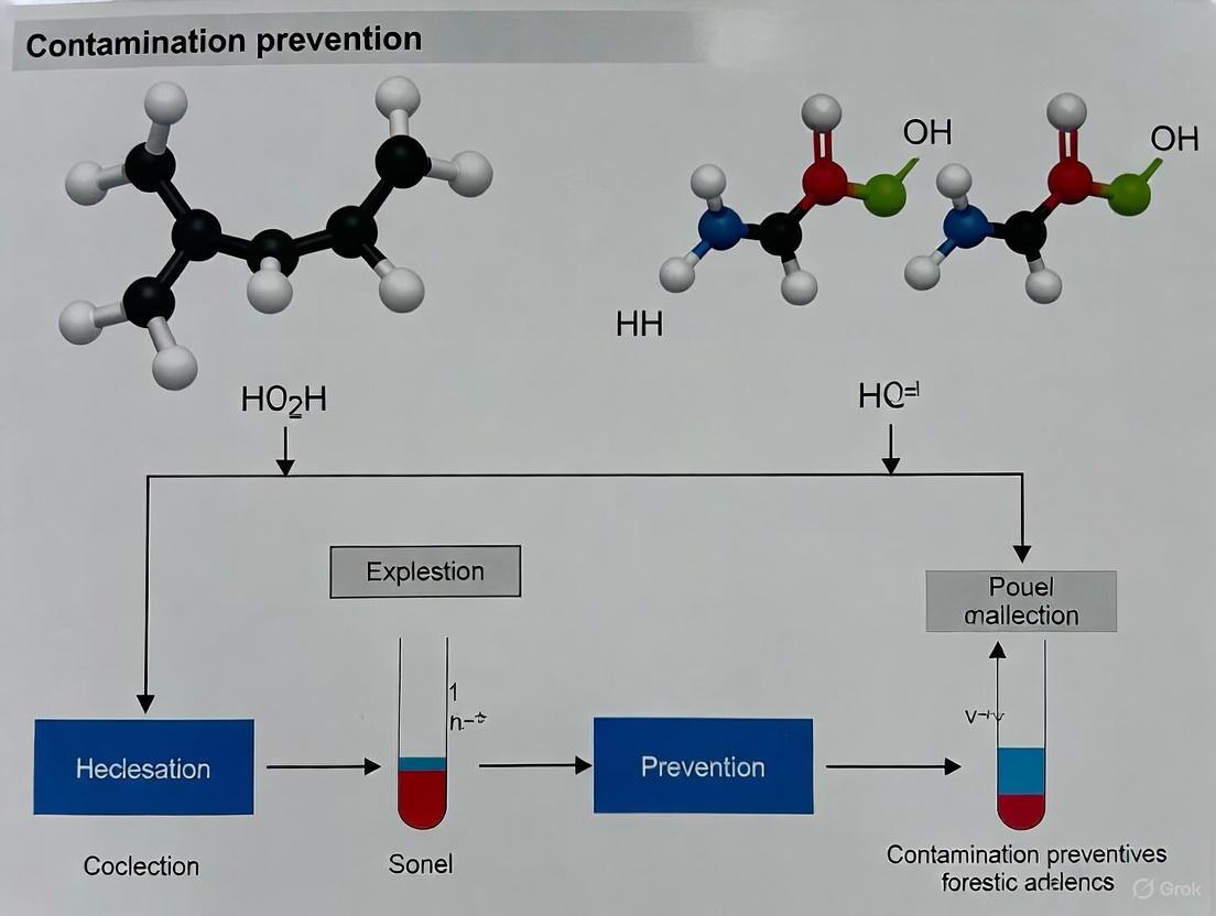
The following diagram illustrates the logical workflow for defining and addressing contamination in a post-blast investigation.
Contamination Management Workflow
The Critical Impact of Contamination on Forensic Evidence Reliability
Troubleshooting Guides
Guide 1: Unexplained Presence of Explosive Traces in Controls
Problem: During the analysis of explosive traces from a case sample, unexpected traces of high explosives like RDX or PETN are detected in your method blanks or negative controls, suggesting laboratory contamination.
Investigation and Resolution:
- Step 1: Confirm the Problem: Repeat the analysis of the blank and control samples. Ensure the signal is consistently present and above the method's detection limit. Check that the instrument signal is correctly identified and not a false positive from interfering compounds [8].
- Step 2: Review Laboratory Practices: Audit your anti-contamination protocols. Confirm that disposable gloves and clean lab coats are worn at all times. Verify that all equipment, including sample collection tools and laboratory work surfaces, was decontaminated with a 10% bleach solution or an appropriate solvent before use [9].
- Step 3: Check Reagents and Equipment: Ensure all solvents and reagents used are high-purity grade and have been tested as blanks. Inspect equipment like swabs and wipes for any pre-existing contamination from manufacturing or packaging processes [10].
- Step 4: Trace the Source: If the contamination persists, systematically test your process. Run blanks through each stage of your workflow—from sample preparation to instrument analysis—to isolate the step where the contamination is introduced.
- Step 5: Implement Corrective Actions: Once the source is found (e.g., a contaminated solvent batch or a specific piece of equipment), replace or thoroughly decontaminate it. Document the incident and update laboratory protocols to prevent recurrence.
Guide 2: Inconsistent Explosive Trace Recovery from Surfaces
Problem: Swab samples collected from the same surface in a scenario test yield highly variable concentrations of explosive residues, leading to unreliable data.
Investigation and Resolution:
- Step 1: Evaluate the Swab and Solvent: Ensure you are using an optimized swabbing system. Research indicates that PU-foam swabs wetted with a mixture of acetonitrile and water (90/10) can provide good recovery for a range of explosives including PETN and TNT [10]. Avoid suboptimal materials.
- Step 2: Standardize the Swabbing Technique: Inconsistency often arises from operator technique. Implement a standardized protocol specifying:
- The pattern and pressure to be used during swabbing.
- The number of times the swab should be passed over the surface.
- The process for swabbing irregular surfaces or edges [10].
- Step 3: Consider the Surface Type: The efficiency of trace recovery is highly dependent on the surface material (e.g., glass, aluminum, plastic). Conduct validation studies on different common surfaces to establish material-specific recovery rates and adjust your expectations accordingly [10].
- Step 4: Optimize the Extraction: After collection, the extraction of explosives from the swab must be efficient. Studies suggest that sonicating the swab in solvent for 10 minutes is an effective method. Ensure the chosen solvent (e.g., acetonitrile/water mixtures) is compatible with your swab type and the target analytes [10].
Frequently Asked Questions (FAQs)
Q1: What is the most critical step in preventing contamination when collecting trace evidence at a crime scene? A1: The most critical step is controlling access and establishing a decontamination zone. The number of personnel at the scene is directly related to contamination risk. A command post should be set up to log all entries, and a decontamination zone should be established where personnel don full personal protective equipment (PPE) and clean all equipment before entering the scene [9].
Q2: How common are high-explosive traces in public places, and what does this mean for my analysis? A2: Current research indicates that the detection of high-explosive traces like TNT, RDX, and PETN in public areas is statistically rare. This low background level makes a finding of these substances highly forensically significant. However, you must remain cautious of innocent sources, such as contamination from military or security training activities, and always compare your findings against the specific context of the case [8].
Q3: What type of swab and solvent should I use for sampling trace explosives? A3: An optimized wet swab sampling procedure suggests using PU-foam swabs wetted with a mixture of acetonitrile and water (90/10). This combination has been shown to provide good recovery for explosives like PETN, TNT, and ammonium nitrate. Subsequent extraction of the swab is typically done with a solvent like acetonitrile/water [10].
Q4: We are analyzing gunshot residue (GSR). What is the risk of finding GSR on someone who hasn't fired a gun? A4: The risk of inorganic GSR (iGSR) transfer from public surfaces is generally considered low. For organic GSR (oGSR), some components can be found in the environment, but the co-detection of specific compounds like nitroglycerine (TNG) with markers like ethyl centralite (EC) provides stronger evidence of a firearms-related event. It is crucial to analyze oGSR compounds in combination and consider the case context [8].
Data Presentation
Table 1: Prevalence of High Explosive Traces in Public Locations
This table summarizes findings on the likelihood of detecting explosive residues in non-military environments, informing the significance of such findings.
| Explosive Compound | Likelihood of Detection in Public Areas | Key Contextual Notes | Primary Analytical Techniques |
|---|---|---|---|
| TNT (Trinitrotoluene) | Rare | Environmental contamination is possible near manufacturing or dumping sites [8] [11]. | LC-MS, GC-MS [8] |
| RDX (Research Department Explosive) | Rare | A very specific military explosive; detection is highly significant [8]. | LC-MS, GC-MS [8] |
| PETN (Pentaerythritol Tetranitrate) | Rare | Used in plastic explosives and detonator cords; a finding is forensically important [8]. | LC-MS, GC-MS [8] |
| Ammonium Nitrate | Common | Dual-use chemical (explosives/fertilizer); interpretation requires caution [8] [10]. | IC, LC-MS [10] |
Table 2: Optimized Swab Sampling for Explosive Traces
This table outlines key parameters for an effective surface sampling protocol based on recent research.
| Parameter | Recommended Specification | Alternative Options | Performance Notes |
|---|---|---|---|
| Swab Type | PU-foam (e.g., Chemtronics CF1050) | Microfiber wipes, Cotton swabs | PU-foam showed good performance in recovery studies [10]. |
| Wetting Solvent | Acetonitrile/Water (90/10) | Acetonitrile/Water (70/30), Methanol/Water | ACN/W (90/10) was identified as an effective mixture [10]. |
| Extraction Method | Sonication for 10 minutes | Shaking | Effective for desorbing traces from the swab matrix [10]. |
| Analysis | LC-Triple Quad MS (for trace levels) | HPLC-UV/VIS (for higher concentrations), Ion Chromatography (for inorganics) | LC-MS provides high sensitivity and selectivity [10]. |
Experimental Protocols
Protocol 1: Surface Sampling for Organic Explosive Traces Using Wet Swabs
Principle: This protocol describes a standardized method for collecting traces of organic explosives (e.g., TNT, PETN) from hard surfaces using solvent-wetted swabs for subsequent LC-MS analysis.
Reagents and Materials:
- PU-foam swabs (e.g., Chemtronics CF1050)
- HPLC-grade acetonitrile and water
- Solvent-resistant gloves
- Clean, disposable forceps
- 10 mL centrifuge tubes
- 0.45 µm nylon membrane syringe filters
- Mechanical pipettes and tips
Procedure:
- Swab Preparation: In a clean area, use a pipette to wet the PU-foam swab with 400 µL of acetonitrile/water (90/10) solution. Do not oversaturate.
- Sampling: Using gloved hands or forceps, systematically wipe the target surface (e.g., 20 cm² area) with the wet swab. Apply consistent, moderate pressure. Swab in one direction initially, then use the other side of the swab for a second pass over the same area in a perpendicular direction.
- Storage: Immediately place the used swab into a clean 10 mL centrifuge tube. Seal the tube tightly to prevent solvent evaporation and loss of analyte.
- Extraction: In the laboratory, add 3 mL of extraction solvent (e.g., acetonitrile/water) to the centrifuge tube. Seal the tube and sonicate for 10 minutes. Remove the swab, squeezing it against the tube walls to recover as much liquid as possible.
- Filtration: Filter the extract through a 0.45 µm nylon syringe filter into a clean vial.
- Analysis: Analyze an aliquot of the filtered extract by LC-MS, diluting with water as necessary for the instrumental method [10].
Protocol 2: Establishing a Decontamination Zone for Crime Scene Work
Principle: To prevent cross-contamination between different scenes or from the laboratory to the scene, a decontamination zone must be established. This is crucial for maintaining the integrity of trace evidence, especially with sensitive DNA and explosives analysis.
Reagents and Materials:
- Barrier tape
- Disposable personal protective equipment (PPE): coveralls, gloves, booties, head cover, mask
- 10% bleach solution or other appropriate disinfectant
- Plastic tarps or small plastic pools
- Biohazard bags for disposable waste
- Decontamination logbook
Procedure:
- Zone Designation: Identify and mark a "safe zone" or "decontamination corridor" at the perimeter of, and leading into, the secured crime scene. This area should be upwind of the main scene if outdoors.
- Equipment Decontamination: Before entering the scene, all equipment (e.g., camera, kits, note pads) should be wiped down with a 10% bleach solution or a suitable disinfectant on a plastic tarp within the decontamination zone.
- PPE Donning: Personnel should don a full set of disposable PPE (mask, jumpsuit, gloves, booties, head cover) within this clean zone before proceeding to the evidence collection area.
- Post-Scene Decontamination: Upon exiting the scene, personnel must enter the decontamination zone to remove their PPE. Booties and outer gloves should be removed last. Equipment should be wiped down again.
- Waste Disposal: All used disposable PPE and cleaning materials should be placed in designated biohazard bags for proper disposal. The use of a small plastic pool can contain runoff during boot decontamination [9].
Workflow and Relationship Visualizations
Evidence Integrity Workflow
Swab Sampling and Analysis Process
The Scientist's Toolkit: Research Reagent Solutions
Essential Materials for Explosives Trace Analysis
| Item | Function/Benefit | Application Note |
|---|---|---|
| PU-Foam Swabs | High recovery efficiency for a broad range of organic explosives from various surfaces. | Preferred over cotton or polyester; use with a solvent. |
| Acetonitrile (HPLC Grade) | Effective wetting and extraction solvent for nitroaromatics and nitramines. | Formulate with water (e.g., 90/10 or 70/30) for swab wetting and extraction. |
| LC-Triple Quadrupole MS | Provides high sensitivity and selective detection of trace-level explosives in complex samples. | Operate in Multiple Reaction Monitoring (MRIM) mode for definitive identification and quantification. |
| Personal Protective Equipment (PPE) | Disposable suits, gloves, booties, and masks prevent contamination from the investigator to the scene and between scenes. | Don in a designated decontamination zone. Considered a biohazard and anti-contamination measure [9]. |
| 10% Bleach Solution | Standard decontamination agent for cleaning equipment and surfaces in the laboratory and decontamination zone. | Effective for neutralizing biological contaminants and reducing cross-contamination risks [9]. |
In the forensic analysis of explosives, three instrumental principles are paramount for developing reliable and legally admissible results: sensitivity, selectivity, and specificity. These parameters define the quality and reliability of trace evidence analysis, which is often present in minute quantities and within complex sample matrices after an explosion or in pre-blast scenarios. For forensic investigations, sensitivity is crucial because explosive residues are typically present at trace levels (e.g., parts-per-million or even parts-per-billion), and false negatives must be avoided. Selectivity allows the method to distinguish the target explosive analyte from a messy mixture of other compounds that may mask its presence. Finally, specificity is essential to unambiguously identify a specific compound, such as identifying nitroglycerin to infer the use of double-base smokeless powder, a conclusion that carries significant scientific and legal weight [6].
The context of contamination prevention is integral to maintaining the integrity of these principles. Uncontrolled contamination can lead to falsely positive or negative results, potentially misdirecting an investigation or compromising its legal standing. Therefore, rigorous procedures, such as those outlined in Good Laboratory Practice (GLP) and standards from the Forensic International Network for Explosives Investigation (FINEX), are necessary to protect evidence from the crime scene through laboratory analysis [1].
Frequently Asked Questions (FAQs)
FAQ 1: Why is sensitivity particularly challenging in post-blast explosives analysis? Post-blast residues of high explosives typically yield extracts with concentrations in the parts-per-billion range, pushing analytical methods to their detection limits. High-order detonations consume nearly all the explosive material, making it difficult to obtain recoverable amounts for analysis. For instance, research into isotopic signatures for source attribution found that obtaining sufficient recoverable amounts of RDX and TNT after a detonation was a key limitation [6].
FAQ 2: How can selectivity be achieved when analyzing complex post-blast debris? Selectivity is often achieved by chemically separating the mixture so individual compounds can be analyzed without interference. Techniques like Gas Chromatography (GC) are coupled with highly selective detectors. In GC, a mixture is vaporized and the components are separated based on their different affinities for the column coating, allowing each compound to be analyzed individually as it exits the column [6].
FAQ 3: What is the practical difference between selectivity and specificity? While both deal with distinguishing the analyte, selectivity is the method's ability to respond to the analyte in a complex mixture without interference from similar compounds. Specificity goes a step further by providing unambiguous identification of the analyte. A highly specific technique can discern small structural differences between similar molecules, which is vital for forming definitive scientific conclusions [6].
FAQ 4: How does contamination risk interact with these analytical principles? Contamination directly undermines all three principles. It can:
- Create false positives, compromising specificity.
- Introduce interferents that mask the true analyte, reducing selectivity.
- Dilute the original trace, making it undetectable and thus reducing effective sensitivity. Studies have shown that TNT has a high contamination potential, underscoring the need for stringent anti-contamination protocols in laboratories [1].
Troubleshooting Guide
This guide addresses common challenges in explosives analysis related to sensitivity, selectivity, and specificity.
| Challenge | Root Cause | Solution |
|---|---|---|
| Low Sensitivity | Analyte concentration below the method's detection limit. | - Optimize sample pre-concentration steps (e.g., applying heat and inert gas to evaporate solvent) [6].- For techniques like GC-VUV, future work is focused on increasing sensitivity to better detect parts-per-billion concentrations [6]. |
| Poor Selectivity | Complex sample matrix with interferents masking the analyte. | - Employ chromatographic separation (e.g., GC) to physically separate compounds before detection [6].- Use selective detectors or reactive techniques. For example, Reactive Desorption Electrospray Ionization (DESI) can use additives in the spray solvent to form specific analyte-adducts, enhancing selectivity [12]. |
| Insufficient Specificity | Inability to differentiate between structural isomers or thermally labile compounds. | - Utilize techniques that provide highly specific spectral data. GC-VUV can overcome limitations of GC/MS by providing unique VUV absorption spectra that differentiate structural isomers [13].- Use tandem mass spectrometry (MS/MS) for confirmatory analysis [12]. |
| Sample Contamination | Uncontrolled introduction of explosives traces during evidence handling or analysis. | - Implement rigorous anti-contamination procedures per GLP and FINEX standards [1].- Conduct regular blank-sampling of laboratory surfaces and equipment to monitor contamination levels [1] [10]. |
Experimental Protocols
Protocol: Optimized Wet Swab Sampling for Surface Contamination
This protocol is designed for the collection of organic and inorganic explosive traces from various surfaces for subsequent laboratory analysis, balancing high recovery with contamination prevention [10].
1. Reagents and Materials:
- Swab: PU-foam swabs (e.g., Chemtronics CF1050).
- Wetting Solvent: Mixture of acetonitrile and water (ACN/W) in a 90/10 ratio.
- Extraction Solvent: Acetonitrile/water (90/10) or other suitable mixtures like methanol/water.
- Labware: Centrifuge tubes, 0.45 µm nylon membrane syringe filters, analytical glass vials.
2. Procedure:
- Swab Wetting: Prior to sampling, wet the PU-foam swab with approximately 400 µL of the ACN/W (90/10) wetting solvent.
- Surface Sampling: Swab the target surface thoroughly. The protocol suggests swabbing a surface twice: using one side of the swab for the first round and the opposite side for the second.
- Sample Drying: After collection, allow the swab to air-dry for approximately 10 minutes at room temperature.
- Extraction: Place the swab in a centrifuge tube and add 3 mL of extraction solvent. Sonicate the closed tube for 10 minutes. Afterwards, squeeze the swab against the tube walls to recover as much liquid as possible, then discard the swab.
- Filtration: Filter the extract through a 0.45 µm nylon membrane syringe filter.
- Analysis: The filtrate can now be analyzed directly or diluted as required for instrumental techniques such as LC-triple quad MS for organic explosives or ion chromatography for inorganic ions like nitrate [10].
Protocol: Analysis of Explosives via GC-VUV
This protocol outlines the use of Gas Chromatography/Vacuum Ultraviolet Spectroscopy (GC-VUV) for the identification of explosive compounds, leveraging its high specificity.
1. Instrument Setup and Calibration:
- GC Conditions: The method can be optimized using a ramped multimode inlet program with a final temperature of 200°C and a GC carrier gas flow rate of 1.9 mL/min [13].
- VUV Conditions: The VUV make-up gas pressure can be set to 0.00 psi. The transfer line/flow cell temperature, while important, was not found to be statistically significant in optimization studies [13].
2. Sample Analysis:
- The sample extract is injected and vaporized in the GC inlet.
- The components are separated as they travel through the GC column based on their interaction with the stationary phase.
- Each separated compound enters the VUV flow cell, where its absorption of vacuum ultraviolet light (in the 120-430 nm range) is measured in real-time. All organic compounds absorb in this region, and their spectra provide a unique fingerprint.
- The resulting VUV spectrum is analyzed. The presence of specific explosives is confirmed by matching the retention time and the spectral signature against a validated library. The technique is particularly useful for differentiating structural isomers and detecting thermally labile compounds that are challenging for GC/MS [13].
Workflow and Relationship Diagrams
Analytical Principles in Contamination Prevention
This diagram illustrates the interconnected relationship between the core analytical principles and the overarching framework of contamination prevention.
The Scientist's Toolkit: Key Research Reagents & Materials
The following table details essential materials used in the collection and analysis of explosive traces, as featured in the cited protocols.
| Item | Function & Application | Example from Protocol |
|---|---|---|
| PU-Foam Swabs | Collection of trace particulates from surfaces. The foam structure is effective at trapping and releasing particles. | Used with ACN/W (90/10) wetting solvent for sampling surfaces in a car mock-up scenario [10]. |
| Acetonitrile (ACN) | A versatile organic solvent used for wetting swabs and extracting a wide range of organic explosives from collection media. | A mixture of ACN/Water (90/10) was determined to be an effective wetting and extraction solvent for TNT and PETN [10]. |
| Basic Yellow 40 (BY40) | A fluorescent dye used to enhance the visibility of latent fingerprints developed with cyanoacrylate (CA) fuming. | Used after CA fuming to develop latent fingerprints on post-blast IED fragments, even after exposure to water [14]. |
| Cyanoacrylate (CA) | The active component in "super glue" fuming. It polymerizes on the moisture and salts in latent fingerprints, creating a white visible polymer. | A key technique for developing latent fingerprints on non-porous surfaces of IEDs, even after neutralization with a waterjet disruptor [14]. |
| Chromatographic Columns | The heart of separation science. These columns separate complex mixtures into individual components for analysis. | A GC column separates explosive compounds before they reach the VUV detector, enabling selective and specific analysis [6]. |
Systematic Approaches to Contamination Control Strategy (CCS) Implementation
Troubleshooting Common CCS Implementation Issues
| Problem Area | Common Issue | Potential Root Cause | Recommended Corrective Action |
|---|---|---|---|
| Evidence Contamination | Unexplained detection of explosive traces (e.g., TNT, RDX) on control samples [1]. | Cross-contamination from laboratory standards or contaminated equipment; poor handling procedures. | Review and reinforce handling SOPs; implement more stringent cleaning validation; use dedicated equipment for standards and evidence [1] [8]. |
| Environmental Monitoring | Recurring high particulate counts in cleanroom or sample preparation area. | Ineffective air filtration; improper gowning; high personnel traffic; inadequate room cleaning. | Check HEPA filter integrity; retrain personnel on aseptic practices; review and optimize cleaning frequency and agents [15] [16]. |
| Personnel & Gowning | Microbial contamination detected on finger plates or settle plates. | Incorrect gowning sequence; use of damaged gowning materials; non-compliance with procedures. | Implement gowning competency assessments; use visible demonstrations and signage; ensure quality of personal protective equipment (PPE) [17]. |
| Data Integrity | Unexplained variability in analytical results for replicate samples. | Low-level microbial or particulate contamination influencing sensitive analytical endpoints [15]. | Enhance environmental monitoring; review sample storage conditions; investigate subclinical contamination in experimental models [15]. |
| Cleaning & Disinfection | Ineffective surface decontamination after a contamination event. | Use of inappropriate disinfectant; incorrect contact time; application error in manual cleaning. | Validate disinfectant efficacy against common lab contaminants; consider automated decontamination (e.g., Vaporized Hydrogen Peroxide) for better reproducibility [18]. |
Frequently Asked Questions (FAQs)
What is the most critical step in preventing contamination during the analysis of explosive traces?
The most critical step is a robust sample handling procedure that strictly separates the analysis of evidence materials from the handling of explosive standards. Contamination with trace amounts of explosives is most likely to occur during the sample preparation stage in the laboratory [1]. Implementing a unidirectional workflow and using disposable equipment where possible are key preventative measures.
Our lab consistently finds traces of TNT in blank samples. Where should we start our investigation?
Start by investigating your standard/reference material handling area. Studies have shown that TNT has the highest contaminant potential of common explosives due to its physical properties and can easily spread in a laboratory environment [1]. Audit your cleaning procedures for surfaces and equipment in contact with TNT standards and ensure these areas are physically separated from evidence processing areas.
How can we justify the significant investment in an automated decontamination system for our facility?
An automated system, such as Hydrogen Peroxide Vapor, provides consistent, validated, and documented decontamination cycles. This reduces the variability inherent in manual cleaning, protects high-value research from loss due to contamination (e.g., irreplaceable forensic evidence or cell therapies), and can reduce downtime between analytical campaigns. The investment should be weighed against the cost of a major contamination event, which could invalidate critical data or evidence [18].
We have strong procedures, but still experience sporadic contamination. What might we be missing?
Review your material and personnel flows. Even with good procedures, contamination can be introduced through raw materials, consumables, or via personnel moving between "clean" and "dirty" zones. Implement a comprehensive Quality Risk Management (QRM) program to identify these less obvious critical control points. A holistic CCS looks at the entire process, from facility design and vendor approval to personnel training and monitoring [19] [17].
What is the key to maintaining a successful CCS long-term?
The key is continuous improvement. A CCS is not a one-time document. It should be a living program that is regularly reviewed and updated based on data from environmental monitoring, investigation reports, and technological advances. Integrating your CCS into the facility's periodic product quality reviews ensures it remains effective and relevant [19] [17].
The Scientist's Toolkit: Essential Reagents & Materials for a Forensic Explosives CCS
| Item | Function & Application | Key Considerations |
|---|---|---|
| HEPA Filters [15] | Provides high-efficiency filtration of airborne particles and microorganisms for supply air in cleanrooms and safety cabinets. | Integrity must be regularly tested and certified. |
| Validated Disinfectants (e.g., alcohols, sporicides) [18] | Used for manual and automated decontamination of surfaces and equipment. | Must be validated for efficacy against a broad spectrum of microbes and for material compatibility. Rotation may be required. |
| Particle Counters [16] | Monitors non-viable particulate contamination in critical environments in real-time. | Critical for ensuring air quality during sensitive analytical procedures. |
| Microbial Air Samplers [16] | Actively samples a known volume of air to quantify viable (living) microbial contamination. | Used for routine environmental monitoring and investigation of contamination events. |
| Sterile, Single-Use Sampling Kits [8] | For collecting samples from surfaces and evidence with minimal risk of introducing contamination. | Should include swabs, wipes, and containers. Use is critical for forensic evidence integrity. |
| Analytical Grade Solvents | Used for extracting trace explosives from evidence samples and for instrument calibration. | High purity is essential to prevent introducing interferents that affect sensitive detection methods like GC/ECD or LC-MS [1] [8]. |
Experimental Workflow for a Holistic CCS
The following diagram illustrates the continuous, lifecycle approach to implementing and maintaining an effective Contamination Control Strategy.
Detailed Investigation Protocol for a Contamination Event
Upon detection of a contamination event (e.g., positive explosive trace in a blank), a systematic investigation is critical.
Exploring the Effects of Blast Conditions on Trace Evidence Survival
Technical Support Center
Troubleshooting Guides & FAQs
This section addresses common experimental challenges in post-blast trace evidence analysis, focusing on contamination prevention and methodology optimization.
FAQ 1: Can latent fingerprints and touch DNA survive on improvised explosive device (IED) components after neutralization or detonation?
Answer: Yes, research confirms that both latent fingerprints and touch DNA can survive destructive conditions. One study demonstrated a 27% fingerprint recovery rate from IEDs neutralized by a waterjet disruptor, with full STR DNA profiles obtainable from touch DNA even after these conditions and subsequent fingerprint development techniques [14]. Success depends on the specific conditions and analytical methods used.
- Recommended Protocol: For latent fingerprints on non-porous surfaces exposed to water or heat, employ Cyanoacrylate (CA) fuming followed by enhancement with a fluorescent dye like Basic Yellow 40 (BY40). This technique has proven effective on surfaces exposed to temperatures up to 500 °C and can be used before DNA analysis without significant interference [14].
- Troubleshooting Tip: If initial fingerprint development fails on wet evidence, ensure substrates are thoroughly dried before CA fuming. For DNA analysis, use extraction kits optimized for low-template and potentially degraded DNA, and interpret results with caution due to potential artefacts like allele drop-in or drop-out [14].
FAQ 2: How can we minimize the risk of laboratory contamination when analyzing trace explosives?
Answer: Contamination prevention requires stringent procedures at every stage, from evidence collection to instrumental analysis [1]. Contaminant potential varies; TNT exhibits the highest risk, followed by RDX and PETN, while Nitroglycerin (NG) presents a lower risk due to its properties [1].
- Critical Control Points: Studies identify the sample preparation room as the most critical location for potential contamination, followed by the laboratory and evidence storage areas [1]. Implement rigorous cleaning protocols for these spaces and use dedicated equipment.
- Best Practices: Adhere to standards like the European Network of Forensic Science Institutes (ENFSI) Best Practice Manual [8]. Use disposable materials where possible, perform regular anti-contamination checks of workspaces, and analyze blank samples alongside casework to monitor for contamination [1].
FAQ 3: What analytical techniques are most effective for identifying explosive residues in complex, degraded post-blast samples?
Answer: A sequential analytical approach using complementary techniques is most effective, especially for homemade explosives (HME) [20].
- Workflow for Complex Samples: Begin with sequential solvent extractions (e.g., ether, acetone, water, sodium hydroxide, pyridine) to isolate different organic and inorganic components [20]. Follow with chemical spot tests and Thin Layer Chromatography (TLC) for preliminary screening. Confirm findings with instrumental techniques like Fourier Transform Infrared Spectroscopy (FTIR) for functional group identification [20].
- Advanced Techniques: For higher sensitivity and specificity, techniques like Gas Chromatography-Mass Spectrometry (GC-MS) and Liquid Chromatography-Mass Spectrometry (LC-MS) are widely used. Ambient Mass Spectrometry and Raman Spectroscopy are emerging for rapid, sensitive detection [8].
The table below summarizes key experimental data on trace evidence survival from controlled IED testing, providing a reference for evaluating your results.
Table: Survival Rates of Forensic Trace Evidence After IED Neutralization
| Evidence Type | Blast/Condition | Recovery/Detection Rate | Key Experimental Findings |
|---|---|---|---|
| Latent Fingerprints | Waterjet Disruptor Neutralization | 27% (31 of 115 deposited prints) [14] | CA-BY40 effective development method; 10 recovered prints were of quality useful for identification [14]. |
| Touch DNA (STR Profiling) | Waterjet Disruptor Neutralization | Full STR profiles obtained [14] | DNA profiles obtained from extremely low amounts of contact DNA; techniques do not always preclude DNA profiling [14]. |
| Inorganic Explosive Residues | Homemade Explosive Detonation | Potassium Nitrate, Ammonium Nitrate confirmed [20] | Detected via chemical tests & FTIR in degraded exhibits 5 months post-blast [20]. |
Detailed Experimental Protocols
Protocol 1: Sequential Solvent Extraction for Post-Blast Residue Analysis
This protocol is designed to maximize the recovery of both organic and inorganic explosive residues from complex, degraded post-bblast debris [20].
- Materials: Diethyl Ether, Acetone (AR), Demineralized Water, Sodium Hydroxide, Pyridine, Syringe Filters (0.22 µm), Whatman-42 filter paper, TLC plates, FTIR instrument [20].
- Procedure:
- Ether Extraction: Treat sample with diethyl ether, filter, and concentrate the filtrate to ~1 ml. Analyze for non-polar organics like diesel oil (e.g., in ANFO) using FTIR [20].
- Acetone Extraction: After ether evaporates, treat the same sample with acetone, filter, and concentrate. Analyze the extract for common organic explosives (e.g., PETN, TNT, RDX) using TLC and FTIR [20].
- Water Extraction: Treat sample with hot distilled water, filter, and concentrate. Analyze the extract for inorganic ions (e.g., Nitrate, Chloride, Ammonium) using chemical tests (see table below) and FTIR [20].
- Alkali Extraction: Treat sample with 2N Sodium Hydroxide, filter, and concentrate. Test for metallic Aluminum and other specific ions [20].
- Pyridine Extraction: Treat sample with Pyridine, filter, and concentrate. Add a drop of Sodium Hydroxide while heating; a color change indicates elemental Sulfur [20].
Table: Chemical Spot Tests for Inorganic Explosive Residues
| Target Ion/Analyte | Chemical Test | Observation for Positive Result |
|---|---|---|
| Nitrite | Griess Test | Presence indicated [20] |
| Nitrate | Griess reagent + Zn dust | Presence indicated [20] |
| Chloride | Silver Nitrate | Presence indicated [20] |
| Chlorate | Aniline sulphate | Absence noted in case study [20] |
Protocol 2: Latent Fingerprint Development on Post-Blast Evidence
This protocol outlines the use of CA fuming for developing latent fingerprints on non-porous surfaces recovered from blast scenes [14].
- Materials: Cyanoacrylate (Super Glue), Basic Yellow 40 (BY40) fluorescent dye, Fuming chamber, Forensic light source (appropriate wavelength for BY40) [14].
- Procedure:
- Ensure the evidence substrate is dry before beginning.
- Place the evidence and a few grams of CA in a fuming chamber.
- Allow the CA to fume and polymerize on the latent fingerprint residues for approximately 20-30 minutes.
- After fuming, the developed prints can be enhanced by applying BY40, which fluoresces under a forensic light source, improving visualization and photography [14].
- Note: This method has been successfully applied to evidence subjected to waterjet disruption and does not preclude subsequent DNA analysis [14].
Evidence Analysis Workflow
The Scientist's Toolkit
Table: Essential Research Reagents and Materials for Explosive Trace Evidence Analysis
| Reagent/Material | Function/Brief Explanation |
|---|---|
| Cyanoacrylate (CA) & Basic Yellow 40 | Develops and enhances latent fingerprints on non-porous surfaces exposed to challenging conditions like water and heat [14]. |
| Sequential Solvents (Ether, Acetone, Water) | Selectively extracts different classes of organic and inorganic explosive residues from complex post-blast samples for analysis [20]. |
| Chemical Spot Test Reagents | Provides rapid, presumptive tests for specific ions indicative of explosives (e.g., Griess test for nitrates/nitrites) [20]. |
| TLC Plates & Developing Solvents | Separates complex mixtures of organic explosive residues for preliminary identification before confirmatory analysis [20]. |
| High-Purity Analytical Standards | Essential for calibrating instruments, confirming identifications, and quantifying trace levels of explosives in environmental samples [8]. |
Advanced Analytical Techniques and Contamination Control Protocols
Gas Chromatography-Vacuum UV Spectroscopy (GC-VUV) for Explosives Traces
FAQs and Troubleshooting Guides
This technical support resource addresses common challenges in GC-VUV analysis of explosive traces, specifically framed within a contamination prevention context for forensic research.
Frequently Asked Questions
Q1: How can I minimize decomposition of thermally labile explosives like NG, PETN, and RDX during GC-VUV analysis?
Thermal decomposition of nitrate esters and nitramines in the GC system manifests as altered VUV spectra and peak broadening. To mitigate this:
- Optimize Inlet Temperature: Lower the final temperature of a ramped multimode inlet program to 200 °C to reduce thermal stress [21].
- Control Transfer Line/Flow Cell Temperature: While one study found this parameter statistically insignificant for peak area, a temperature of 300 °C is commonly used as an optimized setting. Monitor spectra for fine structure indicating breakdown products [21].
- Verify System Cleanliness: Residues in the inlet or column can catalyze decomposition. Perform regular maintenance and use contamination-free liners.
Q2: What are the optimal GC and VUV parameters to achieve the lowest detection limits for trace explosives?
A Central Composite Design (CCD) study for explosive compounds (TATP, DMNB, NG, TNT, etc.) determined the following optimized parameters to maximize peak area and sensitivity [21]:
- GC Carrier Gas Flow Rate: 1.9 mL/min
- VUV Make-up Gas Pressure: 0.00 psi
- Final Ramped Inlet Temperature: 200 °C
Q3: My chromatogram shows co-eluting peaks. Can GC-VUV still identify and quantify the individual explosives?
Yes, a primary advantage of GC-VUV is its ability to deconvolve co-eluting compounds. VUV absorption is additive, and the unique spectral fingerprint of each explosive allows software to resolve overlapping signals [22] [23]. Ensure the VUV spectral library contains reference spectra for the suspected compounds. The goodness of fit (R² >0.999) confirms successful deconvolution and enables accurate quantification of individual analytes [23].
Q4: We are detecting explosive traces in laboratory blanks. What are the critical contamination control points?
Contamination can severely compromise forensic evidence. Key prevention strategies include:
- Personnel Decontamination: Implement strict protocols for personnel handling samples or equipment [8].
- Dedicated Laboratory Spaces: Use separate, designated spaces for trace and bulk explosives analysis [8].
- Disposable Equipment: Utilize single-use, disposable equipment for sample handling where possible [8].
- Regular Proficiency Testing: Participate in internal and external proficiency testing schemes to ensure analytical quality and identify contamination issues [8].
Troubleshooting Guide
The following table outlines common experimental issues, their potential causes, and recommended solutions.
| Problem | Possible Causes | Solutions |
|---|---|---|
| Low Sensitivity/High LODs | Non-optimized carrier gas flow or make-up gas pressure [21]. | Adopt optimized method: 1.9 mL/min flow rate and 0.00 psi make-up gas pressure. |
| Thermal Decomposition of Analytes | Inlet or transfer line temperature too high [21] [24]. | Lower inlet temperature to 200°C. Verify transfer line temperature is not excessively high. |
| Unidentified Peaks in Chromatogram | Contamination from solvents, surfaces, or previous samples [8]. | Implement anti-contamination protocols: use disposable equipment, dedicated lab spaces, and run solvent blanks [8]. |
| Inability to Distinguish Isomers | Reliance on MS alone, which struggles with constitutional isomers [23]. | Use GC-VUV's specific spectral fingerprints for isomer differentiation (e.g., o-, m-, p-xylene) [23]. |
| Poor Peak Shape or Resolution | Incorrect GC column selection or method parameters. | Use columns suitable for explosive compounds. Consider a "chromatographic compression" strategy with faster oven ramps and using VUV deconvolution for any resulting co-elution [23]. |
Experimental Protocol: Optimization of GC-VUV for Explosives
This methodology is adapted from a published optimization study for explosive compounds [21].
1. Scope This protocol describes the use of a Response Surface Methodology (RSM) with a Central Composite Design (CCD) to optimize GC/VUV parameters for the analysis of seven explosive and explosive-related compounds: TATP, DMNB, NG, diphenylamine, TNT, PETN, and RDX.
2. Chemicals and Materials
- Analytes: Standard solutions of NG, PETN, RDX, TNT, TATP, DMNB, and diphenylamine.
- Solvents: Methanol (GC Resolv) and acetone (certified ACS).
- GC Column: Rxi-35Sil MS (30 m × 0.25 mm I.D. × 0.25 μm d_f).
- Internal Standard: 1,3-Dinitrobenzene.
3. Instrumental Configuration
- GC System: Agilent 7890B Gas Chromatograph.
- Detector: VUV Analytics VGA-100 vacuum ultraviolet detector.
- Data Collection: Wavelength range: 120 - 240 nm. Data collection rate: 100 Hz.
4. Optimization Procedure A three-factor, three-level CCD is used to optimize the following parameters, with the response variable being the chromatographic peak area.
- Factor A: GC carrier gas flow rate (1.9, 3.2, 4.5 mL/min)
- Factor B: VUV make-up gas pressure (0.00, 0.15, 0.30 psi)
- Factor C: Final ramped inlet temperature (200, 250, 300 °C)
Additionally, optimize the transfer line/flow cell temperature using a "vary-one-parameter-at-a-time" approach.
5. Data Analysis
- Use statistical analysis software to fit the experimental data from the CCD to a model and find the parameter set that maximizes the peak area response.
- The expected optimal conditions from the cited study are a flow rate of 1.9 mL/min, a make-up gas pressure of 0.00 psi, and a final inlet temperature of 200 °C [21].
Anti-Contamination Workflow for Forensic Explosives Analysis
The following diagram illustrates a systematic workflow to prevent contamination during the analysis of trace explosives, from sample receipt to data reporting.
The Scientist's Toolkit: Key Research Reagent Solutions
Essential materials for GC-VUV analysis of explosive traces are listed below.
| Item | Function/Benefit |
|---|---|
| Rxi-35Sil MS GC Column | A mid-polarity column used for the separation of a wide range of explosive compounds, including nitrate esters and nitroaromatics [21]. |
| VUV Spectral Library | A collection of known VUV absorbance spectra is crucial for compound identification, differentiation of isomers, and deconvolution of co-eluting peaks [22] [23]. |
| Ionic Liquid Stationary Phase GC Column | Specifically designed for analyzing water and other challenging polar compounds, which is useful for characterizing solvents or decomposition products [25]. |
| High-Purity Explosive Standards | Certified reference materials (e.g., NG, PETN, RDX, TNT, TATP) are essential for method development, calibration, and positive identification of unknown traces [21] [8]. |
| Decontamination Solvents | High-purity solvents (e.g., methanol, acetone) for cleaning sampling tools, surfaces, and instrumentation to prevent cross-contamination [21] [8]. |
| Disposable Sampling Kits | Kits containing single-use items (swabs, tweezers, gloves) to prevent the introduction of contaminants and carry-over between samples [8]. |
IR Spectroscopy and Chemometric Methods for Residue Classification
Troubleshooting Guides
Guide 1: Addressing Common FT-IR Spectral Issues in Explosive Residue Analysis
Problem 1: Noisy or Unstable Spectra
- Issue: Spectra show excessive noise, making peak identification difficult.
- Cause: Instrument vibration from nearby equipment or laboratory activity introduces false spectral features [26].
- Solution: Ensure the spectrometer is placed on a stable, vibration-free optical table. Isolate the instrument from pumps, hoods, and heavy foot traffic [26].
Problem 2: Unexpected or Negative Peaks
- Issue: Peaks appear in the spectrum that do not match the sample, or negative absorbance peaks are observed.
- Cause: A dirty Attenuated Total Reflection (ATR) crystal is a common cause of negative peaks. Contamination from previous samples can also cause unexpected features [26].
- Solution: Clean the ATR crystal thoroughly with an appropriate solvent and acquire a fresh background spectrum before measuring your sample [26].
Problem 3: Distorted Baselines or Intensity Changes
- Issue: The spectrum baseline is curved or sloped, or band intensities are distorted.
- Cause: Physical effects like light scattering, reflection, or interference from the sample matrix, especially in complex residues [27].
- Solution: Apply spectral pre-processing. Baseline correction techniques, such as polynomial fitting or multiplicative scatter correction (MSC), can remove these unwanted contributions [27].
Problem 4: Sample Representation Error
- Issue: The spectrum does not represent the bulk material, which is critical for heterogeneous explosive residues.
- Cause: Surface chemistry (e.g., oxidation, additives) may differ from the interior composition [26].
- Solution: For solid materials, collect spectra from both the surface and a freshly cut interior section to confirm you are analyzing the target residue [26].
Guide 2: Overcoming Chemometric Model Performance Issues
Problem 1: Poor Classification Accuracy
- Issue: Machine learning models (PLS-DA, SVM, KNN) fail to classify residues correctly.
- Cause: Inadequate pre-processing leads to models trained on artifacts (e.g., baseline drift) rather than chemical information [27].
- Solution: Implement a robust pre-processing pipeline. This typically includes smoothing (e.g., Savitzky-Golay filter), baseline correction, and normalization to improve signal quality before modeling [28] [27].
Problem 2: Model Fails to Generalize to New Samples
- Issue: The model performs well on training data but poorly on new validation samples.
- Cause: The model is overfitted, or the calibration set lacks sufficient size and diversity (e.g., not covering all expected explosive varieties and environmental conditions) [29] [30].
- Solution: Build models using a large and diverse set of calibration spectra. Validate models with an independent test set and use techniques like cross-validation to ensure robustness [30].
Frequently Asked Questions (FAQs)
FAQ 1: What are the key benefits of using IR spectroscopy combined with chemometrics for explosive residue classification?
IR spectroscopy provides a rapid, non-destructive chemical fingerprint of a sample. When paired with chemometrics, it allows for the automated identification and classification of residues based on their unique spectral signatures. This approach is highly effective for distinguishing between different explosive formulations, even when they are chemically similar, which is crucial for forensic investigations [28] [29].
FAQ 2: How do I choose between NIR and Mid-IR spectroscopy for my analysis?
The choice depends on your sample and application. Mid-IR spectroscopy is highly sensitive and excellent for identifying fundamental molecular vibrations, making it ideal for analyzing homogeneous materials like pure explosives. NIR spectroscopy probes overtone and combination bands, is more rugged, and can be better for analyzing heterogeneous solid samples and for field deployment with portable devices [30].
FAQ 3: What is the single most critical step to prevent contamination in trace explosives analysis?
Adherence to strict anti-contamination protocols is paramount. This includes using disposable equipment and personal protective equipment, decontaminating personnel, and maintaining physically separated laboratory spaces for the analysis of trace and bulk explosives. The highest risk of introducing contamination occurs during the sample preparation stage, so this process requires the most stringent controls [1] [8].
FAQ 4: Which chemometric techniques are most effective for classifying residues?
Both unsupervised and supervised methods are used effectively. Principal Component Analysis (PCA) is excellent for exploratory data analysis and visualizing natural sample groupings. For building predictive classification models, supervised methods like Partial Least Squares-Discriminant Analysis (PLS-DA), Support Vector Machine (SVM), and K-Nearest Neighbors (KNN) have demonstrated high performance in forensic applications [28] [29].
Experimental Protocols for Key Experiments
Protocol 1: ATR-FTIR Analysis and Classification of Paper-Based Evidence
This protocol is adapted from methodologies used for forensic document analysis, which can be applied to paper packaging or containers involved in explosive devices [28].
- Sample Conditioning: Condition all samples for at least 48 hours at 23 ±1°C and 50 ±2% relative humidity.
- Spectra Acquisition:
- Use an FTIR spectrometer equipped with an ATR accessory.
- Collect spectra in the range of 4,000 to 400 cm⁻¹ with a resolution of 4 cm⁻¹.
- Accumulate 32 scans per spectrum to ensure a good signal-to-noise ratio.
- Use an air background scan as a reference.
- Data Pre-processing:
- Select the most informative spectral range (e.g., 1,800–800 cm⁻¹).
- Apply pre-processing to remove scatter effects. Common steps include:
- Chemometric Modeling:
- Use a dataset of at least 140 spectra for robust modeling.
- Develop classification models (e.g., PLS-DA, SVM, KNN) using the pre-processed spectral data and known sample classes.
- Validate model performance using an independent test set or cross-validation.
Protocol 2: Chemometric Workflow for Spectral Data Analysis
This general workflow outlines the steps for translating raw spectral data into a classification result [27].
Protocol 3: Contamination Control During Sample Handling
A critical protocol for ensuring the integrity of trace evidence in forensic explosives analysis [1].
- Workspace Preparation: Use a dedicated, clean workspace. Wipe all surfaces with a solvent like methanol before and after analysis.
- Personal Protective Equipment (PPE): Always wear powder-free gloves and a lab coat. Change gloves between handling different samples.
- Tool Handling: Use disposable tools whenever possible. If reusable tools are necessary, clean them thoroughly in an ultrasonic bath with solvent and allow them to dry completely before use.
- Sample Storage: Store evidence samples in separate, clean containers to prevent cross-contamination.
- Blanks Analysis: Regularly run procedural blanks (e.g., a clean swab taken through the entire extraction and analysis process) to monitor for laboratory-borne contamination.
Table 1: Comparison of IR Spectroscopy Techniques in Forensic Analysis
| Technique | Advantages | Limitations | Best for Explosive Analysis |
|---|---|---|---|
| FTIR | High-resolution molecular fingerprinting; well-established method [29]. | Requires sample preparation; can be sensitive to environmental contaminants [29]. | Laboratory-based confirmation of bulk explosive materials. |
| ATR-FTIR | Minimal sample prep; high surface sensitivity; ideal for solids [28] [29]. | Limited penetration depth; sensitivity depends on sample homogeneity [29]. | Rapid analysis of solid residues, particles, and surfaces. |
| NIR Spectroscopy | Portable, rapid on-site detection; non-destructive; reagent-free [29] [30]. | Lower spectral resolution; requires robust chemometric models for interpretation [29] [30]. | Field-screening and initial classification of unknown materials. |
Table 2: Performance of Chemometric Models for Residue Classification
Data based on a study classifying 140 copy-paper samples using ATR-IR, demonstrating the model efficacy applicable to forensic residue analysis [28].
| Model | Key Principle | Reported Performance (Example Study) |
|---|---|---|
| PLS-DA | Finds a multi-dimensional direction in the data that maximizes covariance between the spectral data and the class membership [28]. | Demonstrated good performance in classifying paper samples, highlighting its potential for forensic analysis [28]. |
| SVM | Finds an optimal hyperplane in a high-dimensional space to separate different classes of samples [28]. | Used successfully in conjunction with IR spectroscopy for accurate classification tasks [28]. |
| KNN | Classifies a new sample based on the majority class among its 'k' most similar samples in the training set [28]. | An effective model used for classifying materials based on their infrared spectral data [28]. |
The Scientist's Toolkit: Essential Research Reagents & Materials
Table 3: Key Materials for IR Spectral Analysis
| Item | Function | Application Note |
|---|---|---|
| ATR Crystal (Diamond, ZnSe) | Enables sample measurement by internal reflection without preparation [28] [31]. | Diamond is durable and chemically inert. ZnSe has a good spectral range but is brittle and attacked by acids [31]. |
| IR-Compatible Solvents (e.g., CHCl₃, ACN) | Used for cleaning optics and for liquid sample analysis. | Must be spectrally pure and transparent in the spectral region of interest. |
| Background Material | A substance used to collect a reference spectrum (e.g., clean air, solvent). | Critical for generating a valid sample spectrum. Must be free of contaminants [26]. |
| Pellet Dies | Used to prepare solid samples as KBr pellets for transmission analysis. | KBr is hygroscopic; pellets must be prepared quickly and kept dry [31]. |
| Certified Reference Materials | Pure explosive standards for instrument calibration and model training. | Essential for validating methods and ensuring accurate identification [8]. |
DNA Recovery and STR Profiling from Post-Blast IED Components
Troubleshooting Guides
FAQ: Addressing Common Experimental Challenges
Q1: Our STR profiles from post-blast samples show allele drop-out and peak height imbalance. What could be causing this and how can we mitigate it?
A: Allele drop-out and peak imbalance in STR profiles from post-blast IED components are frequently caused by DNA degradation and the presence of PCR inhibitors introduced during the explosion [32]. The destructive conditions of an explosion, including extreme heat and pressure, can fragment DNA molecules, making full amplification difficult. Furthermore, soot and residues from the explosive material itself can act as potent inhibitors in the PCR process.
- Solution A: Implement Direct PCR Protocols. Several studies indicate that bypassing the DNA extraction step via direct PCR can significantly improve STR profiles from low-template, touched DNA samples [33]. This method avoids the inevitable DNA loss that occurs during extraction and purification, thereby retaining more of the already limited genetic material for amplification.
- Solution B: Optimize Sample Collection with Fluorescent Visualization. Using fluorescent dyes such as Diamond Nucleic Acid Dye (DD) to visually identify cellular material on post-blast fragments before collection can greatly improve your success rate [33]. This technique allows for targeted sampling of swabs from areas with the highest concentration of cellular debris, ensuring that the most promising material is selected for downstream analysis.
Q2: After a controlled detonation, we are left with numerous fragmented components. How can we efficiently triage these items for DNA analysis?
A: Triage is a critical step, especially when dealing with a large volume of fragmented evidence. The most effective method is to use latent DNA detection to guide your process.
- Recommended Protocol: Implement a workflow where all recovered fragments are first screened with a fluorescent nucleic acid stain. Research demonstrates that cellular material deposited by touch can be visualized even after exposure to a controlled explosion [33]. By recording the location of fluorescent spots, you can prioritize fragments for swabbing and analysis. This focused approach saves time and resources by excluding items with no detectable biological material.
Q3: We are concerned about contamination during the analysis of low-template DNA from IED components. What controls are essential?
A: Stringent contamination controls are non-negotiable in forensic DNA analysis, particularly with sensitive, low-template samples [34].
- Extraction Blank Control: This control, processed alongside your evidence samples, monitors for contamination introduced from reagents or during the DNA extraction process itself [34].
- Amplification Negative Control: This control, which contains all PCR reagents but no DNA template, is essential for identifying contamination within the amplification kit reagents or during the PCR setup [34].
- Amplification Positive Control: A sample of known DNA quantity and profile verifies that the amplification process is functioning correctly [34].
Experimental Protocols for Key Workflows
Protocol 1: Fluorescent Visualization and Direct PCR for Post-Blast Samples
This protocol is adapted from research that successfully generated STR profiles from post-detonation IED components [33].
- Post-Blast Fragment Collection: Collect all potential IED fragments from the blast scene.
- Staining and Visualization:
- Treat the fragments with Diamond Nucleic Acid Dye (DD).
- Examine the fragments under a suitable fluorescent light source to identify areas with concentrated cellular material.
- Document the location of these fluorescent spots.
- Targeted Sampling:
- Use a moistened swab to collect DNA from the visualized areas.
- Allow the swab to dry.
- Direct PCR Amplification:
- Introduce a small portion of the swab directly into the PCR reaction mix.
- Key Consideration: While this method avoids DNA loss from extraction, it may introduce PCR inhibitors. The trade-off between template retention and potential inhibition must be evaluated.
- STR Analysis: Proceed with standard capillary electrophoresis and profile interpretation.
Protocol 2: Latent Fingerprint and DNA Recovery from Neutralized IEDs
This protocol is based on a study that sequentially recovered fingerprints and DNA from IEDs neutralized by a waterjet disruptor [32].
- Component Retrieval: After neutralization, retrieve all IED components. Note that a waterjet will cause destruction, relocation of parts, and humidification.
- Fingerprint Development:
- Dry all components thoroughly.
- Develop latent fingerprints using Cyanoacrylate (CA) fuming followed by staining with Basic Yellow 40 (BY40). This combination was identified as the most effective technique for samples exposed to water [32].
- Photograph and lift developed prints for dactyloscopic analysis.
- DNA Sampling:
- Following fingerprint processing, swab the same components for touch DNA.
- DNA Extraction and Quantification: Extract DNA using a method validated for low-yield samples. Quantify the DNA, expecting very low amounts (picogram range).
- STR Profiling: Perform STR amplification with an increased cycle number if validated for low-copy-number DNA, and analyze the profile.
Data Presentation
Table 1: Survival of Forensic Evidence After IED Neutralization and Detonation
Data derived from experimental research on IED components [32].
| Experimental Condition | Evidence Type | Success Metric | Result | Key Finding |
|---|---|---|---|---|
| Waterjet Disruptor (Neutralization) | Latent Fingerprints | FP Recovery Rate | 27% (31/115 FPs) | CA-BY40 effective despite water exposure [32]. |
| Waterjet Disruptor (Neutralization) | Touch DNA | STR Profile Success | Full profiles obtained in some cases | Full STR profiles possible even after neutralization [32]. |
| Controlled Detonation | Touch DNA | STR Profile Success | Full profile 250mm from charge; fewer alleles at 100mm | Distance from explosive charge is a critical factor [33]. |
Table 2: Research Reagent Solutions for DNA Recovery from IEDs
Essential materials and their functions for post-blast forensic analysis [32] [33].
| Reagent / Material | Function in the Workflow | Specific Application Note |
|---|---|---|
| Diamond Nucleic Acid Dye (DD) | Latent DNA visualization for triage | Stains cellular material on post-blast fragments; allows for targeted sampling [33]. |
| Cyanoacrylate (Basic Yellow 40) | Latent fingerprint development | Preferred method for developing fingerprints on non-porous surfaces exposed to water or heat [32]. |
| Direct PCR Kits | DNA amplification | Bypasses extraction to minimize DNA loss; requires validation for inhibitor tolerance [33]. |
| PowerQuant System | DNA quantification & QC | Assesses DNA concentration and degradation index to guide amplification strategy [35]. |
Workflow and Relationship Diagrams
Post-Blast Evidence Analysis Workflow
Factors Affecting STR Profile Quality
Latent Fingerprint Development Techniques After Destructive Conditions
Technical Support Center
Troubleshooting Guides
Guide 1: Developing Latent Fingerprints After Water Exposure or Neutralization
Problem: Low fingerprint recovery rate on IED components after deployment of waterjet disruptors or exposure to water.
- Issue: Water exposure degrades ridge detail and washes away eccrine secretions.
- Solution: Implement cyanoacrylate fuming followed by Basic Yellow 40 (BY40) staining.
- Protocol:
- Allow components to dry completely at room temperature in a fume hood [14].
- Perform cyanoacrylate (CA) fuming in a dedicated fume chamber according to standard procedures [14].
- Apply Basic Yellow 40 fluorescent dye to enhance developed prints [14].
- Examine under appropriate forensic light source [14].
- Expected Outcome: Study data demonstrates 27% fingerprint recovery (31 of 115 deposited prints) after waterjet disruptor deployment using this method [14].
Problem: Insufficient contrast on colored or patterned surfaces.
- Issue: Traditional powders provide inadequate contrast against multicolored backgrounds.
- Solution: Apply color-tunable BODIPY dyes via spraying method [36].
- Protocol:
- Advantage: Four different colored BODIPY dyes (green, yellow, orange, red) available to contrast with various background colors [36].
Guide 2: Developing Latent Fingerprints After Explosive Detonation
Problem: Extreme thermal and pressure conditions degrade fingerprint quality.
- Issue: High explosives release heat up to 5227°C and create destructive shock waves [14].
- Solution: Apply fluorescent small particle reagents (SPRs) after initial visual examination [37].
- Protocol:
- Document and photograph fragments before processing [14].
- Prepare titanium dioxide (TiO₂), zinc carbonate (ZnCO₃), or zinc oxide (ZnO) based fluorescent SPRs [37].
- Apply SPR suspension to substrate using immersion or brushing technique [37].
- Rinse gently with distilled water [37].
- Examine under forensic light source [37].
- Performance Data: TiO₂-based SPRs show superior efficiency, followed by ZnCO₃ and ZnO [37].
Problem: Simultaneous need for fingerprint and DNA recovery from valuable evidence.
- Issue: Some development techniques compromise subsequent DNA analysis.
- Solution: Use CA fuming which does not interfere with subsequent STR DNA profiling [14].
- Key Finding: Full STR profiles can be obtained even after fingerprint development techniques are applied [14].
Guide 3: Developing Latent Fingerprints on Cartridge Cases
Problem: Fired cartridge cases exposed to high temperatures and friction during firing process.
- Issue: Smudged, smeared, or partial fingermarks deteriorated by firing process [38].
- Optimal Solution: Sequential application of cyanoacrylate fuming, gun bluing, and Basic Yellow 40 [38].
- Alternative Solution: Cyanoacrylate fuming followed by palladium deposition [38].
- Protocol:
Frequently Asked Questions (FAQs)
Q1: What is the expected fingerprint recovery rate after destructive conditions like explosion or water exposure?
A1: Recovery rates vary by condition:
- After waterjet disruptor deployment: Approximately 27% recovery rate observed in controlled studies [14].
- After detonation: Variable but possible; success depends on explosive type and proximity to blast [14].
- On fired cartridge cases: Challenging but achievable with proper sequential processing [38].
Table 1: Fingerprint Recovery Rates After Destructive Conditions
| Condition | Development Method | Recovery Rate | Key Factors |
|---|---|---|---|
| Waterjet Disruptor | CA fuming + BY40 | 27% (31/115 prints) | Water pressure, exposure duration [14] |
| Detonation | Fluorescent SPRs (TiO₂) | Variable | Explosive type, distance from blast [37] |
| Fired Cartridge Cases | CA + Gun Bluing + BY40 | Successful case reports | Chamber friction, ejection mechanism [38] |
Q2: Can both fingerprints and DNA evidence be recovered from the same item after destructive conditions?
A2: Yes, sequential recovery is possible. Studies demonstrate that:
- Cyanoacrylate fuming does not prevent subsequent DNA analysis [14].
- Full STR profiles can be obtained from touch DNA even after fingerprint development techniques [14].
- Despite extremely low DNA amounts post-detonation or neutralization, viable STR profiles can be generated [14].
Q3: What contamination prevention procedures are essential when processing evidence for trace explosives and fingerprints?
A3: Implement a dual protection system:
- Inner Protection: Use "disposable laboratory" concept with disposable work surfaces, gloves, oversuits, and glassware [39].
- Outer Protection: Control personnel entry, maintain positive pressure, and filter air entering laboratory [39].
- Quality Assurance: Regular surface sampling to monitor contamination levels [39].
Q4: Are there non-destructive techniques to visualize fingerprints while preserving chemical history?
A4: Yes, several advanced imaging techniques:
- Infrared Spectroscopic Imaging: Distinguishes fingerprints with explosive residues without sample preparation [40].
- DESI-MS (Desorption Electrospray Ionization-Mass Spectrometry): Detects exogenous compounds like explosives while imaging ridge detail [41].
- ToF-SIMS (Time-of-Flight Secondary Ion Mass Spectrometry): Provides high-resolution images while characterizing chemical contaminants [41].
Q5: What alternative techniques exist for challenging surfaces like curved bullet casings?
A5: Advanced instrumental methods can overcome these challenges:
- ToF-SIMS with rotation stage: Successfully images high-resolution fingerprints on curved bullet casings [41].
- Infrared chemical imaging: Detects trace explosive residues between fingerprint ridges with spatial resolution [40].
Experimental Protocols
Protocol 1: Sequential Processing for Fired Cartridge Cases
Table 2: Sequential Processing Steps for Cartridge Cases
| Step | Reagent/Method | Purpose | Implementation Notes |
|---|---|---|---|
| 1 | Cyanoacrylate (CA) Fuming | Polymerizes with fingerprint residues to create stable structure | Standard fuming chamber procedures; does not interfere with DNA [38] |
| 2 | Gun Bluing | Creates contrast on metal surfaces | Commercial formulations; follow manufacturer specifications [38] |
| 3 | Basic Yellow 40 (BY40) | Fluorescent dye for enhanced visualization | Apply after CA fuming; examine with appropriate forensic light source and filters [38] |
Protocol 2: Fluorescent Small Particle Reagent Application
For surfaces exposed to fire, water, or burial:
- SPR Preparation: Formulate TiO₂, ZnCO₃, or ZnO-based suspensions [37].
- Application: Immerse item or brush suspension onto surface [37].
- Rinsing: Gently rinse with distilled water to remove excess reagent [37].
- Visualization: Examine under forensic light source with appropriate filters [37].
- Documentation: Photograph with forensic photography system [37].
Workflow Visualization
Fingerprint Development Decision Workflow
Research Reagent Solutions
Table 3: Essential Reagents for Latent Fingerprint Development
| Reagent | Primary Function | Application Context | Key Features |
|---|---|---|---|
| Cyanoacrylate (CA) | Polymerizes with fingerprint residues | General use, water-exposed evidence | Forms white polymer deposit; doesn't interfere with DNA [14] |
| Basic Yellow 40 (BY40) | Fluorescent dye | Post-CA fuming enhancement | Binds to CA polymer; requires forensic light source [14] [38] |
| Titanium Dioxide (TiO₂) SPR | Fluorescent small particle reagent | Destructive conditions (fire, water, burial) | Hydrophobic interaction with residues; superior efficiency [37] |
| BODIPY Dyes | Color-tunable fluorescent dyes | Complex colored backgrounds | Four colors available; spray application; high fluorescence quantum yield [36] |
| Gun Bluing Solution | Metal surface treatment | Cartridge cases and metal components | Creates contrast on brass surfaces [38] |
Isotopic Signature Analysis for Explosive Material Attribution
Frequently Asked Questions (FAQs)
Q1: What is an isotopic signature and how can it be used for explosive material attribution? An isotopic signature (or isotopic fingerprint) is a ratio of stable or radioactive isotopes of particular elements in an investigated material. These ratios are measured by isotope-ratio mass spectrometry against isotopic reference materials in a process called isotope analysis [42]. For explosive material attribution, these signatures can provide a "fingerprint" that may link explosive materials to their manufacturing source, even after detonation, by identifying geographic or manufacturer-specific characteristics [43] [6].
Q2: Why is contamination prevention so critical in isotopic analysis of explosives? Contamination prevention is paramount because modern analytical instruments can detect picogram quantities of substances. Uncontrolled contamination during evidence handling can lead to falsely positive or negative results, which could result in wrongful accusations or suspects escaping liability [1]. Proper anti-contamination procedures are essential at all stages - from crime scene investigation to laboratory analysis - to ensure evidentiary integrity [1] [8].
Q3: Which isotopic signatures are most useful for explosive attribution and why? The most useful isotopic signatures for explosive attribution include:
- Nitrogen isotopes (δ¹⁵N): Many explosives contain nitrogen; this signature can help trace manufacturing sources [42]
- Carbon isotopes (δ¹³C): Can distinguish between different carbon sources in organic explosives [42]
- Oxygen isotopes (δ¹⁸O): Particularly useful for oxidizers like ammonium nitrate [42] [6]
- Sulfur isotopes (δ³⁴S): Valuable for sulfur-containing explosives [42]
Q4: What are the main challenges in obtaining reliable isotopic signatures from post-blast residues? Key challenges include:
- Low analyte concentrations: High-order detonations consume nearly all explosive material, leaving minimal residue [6]
- Environmental degradation: Useful chemical signatures may degrade over time [6]
- Complex matrices: Post-blast debris contains many interfering substances [6] [44]
- Sample contamination: Risk of contamination during collection and analysis [1]
Troubleshooting Common Experimental Issues
Problem: Inconsistent Isotopic Measurements
Possible Causes and Solutions:
- Cause: Sample contamination during collection or storage
- Solution: Implement strict anti-contamination protocols including use of disposable equipment, designated laboratory spaces, and personnel decontamination [1] [8]
- Cause: Instrument calibration drift
- Solution: Regular calibration with certified reference materials and participation in proficiency testing schemes [8]
Problem: Low Signal from Post-Blast Residues
Possible Causes and Solutions:
- Cause: Inefficient extraction from complex matrices
- Solution: Optimize solid-phase extraction methods; research shows Oasis HLB and Isolute ENV+ sorbents yield superior recoveries for many explosives [44]
- Cause: Sample degradation during storage
- Solution: Implement proper storage conditions and analyze samples as quickly as possible; some studies show isotopic ratios can change with improper storage [42]
Possible Causes and Solutions:
- Cause: Overlapping isotopic signatures from different sources
- Solution: Use mixing models like IsoSource or IsoError developed by EPA that can handle multiple sources and provide confidence intervals for source proportional contributions [45]
Experimental Protocols for Isotopic Analysis of Explosives
Sample Collection Protocol
- Personal Protective Equipment: Wear disposable gloves, protective clothing, and hair nets to prevent contamination [1]
- Surface Sampling: Use separate clean swabs for each sample area; focus on surfaces likely to have retained explosive residues [46]
- Control Samples: Collect control samples from adjacent areas to establish background levels [8]
- Documentation: Photograph and document sampling locations with detailed notes
- Packaging: Use clean, sealed containers specifically designed for trace evidence
Sample Preparation and Extraction
- Solid-Phase Extraction (SPE)
- Use dual sorbent SPE cartridges (Oasis HLB and Isolute ENV+) for improved recovery [44]
- Condition cartridges with appropriate solvents before sample loading
- Elute with optimized solvent mixtures for different explosive classes
- Concentration
- Gently evaporate extracts under purified nitrogen stream
- Reconstitute in compatible solvent for instrumental analysis
- Cleanup
- Use appropriate cleanup steps to remove co-extracted interferents
- Validate recovery rates for each explosive type
Instrumental Analysis Parameters
- Isotope-Ratio Mass Spectrometry (IRMS)
- Optimize instrument parameters for specific elements (C, N, O, S)
- Use certified isotopic standards for calibration
- Maintain consistent operating conditions throughout analysis
- Quality Control
- Include method blanks with each batch to monitor contamination
- Analyze reference materials to ensure accuracy
- Perform duplicate analyses to assess precision
Background Contamination Data for Common Explosives
Table: Probability of Environmental Contamination with Explosives in Public Spaces [8]
| Explosive Compound | Probability in Public Spaces | Notes |
|---|---|---|
| TNT | Very low | Rarely detected in public areas with no military context |
| RDX | Very low | Statistically rare in typical environments |
| PETN | Very low | Minimal detection in public locations |
| Nitroglycerin | Low but variable | Stronger evidentiary value when detected with markers like ethyl centralite |
Table: Relative Contamination Potential of Explosives During Laboratory Procedures [1]
| Explosive | Contamination Potential | Key Properties Affecting Transfer |
|---|---|---|
| TNT | Highest | Solid, good adhesion to surfaces, electrostatic properties |
| RDX | Medium | Solid, transfers less readily than TNT |
| PETN | Medium | Solid, similar transfer to RDX |
| Nitroglycerin (NG) | Lowest | Liquid, transfers mainly by evaporation/condensation |
Research Reagent Solutions and Essential Materials
Table: Essential Materials for Isotopic Analysis of Explosives
| Item | Function/Benefit |
|---|---|
| Oasis HLB SPE Cartridges | Superior recovery of multiple explosive classes from complex matrices [44] |
| Isolute ENV+ SPE Cartridges | Complementary chemistry to HLB; effective in dual-sorbent approaches [44] |
| Certified Isotopic Standards | Essential for instrument calibration and quality assurance [42] [8] |
| Disposable Sampling Kits | Prevents cross-contamination during evidence collection [1] [8] |
| IsoSource Software | Calculates ranges of source proportional contributions when number of sources is large [45] |
| High-Purity Solvents | Minimizes introduction of interfering compounds during extraction |
Experimental Workflow Diagram
Isotopic Analysis Workflow
Solving Common Contamination Challenges in Explosives Analysis
Implementing Forensic DNA Elimination Databases
A Forensic DNA Elimination Database is a curated collection of DNA profiles from personnel who are likely to have legitimate access to forensic evidence, including crime scene investigators, laboratory staff, and first responders. Its primary function is to distinguish between DNA evidence relevant to a case and DNA introduced incidentally through handling during investigation or analysis. Within the specialized field of explosives forensic analysis, where trace evidence is minute and easily contaminated, such databases are critical for ensuring the integrity of analytical results. By comparing evidentiary DNA profiles against the elimination database, researchers and forensic experts can filter out known contaminant profiles, thereby focusing on the unknown profiles potentially linked to a suspect or the explosive event itself [47] [48].
The implementation of these databases is a cornerstone of a robust contamination prevention strategy. In explosives research, where samples may contain nanogram amounts of high explosives like RDX or PETN, the risk of contamination from laboratory staff can compromise not only the DNA analysis but also the chemical analysis of the explosives themselves [8] [29]. Cross-contamination can lead to false positives, misinterpretation of evidence, and ultimately, a miscarriage of justice. Therefore, an elimination database serves as a foundational quality control measure, safeguarding the validity of forensic conclusions in both pre-blast and post-blast investigations.
Core Concepts and Implementation Strategy
Key Definitions and Objectives
- Forensic DNA Elimination Database: A secure, reference database containing DNA profiles of non-suspect individuals whose DNA is likely to be found at crime scenes or on evidence due to their professional duties. Its purpose is to exclude their profiles from the evidential sample, thus refining the investigative focus [48].
- Contamination: The inadvertent introduction of exogenous DNA into a forensic sample. In the context of explosives analysis, this can occur during sample collection from a blast scene, transport, or laboratory processing, and can originate from skin cells, hair, or saliva of personnel [48].
- Touch DNA: Microscopic amounts of DNA transferred through skin contact with a surface. This is a significant vector for contamination, as studies have shown that secondary transfer (e.g., Person A shaking hands with Person B, then handling an object) can falsely place an individual's DNA on evidence they never touched [48].
The primary objective of implementing a DNA elimination database is to enhance the reliability and accuracy of forensic DNA evidence. For researchers and scientists in explosives analysis, this translates to:
- Improving Evidentiary Value: By eliminating known profiles, the remaining DNA in a sample is more likely to be associated with the criminal act or the handling of the explosive device.
- Upholding Quality Assurance: The database is a proactive measure that forms part of a laboratory's accreditation and quality management system, demonstrating a commitment to producing defensible results.
- Facilitating Complex Analysis: In cases involving mixed DNA profiles from multiple individuals, the removal of known contributor profiles simplifies the interpretation of results.
Step-by-Step Implementation Guide
Establishing a DNA elimination database requires careful planning and execution. The following workflow outlines the core process, from planning and profiling to integration and use.
Implementation Workflow for a DNA Elimination Database
Phase 1: Planning and Policy Development The foundation of a successful database is robust policy. Engage with all stakeholders, including laboratory management, legal advisors, and human resources. Critically define which personnel roles require profiling (e.g., crime scene technicians, evidence room staff, forensic analysts). Develop a clear legal framework that addresses informed consent, data privacy, storage duration, and permissible uses of the profiles, ensuring compliance with local regulations like GDPR [49].
Phase 2: Database Population With policies in place, initiate the collection process. Begin with comprehensive staff training on the purpose and importance of the database to ensure cooperation and proper procedure adherence. Collect reference samples using approved kits under a strict chain of custody to maintain sample integrity. Generate DNA profiles in an accredited laboratory and upload them securely to the database, with access restricted to authorized personnel only [50] [48].
Phase 3: Integration and Operational Use Integrate the database into the standard evidence screening protocol. When DNA is recovered from an explosives-related sample (e.g., swabs from a circuit board or bomb casing), the profile is automatically compared against the elimination database. Matching profiles are flagged as potential contaminants and filtered from the results. Any unknown profiles that remain become the focus of the criminal investigation.
Phase 4: Database Maintenance The database is a dynamic tool that requires ongoing management. Establish procedures for adding new employees and removing profiles of staff who have left. Schedule regular audits to review database security, usage protocols, and overall effectiveness. This ensures the system remains current, secure, and fit for purpose [49].
Contamination Prevention and Best Practices
Preventing contamination at its source is more effective than relying solely on the database to identify it post-discovery. A multi-layered approach is essential.
At the Crime Scene
First responders and crime scene investigators are often the first potential source of contamination. The following protocols are critical:
- Personal Protective Equipment (PPE): Wear disposable gloves, masks, shoe covers, and full-body suits. Gloves should be changed frequently, especially after handling different items of evidence [50].
- Minimal Handling: Avoid touching areas where DNA may exist. Refrain from talking, sneezing, or coughing over evidence [48].
- Dedicated Equipment: Use disposable collection tools wherever possible. Reusable instruments must be thoroughly decontaminated before and after each sample is taken [50].
In the Laboratory
The laboratory environment, where sensitivity to trace DNA and explosives is highest, requires stringent controls. The following diagram illustrates the key zones and the unidirectional workflow necessary to prevent cross-contamination.
Laboratory Zoning and Workflow to Prevent Contamination
Environmental and Procedural Controls:
- Physical Separation: Maintain separate, dedicated rooms for pre-PCR (DNA extraction and setup) and post-PCR (amplified DNA analysis) activities to prevent amplified DNA from contaminating sensitive evidence samples [49].
- Unidirectional Workflow: Personnel and samples must move in one direction only: from pre-PCR to post-PCR areas. Re-entry into a clean pre-PCR area after being in a post-PCR area must be prohibited without a complete change of clothing and decontamination.
- Airflow Management: Pre-PCR laboratories should be maintained at a positive air pressure relative to corridors and post-PCR areas to prevent the influx of airborne contaminants. Conversely, post-PCR areas should be under negative pressure [49].
- Rigorous Cleaning: Surfaces must be decontaminated frequently with reagents proven to destroy DNA. Ultraviolet (UV) light irradiation can also be used to cross-link and destroy DNA on surfaces and in cabinets [49].
Efficacy of Common Cleaning Reagents
The choice of cleaning reagent is crucial, as not all disinfectants effectively remove amplifiable DNA. A recent study tested the efficiency of various protocols used by ten European forensic genetic laboratories. The table below summarizes the quantitative results of DNA recovery after cleaning.
Table: Quantitative Efficacy of DNA Decontamination Reagents [49]
| Cleaning Reagent | Active Ingredient | DNA Recovered (%) | Efficacy for DNA Removal |
|---|---|---|---|
| Positive Control | - | 100 ± 10.3 | Ineffective |
| 1% Bleach | Hypochlorite (NaClO) | 0 | Fully Effective |
| 3% Bleach | Hypochlorite (NaClO) | 0 | Fully Effective |
| 1% Virkon | Oxidation (KHSO₅) | 0 | Fully Effective |
| DNA AWAY | Alkaline (NaOH) | 0.03 ± 0 | Highly Effective |
| 0.1% Bleach | Hypochlorite (NaClO) | 1.36 ± 0.3 | Partially Effective |
| 70% Ethanol | Ethanol | 4.29 ± 1.2 | Ineffective |
| Isopropanol Wipe | Isopropanol | 9.23 ± 0.5 | Ineffective |
| Liquid Isopropanol | Isopropanol | 87.99 ± 7.4 | Ineffective |
Key Insight: While ethanol and isopropanol are common disinfectants for microbes, they are largely ineffective for removing DNA. Freshly prepared dilute bleach (≥1%) and Virkon are the most reliable reagents for DNA decontamination of laboratory surfaces [49].
Troubleshooting Guides and FAQs
This section addresses specific challenges researchers may encounter during experiments involving explosives and DNA evidence.
Frequently Asked Questions
Q1: Our laboratory is setting up a new DNA elimination database. Which personnel should be prioritized for profiling? Prioritize all personnel who physically handle evidence or are present in areas where evidence is unsealed and processed. This includes crime scene technicians, forensic analysts working in the biology and explosives sections, evidence custodians, and cleaning staff in laboratory areas. The goal is to cover all potential vectors of inadvertent contamination [48].
Q2: We detected a staff profile on a swab from an unexploded IED component. Does this automatically indicate contamination? Not necessarily. While it is a strong indicator, it is crucial to conduct an administrative review. Confirm the individual's legitimate access to the item through case records and evidence handling logs. Cross-check the specific work assignments and evidence handling procedures for that day. The elimination database flags a potential contaminant, but a thorough investigation is required to determine if the transfer was incidental or has evidentiary significance [48].
Q3: How should we package explosives evidence to minimize contamination and preserve both explosive and DNA integrity? To prevent degradation and mold, air-dry wet evidence thoroughly before packaging. Place evidence in new paper bags or envelopes; do not use plastic bags as they retain moisture and accelerate DNA degradation [50]. Seal packages with evidence tape and avoid staples, which pose a contamination risk and can damage evidence. Always maintain a documented chain of custody [50] [48].
Q4: A key instrument in our explosives trace analysis lab (e.g., GC-MS) needs servicing. How can we prevent DNA contamination during maintenance? Develop a specific protocol for non-laboratory personnel entering controlled spaces. This should include mandatory PPE (lab coats, gloves, masks), decontamination of the instrument and surrounding workspace by lab staff before service begins, and escorting service personnel at all times. If possible, create a log for all equipment service, documenting the decontamination procedures performed beforehand.
Troubleshooting Common Problems
Table: Troubleshooting Common DNA Contamination Issues
| Problem | Potential Cause | Corrective & Preventive Actions |
|---|---|---|
| Consistent detection of lab staff DNA on negative controls. | Widespread laboratory contamination, likely from amplified DNA (amplicon) or contaminated reagents. | Review workflow to ensure strict unidirectional movement [49]. Decontaminate workspaces with 1% bleach or Virkon [49]. Use UV light in biosafety cabinets when not in use. Prepare and aliquot reagents in a dedicated, clean environment. |
| A single staff member's profile appears on multiple, unrelated evidence items. | Contamination from a specific individual, potentially due to improper technique or a contaminated workspace. | Retrain the staff member on contamination prevention and proper PPE use [50]. Audit their workspace and equipment for contamination and decontaminate thoroughly. Review their sample handling records. |
| Low-level, mixed DNA profiles are obtained from explosives swabs, making interpretation difficult. | "Touch DNA" transfer from multiple handlers, or degradation of the original DNA due to environmental factors or the explosive compound itself. | Enhance scene collection protocols to minimize handling. Use the elimination database to subtract known profiles. Optimize DNA extraction methods for low-yield and inhibited samples. Consider Next-Generation Sequencing (NGS) for complex mixtures [51]. |
| An explosive residue sample was compromised at the scene by first responder activity. | Lack of awareness or training among first responders (e.g., paramedics, patrol officers) about evidence preservation. | Provide joint training sessions for law enforcement and emergency services on basic crime scene preservation, emphasizing the establishment of a common approach path and minimizing touch/contact with potential evidence [50]. |
Experimental Protocols and Reagents
Detailed Protocol: Surface Decontamination Validation
This protocol is designed to test and validate the efficacy of decontamination procedures in a laboratory setting, based on established methodologies [49].
Objective: To verify that a specific cleaning procedure effectively removes amplifiable DNA from laboratory surfaces. Principle: Surfaces are intentionally contaminated with a known DNA sample, subjected to the standard cleaning procedure, and then swabbed to quantify any residual DNA.
Materials:
- Known DNA source (e.g., quantified human DNA, saliva sample, or MPS library)
- Clean, disposable surface for testing (e.g., laminate sheet)
- Absorbent wipes (e.g., Sitrix V1)
- Cleaning reagents (e.g., 1% fresh household bleach, 70% ethanol, 1% Virkon)
- Sterile cotton-tipped swabs
- Molecular grade water
- DNA extraction kit (e.g., QIAamp DNA Blood Mini Kit)
- Real-time PCR instrument and quantification kit
Methodology:
- Contamination: Pipette 10 µL of the DNA solution (e.g., 0.5 ng/µL) onto the clean test surface. Mark the area (e.g., a 2 cm² square). Allow the droplet to air-dry completely (approximately 45 minutes).
- Cleaning: Apply the test cleaning reagent to an absorbent wipe. Rub the contaminated surface area thoroughly with the wet wipe. Allow the surface to air-dry for approximately 30 minutes.
- Sampling: Moisten a sterile cotton swab with 20 µL of molecular grade water. Swab the entire test surface area thoroughly.
- Extraction: Extract DNA from the cotton swab using the commercial extraction kit, following the manufacturer's protocol for buccal swabs.
- Quantification: Quantify the extracted DNA using a sensitive real-time PCR assay. Compare the results to a positive control (a contaminated surface that was not cleaned) and a negative control (a clean surface swabbed without contamination).
Interpretation: A cleaning protocol is considered effective if the amount of DNA recovered post-cleaning is reduced to zero or to a negligible level (e.g., <0.1%) compared to the positive control. Protocols using 1% bleach or 1% Virkon should yield no amplifiable DNA [49].
The Scientist's Toolkit: Essential Research Reagents
Table: Key Reagents for DNA Decontamination and Forensic Analysis
| Reagent / Material | Function / Purpose | Key Considerations |
|---|---|---|
| Household Bleach (≥1%) | DNA decontamination of surfaces and equipment. | Highly effective and inexpensive. Active ingredient is hypochlorite. Can be corrosive to metals; surfaces may need to be wiped with water or ethanol afterward to prevent damage [49]. |
| Virkon | DNA decontamination and general disinfection. | A potent oxidizing agent effective against DNA and microbes. Less corrosive than bleach. Requires preparation of a fresh solution for optimal efficacy [49]. |
| DNA AWAY | Commercial product for surface DNA decontamination. | Alkaline-based solution designed to degrade DNA. Highly effective, though may leave trace DNA; follow manufacturer's instructions [49]. |
| Ethanol (70%) | General disinfection and routine cleaning. | Effective against many microbes but ineffective for reliable DNA removal. Should not be used as the sole agent for DNA decontamination in critical areas [49]. |
| Isopropanol | General disinfection and cleaning. | Similar to ethanol, it is ineffective for reliable DNA removal and should not be relied upon for DNA decontamination [49]. |
| QIAamp DNA Blood Mini Kit | Silica-membrane based DNA extraction from swabs and other samples. | Provides a reliable method for purifying DNA from complex samples, removing inhibitors that can affect downstream analysis like PCR [49]. |
| Next-Generation Sequencing (NGS) Kits | Advanced DNA profiling of degraded or mixed samples. | Can sequence entire genomes from trace DNA and distinguish between multiple contributors in a mixture, overcoming limitations of traditional methods [51]. |
Optimizing Sample Collection from Complex Post-Blast Matrices
Troubleshooting Guides and FAQs
Frequently Asked Questions
FAQ 1: What is the single biggest risk to sample integrity during post-blast collection? The most significant risk is cross-contamination, both from sample-to-sample and from the introduction of contaminants during the collection process itself. This can occur via improperly cleaned tools, impurities in reagents, or environmental particulates. Studies indicate that up to 75% of laboratory errors originate in the pre-analytical phase, often due to improper handling or suboptimal sample collection [52].
FAQ 2: How can I tell if my sample has been contaminated during collection? The use of control samples is essential for identifying contamination. By running blank swabs or substrate samples (e.g., clean sand) through your entire collection and analysis workflow, you can establish a baseline. Deviations in this baseline, such as the detection of explosive compounds in the blank, indicate contamination has occurred [52] [53].
FAQ 3: Are some surfaces more challenging to sample from than others? Yes, the sampling surface greatly impacts efficiency. Research shows that swabbing recovery rates can vary based on the substrate. One study achieved a 27% fingerprint recovery rate from IED components after neutralization with a waterjet, demonstrating that post-blast conditions and surface type directly affect the recoverable trace amount [14]. Porous, irregular, or wet surfaces present greater challenges.
FAQ 4: Can I use the same swab for organic and inorganic explosives? Yes, but the swab type and wetting solvent must be optimized for a broad spectrum of analytes. One optimized procedure uses PU-foam swabs wetted with acetonitrile/water (90/10). These swabs were successfully used for the simultaneous collection of PETN, TNT, and ammonium nitrate, and were subsequently analyzed via LC-MS and ion chromatography [10].
Troubleshooting Common Problems
Problem 1: Inconsistent or non-reproducible results between samples.
- Potential Cause: Cross-contamination between samples during collection or handling. This is often due to reused tools that are not thoroughly decontaminated [52].
- Solution: Implement single-use disposable tools wherever possible, such as plastic homogenizer probes or swabs [52]. For reusable tools, validate cleaning procedures by running a blank solution after cleaning to confirm the absence of residual analytes [52].
Problem 2: Low recovery of explosive traces from swabs.
- Potential Cause: Suboptimal swab type or wetting solvent for the target explosive [10].
- Solution: Follow validated protocols. For a range of high explosives, PU-foam swabs wetted with acetonitrile/water (90/10) have shown good performance for PETN, TNT, and ammonium nitrate. Subsequent solvent extraction (e.g., sonication for 10 minutes) is then required to recover the analytes from the swab [10].
Problem 3: High background interference in analysis.
- Potential Cause: Inefficient sample clean-up after collection, failing to remove interfering compounds from the complex post-blast matrix [53].
- Solution: Integrate a clean-up step into your protocol. Solid-Phase Extraction (SPE) is widely used for this purpose to purify the sample extract before instrumental analysis, which is critical for achieving reliable results with techniques like GC-MS [53].
Problem 4: Failure to detect traces after a significant blast.
- Potential Cause: The destructive conditions of an explosion (heat, pressure) can degrade forensic evidence, leaving only minimal quantities of unreacted material [53] [14].
- Solution: Use highly sensitive analytical techniques and robust collection methods. Even after detonation, touch DNA and latent fingerprints can survive on IED fragments. Techniques like cyanoacrylate fuming for fingerprints and subsequent DNA analysis have yielded identifiable profiles post-blast [14].
Optimized Methods & Data
The table below summarizes key parameters from recent studies for the collection and extraction of explosive residues.
Table 1: Optimized Collection and Extraction Parameters for Explosive Residues
| Explosive Analyte | Recommended Swab Type | Optimal Wetting Solvent | Extraction Method & Solvent | Reported Recovery / Findings |
|---|---|---|---|---|
| PETN, TNT, ANFO [10] | PU-foam | Acetonitrile/Water (90/10) | Sonication for 10 min | Effective for ng to µg scale surface contamination |
| Organic Explosives (Post-blast) [53] | N/S (Sand matrix) | Acetone | Soaking with agitation (2h) | Successful extraction from post-blast sand |
| TNT Metabolites (Bile/Urine) [11] | N/A (Liquid matrix) | N/A | LC-MS/MS Analysis | Detected in fish bile & urine (up to 95.9 ng/mL 4-ADNT) |
Abbreviations: N/S - Not Specified; N/A - Not Applicable
The Scientist's Toolkit: Essential Research Reagent Solutions
Table 2: Key Materials for Explosives Trace Collection and Analysis
| Item | Function / Application |
|---|---|
| PU-Foam Swabs | Effective for simultaneous sampling of organic and inorganic explosives from surfaces [10]. |
| Acetonitrile (ACN) | High-purity solvent used for wetting swabs and extracting a wide range of organic explosives [10]. |
| Solid-Phase Extraction (SPE) Cartridges | For sample clean-up to remove interfering compounds from complex post-blast extracts before instrumental analysis [53]. |
| Sodium Hydroxide (NaOH) & Griess Reagent | Classical chemical reagents used in spot tests and for visualization on Thin-Layer Chromatography (TLC) plates for explosive screening [53]. |
| Cyanoacrylate (CA) + Basic Yellow 40 (BY40) | Used for developing latent fingerprints on non-porous IED components, even after exposure to water or other disruptive conditions [14]. |
Experimental Workflow for Optimized Sample Collection
The following diagram illustrates a generalized and optimized workflow for collecting samples from complex post-blast scenes, integrating steps for contamination prevention.
Addressing Interference from Environmental Contaminants
Frequently Asked Questions (FAQs)
1. What are the most common sources of environmental contamination in explosives analysis? Environmental contamination can arise from many everyday sources. Inorganic explosive residues are particularly challenging because they consist of common ions (e.g., nitrate, ammonium, chloride) also found in fertilizers, cleaning products, and brake pad dust [8] [54]. For organic gunshot residue, some components like 2,6-dinitrotoluene (2,6-DNT) can be found in non-shooting environments, though others like nitroglycerin in conjunction with ethyl centralite hold stronger evidentiary value [8].
2. How can I minimize false positives from my sampling materials? Sampling materials like swabs and filters can be a significant source of ionic interferents. A systematic evaluation found that many commercial swabs contain non-negligible levels of ions like chloride, sulfate, and nitrate. To address this, you can:
- Pre-clean materials: Implement a washing procedure for swabs before use. Studies show that washing with ultrapure water can significantly reduce the concentration of contaminating ions [54].
- Select materials carefully: Choose sampling materials with low inherent levels of interferents. Some syringe filters and certain cotton swabs have been shown to have lower total ion content [54].
3. My samples have complex matrices. What analytical techniques help distinguish explosives from interferents? Chromatography coupled with selective detectors is key for separating analytes from complex backgrounds.
- GC-VUV (Gas Chromatography-Vacuum Ultraviolet Spectroscopy): This technique offers high specificity because every compound has a unique absorption spectrum in the VUV range. It can differentiate between explosives and chemically similar interferents based on their distinct spectral fingerprints [6].
- LC-MS (Liquid Chromatography-Mass Spectrometry): The combination of chromatographic separation and mass spectral detection provides a high degree of confidence in identifying organic explosives and gunshot residue, even in messy samples [8] [10].
- IC-MS (Ion Chromatography-Mass Spectrometry): For inorganic explosives, this technique adds selective mass detection to the ionic separation of IC, helping to confirm the identity of common anions and cations in the presence of background interferents [54].
4. Are there portable techniques for on-site analysis that are robust against contamination? Yes, portable techniques are advancing. Near-Infrared (NIR) spectroscopy combined with robust chemometric models (like Linear Discriminant Analysis) allows for rapid, on-site identification of intact energetic materials with minimal sample preparation [29] [55]. However, its sensitivity is lower than laboratory techniques, typically requiring analytes to be present at percentages by weight [55].
Troubleshooting Guides
Problem: High Background in Inorganic Explosives Analysis
Potential Cause: Contamination from sampling materials or the environment introducing target ions (e.g., NO₃⁻, NH₄⁺, Cl⁻).
Solutions:
- Blank Analysis: Always run procedural blanks alongside your samples. This involves taking a swab through your entire collection and analysis process without sampling a surface. Any ions detected in the blank are likely from your materials or process [54].
- Material Pre-treatment: Pre-wash swabs and filters with ultrapure water. Research demonstrates this can reduce interferent ion concentrations to acceptable levels. The table below summarizes findings from one study [54]:
| Material Type | Code | Key Interferent Ions Found | Total Ion Content (approx.) | Recommendation |
|---|---|---|---|---|
| Forensic Swab | FS-1 | Cl⁻, SO₄²⁻ | >4.0 mg/L | Pre-wash before use |
| Forensic Swab | FS-2 | Cl⁻, SO₄²⁻ | >4.0 mg/L | Pre-wash before use |
| Syringe Filter | SF-1 | Cl⁻, NO₃⁻ | 0.4 mg/L | Use with caution |
| Syringe Filter | SF-2 | None significant | <0.2 mg/L | Recommended |
| Cotton Ball | CB-1 | Cl⁻, SO₄²⁻ | 0.8 mg/L | Pre-wash before use |
| Cotton Disc | CD-1 | Cl⁻ | 0.2 mg/L | Recommended |
- Establish Thresholds: Determine the typical background levels of ions in your specific operating environment (e.g., a police laboratory vs. a training facility) to set realistic detection thresholds and avoid false positives from ambient contamination [8] [10].
Problem: Low or Inconsistent Recovery of Organic Explosive Traces from Surfaces
Potential Cause: Inefficient swabbing technique or suboptimal solvent choice for extraction.
Solutions:
- Optimize Swab and Solvent: Research indicates that the combination of swab type and wetting solvent significantly impacts recovery. One optimized protocol for PETN, TNT, and ammonium nitrate suggests:
- Swab Type: Use PU-foam swabs.
- Wetting Solvent: A mixture of acetonitrile and water (90/10).
- Extraction: Subsequent solvent extraction of the swab, followed by analysis with LC-MS/MS or IC [10].
- Validate Recovery Efficiency: Test your entire sampling and extraction process on contaminated control surfaces (e.g., glass, metal, plastic) with known amounts of analyte. This allows you to calculate and report your method's specific recovery rate [10].
- Systematic Sampling: In a scenario-based study, significant spreading of explosive traces beyond direct contact was not observed. This underscores the importance of systematic and thorough sampling of all surfaces with which the explosive or handler had direct contact [10].
Experimental Protocol: Optimized Swab Sampling for Surface Contamination
This protocol is adapted from research investigating explosives traces in a simulated car loading scenario [10].
1. Goal: To reliably collect organic (e.g., TNT, PETN) and inorganic (e.g., ammonium nitrate) explosive residues from various hard surfaces.
2. Materials:
- Swabs: PU-foam swabs (e.g., Chemtronics CF1050)
- Solvent: HPLC-grade acetonitrile and ultrapure water
- Wetting Solution: Acetonitrile/Water (90/10 v/v)
- Collection Vials: Clean glass vials or centrifuge tubes
- Syringe Filters: 0.45 µm nylon membrane filters (ensure low interferent levels, e.g., Type SF-2 from [54])
- Analytical Instruments: LC-triple quad MS for organics, Ion Chromatography for inorganics.
3. Procedure: 1. Preparation: Lightly wet the PU-foam swab with the acetonitrile/water (90/10) solution. 2. Sampling: Firmly swab the target surface using a systematic pattern (e.g., an "S" pattern). Rotate the swab to use all sides. 3. Storage: Immediately place the used swab into a clean collection vial and seal it to prevent evaporation and contamination. 4. Extraction: In the lab, add 3 mL of extraction solvent (e.g., acetonitrile/water) to the vial. Sonicate for 10 minutes. 5. Recovery: Squeeze the swab against the vial walls to recover as much liquid as possible, then discard the swab. 6. Filtration: Filter the extract using a 0.45 µm syringe filter into a clean vial. 7. Analysis: Proceed with analysis by LC-MS for organic explosives or IC for inorganic ions. For LC-MS, dilute an aliquot of the filtrate 1:1 with water prior to injection [10].
Workflow Diagram for Contamination Prevention
The following diagram outlines a logical workflow for addressing contamination in a forensic explosives lab, integrating preventative measures and analytical checks.
Research Reagent Solutions
This table details key materials and their functions in forensic explosives analysis, as cited in recent research.
| Research Reagent / Material | Function in Analysis | Key Consideration |
|---|---|---|
| PU-Foam Swabs | Collection of explosive residues from surfaces. | Shown to be effective for a range of organic and inorganic explosives when used with an optimized wetting solvent [10]. |
| Acetonitrile/Water (90/10) | Wetting solvent for swabs to enhance collection efficiency. | Optimized mixture for recovering PETN, TNT, and ammonium nitrate from surfaces [10]. |
| Oasis HLB / Isolute ENV+ Sorbents | Solid-phase extraction (SPE) for sample clean-up and concentration. | Found to yield the best quantitative recoveries for a wide range of explosives across different matrices, reducing interferents [44]. |
| Low-Interferent Syringe Filters | Filtration of sample extracts prior to instrumental analysis. | Critical to select filters that do not leach ions like chloride or nitrate; some brands show negligible interferent levels [54]. |
| Certified Reference Materials | Calibration and qualitative identification of analytes via MS or spectroscopy. | Essential for definitive identification, as per the European Network of Forensic Science Institutes Best Practice Manual [8]. |
Enhancing Sensitivity for Low-Abundance Post-Blast Residues
Technical Support Center
Frequently Asked Questions (FAQs)
FAQ 1: What are the most sensitive analytical techniques for detecting trace explosive residues? Modern analytical techniques combine high-resolution instrumentation with ambient ionization methods to maximize sensitivity. Direct Analysis in Real TimeMass Spectrometry (DART-MS) is particularly effective as it allows for rapid, direct analysis of specimens with minimal sample preparation, simultaneously thermally desorbing and ionizing substances for highly sensitive detection [56]. Gas Chromatography-Mass Spectrometry (GC-MS) and Liquid Chromatography-Mass Spectrometry (LC-MS) provide exceptional selectivity and sensitivity for a wide range of organic explosives and are widely accepted in courts of law [8] [57]. Furthermore, Infrared (IR) Spectroscopy, especially Attenuated Total Reflectance FTIR (ATR-FTIR), offers a non-destructive method with high specificity for molecular fingerprinting, achieving high classification accuracy when combined with chemometric analysis [29].
FAQ 2: How can I minimize false positives from environmental contamination? False positives are a significant challenge, but several strategies can mitigate this risk. Firstly, understanding that high explosives like RDX, PETN, and TNT are statistically rare in public areas without a military context reduces the likelihood of innocent contamination [8]. Secondly, employing high-resolution mass spectrometry can reduce isobaric matrix interference by providing superior confidence in mass assignment compared to techniques like Ion Mobility Spectrometry (IMS) [56] [8]. Finally, integrating chemometric approaches such as Principal Component Analysis (PCA) can help distinguish explosive components from environmental contaminants with improved precision by modeling complex data patterns [29].
FAQ 3: Can latent fingerprints and DNA be recovered from post-blast debris? Yes, trace evidence can survive explosive and neutralization events. Research demonstrates that latent fingerprints can be successfully developed using techniques like Cyanoacrylate fuming with Basic Yellow 40 (CA-BY40) after exposure to water jets and destructive conditions, with one study reporting a fingerprint recovery rate of 27% [14]. Furthermore, touch DNA can be recovered from post-blast fragments, and despite low quantities, full Short Tandem Repeat (STR) profiles can often be constituted, providing powerful individualizing evidence [14].
FAQ 4: What is the best sampling method for post-blast residues? DART-MS has been verified to identify trace explosive residues using multiple sampling techniques, providing flexibility. You can perform direct analysis of post-blast IED fragments themselves. Alternatively, indirect sub-sampling using both dry and wet swabs is effective and compatible with standard crime scene collection procedures [56]. The adaptability to swabs is crucial as they are commonly used to sample bomb craters, vehicles, and large objects [56].
Troubleshooting Guides
Problem: Low or No Signal for Target Explosive Compounds
- Potential Cause 1: Suboptimal Ion Source Parameters.
- Solution: Systematically optimize your DART ion source settings. The DART probe temperature is critical; it should be varied between 50°C and 400°C to find the ideal thermal desorption point for your target compounds. Ensure the mass spectrometer is configured to detect electronegative compounds typical of explosives [56].
- Potential Cause 2: Inefficient Sample Recovery.
- Solution: Validate your swabbing protocol. Ensure swabs are clean and the swabbing technique is consistent. For porous surfaces, consider using a wet swab with a solvent like acetone or methanol to improve residue recovery, as this has been shown to be compatible with DART-MS analysis [56].
- Potential Cause 3: Excessive Matrix Interference.
- Solution: Incorporate a sample clean-up step or leverage the separation power of chromatography. While DART-MS is an ambient technique, complex samples may require Gas Chromatography (GC) or Liquid Chromatography (LC) front-end separation to isolate target analytes from the background matrix before mass spectrometric detection [8] [57].
Problem: Inconsistent Results Between Replicates
- Potential Cause 1: Uncontrolled Contamination.
- Solution: Implement rigorous anti-contamination protocols. This includes using disposable equipment, decontaminating work surfaces between samples, and designating separate laboratory spaces for trace and bulk explosives analysis. Adhere to best practice manuals for forensic recovery to maintain evidence integrity [8] [14].
- Potential Cause 2: Instrument Drift or Calibration Issues.
- Potential Cause 3: Degradation of Labile Compounds.
- Solution: Be aware that residues of organic peroxide Homemade Explosives (HMEs) like TATP can degrade rapidly [56]. Prioritize rapid analysis after collection and consider stabilizing solvents if storage is necessary.
Experimental Protocols for Key Techniques
Protocol 1: DART-MS Analysis of Post-Blast Swabs This protocol is adapted from methods validated for identifying explosive residues from genuine post-blast debris [56].
- Sample Collection: Use a clean cotton or synthetic fiber swab. For dry swabbing, firmly wipe the surface of interest. For wet swabbing, moisten the swab with a minimal amount of a high-purity solvent like acetone or methanol before sampling.
- Sample Introduction: Secure the swab using long-forceps or a dedicated sampling holder.
- DART Parameter Setup:
- Ionization Mode: Negative ion mode is typically used for electronegative explosive compounds.
- Gas Temperature: Set the helium gas stream temperature between 350°C and 400°C, optimized for thermal desorption of your target explosives.
- Grid Voltage: Adjust to maximize ion transmission for your specific instrument.
- MS Parameter Setup:
- Configure the mass spectrometer for high-resolution analysis if available to reduce isobaric interferences.
- Set the mass range to scan from m/z 50 to 500 to cover common explosives and their degradation products.
- Data Analysis: Compare the acquired mass spectra against a library of known explosive compounds using high-resolution accurate mass (HRAM) data for confident identification.
Protocol 2: Development of Latent Fingerprints on Post-Blast Fragments This protocol is based on research demonstrating the survival of fingermarks after neutralization and detonation [14].
- Safety and Documentation: Ensure the fragment is safe to handle and photograph the item under examination.
- Drying: If the fragment is wet from rain or neutralization efforts, allow it to air-dry completely in a clean environment.
- Cyanoacrylate (CA) Fuming: Place the fragment in a sealed fuming chamber with Cyanoacrylate glue. Heat the glue to approximately 120°C to generate fumes. Expose the fragment to the fumes for 15-30 minutes, or until a white polymeric deposit is visible on the latent print residues.
- Dye Staining (Enhancement): After fuming, treat the fragment with a fluorescent dye solution, such as Basic Yellow 40 (BY40).
- Visualization: Examine the fragment under forensic light sources in the blue wavelength range (around 450 nm). Developed fingerprints will fluoresce and can be photographed for comparison.
The tables below consolidate key quantitative data from research to inform your experimental design and expectations.
Table 1: Analytical Technique Performance for Explosive Residue Detection
| Technique | Target Analytes | Specificity | Typical Limit of Detection (LOD) | Key Advantages |
|---|---|---|---|---|
| DART-MS [56] | Organic explosives (e.g., HMEs) | High (with HRMS) | Picogram to Nanogram (pg–ng) | Minimal sample prep; rapid, direct analysis |
| GC-MS/LC-MS [8] [57] | Organic explosives, additives | Medium to High | Picogram to Nanogram (pg–ng) | High selectivity; established legal acceptance |
| IR Spectroscopy [29] | Explosives, precursors | High (molecular fingerprint) | Microgram (μg) | Non-destructive; minimal sample preparation |
| Ion Mobility Spectrometry (IMS) [8] | Organic explosives | Medium | Picogram to Nanogram (pg–ng) | Rapid field deployment; portable |
| Scanning Electron Microscopy/Energy Dispersive X-ray (SEM/EDX) [8] [57] | Inorganic elements (Pb, Sb, Ba) | High (elements) | Picogram (pg) | Gold standard for inorganic GSR; morphological data |
Table 2: Survival Rates of Forensic Trace Evidence Post-Blast
| Evidence Type | Destructive Condition | Recovery/Development Technique | Success Rate / Key Finding | Reference |
|---|---|---|---|---|
| Latent Fingerprints | Waterjet Disruptor (Neutralization) | CA-BY40 | 27% of deposited prints recovered | [14] |
| Touch DNA | Waterjet Disruptor (Neutralization) | STR Profiling | Full STR profiles possible from low template DNA | [14] |
| Latent Fingerprints | Detonation | CA-BY40 | Successful development possible on fragments | [14] |
| Touch DNA | Detonation | STR Profiling | Full STR profiles constituted despite low amounts | [14] |
Experimental Workflow and Signaling Pathways
Post-Blast Residue Analysis Workflow
Contamination Prevention Decision Guide
The Scientist's Toolkit: Research Reagent Solutions
Table 3: Essential Materials for Low-Abundance Explosives Analysis
| Reagent / Material | Function | Application Notes |
|---|---|---|
| High-Purity Solvents (Acetone, Methanol, Acetonitrile) [56] | Swab moistening, sample extraction, and instrument calibration. | Ensure ACS grade or better to minimize background contamination. |
| Cotton or Synthetic Swabs [56] | Collection of explosive residues from surfaces post-blast. | Use clean, dedicated swabs. Compatibility with DART-MS has been demonstrated. |
| Cyanoacrylate & Basic Yellow 40 [14] | Development of latent fingerprints on non-porous post-blast fragments. | Effective after exposure to water and destructive conditions; does not interfere with subsequent DNA analysis. |
| Analytical Standards (e.g., TNT, RDX, PETN, TATP) [8] [29] | Instrument calibration, method validation, and quality control. | Critical for accurate identification and quantification. High purity is essential. |
| Certified Reference Materials (CRM) | Quality assurance and method accreditation. | Used for regular proficiency testing and ensuring analytical method reliability. |
Quality Risk Management for Holistic Contamination Control
In forensic explosives analysis, the detection of trace compounds is critical for criminal investigations. Contamination during analytical procedures can compromise evidence, leading to falsely positive or negative results with serious legal implications [1]. A holistic Contamination Control Strategy (CCS) based on Quality Risk Management (QRM) principles provides a systematic framework to identify, evaluate, and control contamination risks throughout the entire analytical process. This technical support center provides troubleshooting guidance and protocols to maintain analytical integrity in explosives forensic research.
Troubleshooting Guide: Common Contamination Issues
Table: Troubleshooting Common Contamination Problems
| Problem | Potential Root Cause | Investigation Steps | Corrective & Preventive Actions |
|---|---|---|---|
| High procedural blanks for NG, TNT, RDX, or PETN [1] | - Contaminated solvents or labware- Carryover from previous sample- Ineffective analyst decontamination | - Analyze clean solvents directly- Use swabs to sample analyst gloves and work surfaces- Review sample preparation workflow for segregation | - Implement rigorous cleaning validation for labware- Establish unidirectional workflow from clean to dirty areas- Use dedicated, disposable tools for high-risk samples |
| Unexplained peaks in chromatographic baseline [58] [59] | - Degraded mobile phase or HPLC column- Contaminated detector flow cell- Microbial growth in water system | - Replace mobile phase with fresh solvents- Inspect and clean detector flow cell for particulates- Perform microbial testing of in-house water | - Establish expiry dates and proper storage for mobile phases |
| Inconsistent recovery of explosives traces during method validation | - Variable environmental conditions (humidity, temperature)- Inefficient swabbing technique or solvent- Loss of analyte on container walls | - Monitor and record lab environment during sampling- Compare recovery rates across different swab materials and extraction solvents | - Standardize and validate the entire evidence recovery protocol- Use internal standards to correct for analyte loss |
Frequently Asked Questions (FAQs)
What is a Contamination Control Strategy (CCS) and why is it required?
A Contamination Control Strategy (CCS) is a systematic, holistic approach designed to define all critical control points and assess the effectiveness of all controls and monitoring measures employed to manage contamination risks [19]. It is a regulatory expectation in many fields and is required by the revised EU GMP Annex 1 for sterile manufacturing [61] [60]. In forensic explosives analysis, a CCS is mandatory to ensure the integrity and legal defensibility of analytical results by providing documented proof that contamination was prevented throughout evidence handling [1].
Which analytical procedure step poses the highest contamination risk?
Research indicates that the sample preparation stage carries the highest risk of contamination with explosives like TNT, RDX, and PETN [1]. This stage involves activities such as transferring, weighing, and extracting samples, which can generate airborne particulates or lead to direct contact contamination. Adhering to a "one thing at a time" troubleshooting principle helps isolate the exact cause without introducing additional variables [58].
How can we control contamination from personnel?
Personnel are a primary contamination source. Control measures include [61] [19] [60]:
- Comprehensive Training: Implement continuous, engaging training programs on proper gowning, aseptic techniques, and contamination control principles.
- Strict Gowning Protocols: Use dedicated protective suits, gloves, and hair covers appropriate for the laboratory grade.
- Contamination Control Ambassadors: Appoint trained role models to promote best practices and ensure protocol adherence.
Our laboratory is aging. How can we mitigate inherent contamination risks?
Aging facilities require increased vigilance. Key actions include [61] [60]:
- Increased Audits: Conduct more frequent and surprise audits to identify and rectify damage or wear that could harbor contaminants.
- Infrastructure Maintenance: Pay close attention to maintaining pressure cascades and the integrity of air filtration systems.
- Process Mapping: Physically walk through the facility and process with subject matter experts to identify "hot-spots" where theory and reality may not match, especially after years of ad-hoc changes [61].
What is the most effective way to manage disinfectants in the lab?
A robust disinfectant program is a key remediation pillar [19]. Key considerations are [62]:
- Sporicidal Frequency: Base the frequency of sporicidal use on environmental monitoring data, starting with a defined schedule (e.g., end of the month) and adjusting based on results.
- Validation: Validate the efficacy of disinfectants against relevant environmental isolates, not just standard ATCC strains.
- Residue Management: If residue is a concern, a rinse step with a suitable solvent like 70% IPA can be performed after disinfection.
The Scientist's Toolkit: Essential Research Reagents & Materials
Table: Key Materials for Contamination Control in Explosives Analysis
| Item | Function & Importance | Contamination Control Consideration |
|---|---|---|
| Polymeric Control Mats (e.g., Dycem) [60] | Captures up to 99.9% of foot and wheel-borne particles at the room entrance. | Superior particle capture and antimicrobial properties reduce the transfer of contaminants from shoes and carts. |
| Sporicidal Agents [62] | Remediation; used to eliminate bacterial and fungal spores from surfaces. | Frequency of use should be risk-based. Efficacy must be validated against relevant environmental isolates. |
| Rapid Microbiological Methods (RMM) [60] | Sensitive, precise, and fast detection of microorganisms in the environment. | Enables faster, more precise monitoring, allowing for proactive intervention. Can be qualitative (e.g., DNA probes) or quantitative. |
| Solvent Reservoirs & Mobile Phases [59] | The liquid medium that carries the sample through the HPLC system. | Must be high-purity and prepared correctly. Contamination here can cause baseline drift, ghost peaks, and column damage. |
| Guard Column [59] | A short column placed before the analytical column to trap contaminants. | Protects the much more expensive analytical column from particulates and irreversibly adsorbed compounds, extending its life. |
| Analytical Column [59] | The core component where separation of explosive compounds occurs. | Performance is critical. Must be compatible with the analytes and stored properly. High pressure can indicate a blockage from contaminants. |
Experimental Workflow: Contamination Control in Trace Explosives Analysis
The following diagram illustrates the logical workflow and key control points for preventing contamination during the analysis of trace explosives evidence.
Contamination Control Evidence Analysis Workflow
This workflow emphasizes that contamination control is a continuous process supported by foundational elements (Environment, Personnel, Materials). The Sample Preparation step is identified as the highest risk activity [1]. The process is underpinned by continuous monitoring, with any deviations triggering a Corrective and Preventive Action (CAPA) feedback loop to ensure ongoing improvement and a sustainable state of control [19].
Evaluating and Validating Contamination Prevention Methods
Infrared (IR) spectroscopy is a fundamental analytical technique used to identify materials based on their interaction with infrared light, which excites molecular vibrations. The resulting spectrum serves as a unique "chemical fingerprint" for the substance being analyzed [63]. In forensic science, particularly in the analysis of explosives, these techniques are vital for identifying unknown materials and detecting trace residues while maintaining sample integrity and preventing contamination [29] [64]. This technical support center focuses on three principal IR techniques: Fourier-Transform Infrared (FTIR), Attenuated Total Reflectance FTIR (ATR-FTIR), and Near-Infrared (NIR) spectroscopy.
The table below summarizes the core characteristics, strengths, and weaknesses of each technique relevant to explosives analysis:
Table 1: Core Characteristics of IR Spectroscopy Techniques
| Feature | FTIR (Transmission) | ATR-FTIR | NIR |
|---|---|---|---|
| Fundamental Principle | Measures light passing through a thin sample [63] | Measures interaction of evanescent wave with sample in contact with a crystal [65] [66] | Measures overtone and combination vibrations of C-H, N-H, O-H bonds [63] |
| Typical Sample Preparation | Extensive (grinding with KBr, pressing pellets, or microtoming) [63] | Minimal (simply placing sample on crystal) [63] [65] | Minimal; can analyze intact samples through some packaging [29] |
| Sample Depth/Destructive? | Analyzes entire volume / Often destructive [63] | Analyzes surface (0.5-5 µm) / Non-destructive [65] [67] | Penetrates deeply / Non-destructive [29] |
| Primary Forensic Application | Traditional method for pure analytes; FTIR microscopy for trace evidence [63] | Analysis of solids, liquids, pastes; identification of pure explosives [29] [65] | Rapid, on-site screening of intact energetic materials [29] |
| Key Advantage | High-resolution spectra; extensive library compatibility | Minimal preparation reduces contamination risk; robust for various samples | High speed and portability for field deployment |
| Key Disadvantage | Time-consuming preparation risks contamination; not for heterogeneous samples | Limited penetration depth; requires good crystal contact | Indirect chemical information; requires complex chemometrics for interpretation [29] |
Troubleshooting Guides and FAQs
This section addresses common practical challenges to ensure data reliability and prevent analytical errors in a sensitive field like explosives forensics.
General FTIR System Issues
Problem: The baseline is not stable.
- Possible Cause & Solution: This is often caused by instrumental or environmental factors.
- Purge Fluctuations: Lower the purge flow rate to minimize acoustic noise inside the instrument [68].
- Moisture: Check the humidity indicator. If it indicates high humidity, replace the desiccant [68].
- Warm-up Time: Ensure the instrument and any cooled detectors have been powered on and cooled for sufficient time (typically 15 minutes to one hour) for temperature stabilization [68].
- Possible Cause & Solution: This is often caused by instrumental or environmental factors.
Problem: The system scans normally but the signal intensity is very low.
- Possible Cause & Solution:
- Misalignment: Perform an instrument alignment procedure [68].
- Accessory Issues: Ensure any accessories (e.g., ATR) are installed and aligned correctly [68].
- Aperture Setting: Verify the aperture setting in the software is appropriate for your detector (e.g., High Resolution for MCT detectors) [68].
- Fogged Windows: Check the sample compartment windows; fogged windows may need replacement [68].
- Possible Cause & Solution:
ATR-FTIR Specific Issues
Problem: Strange negative peaks or distorted baselines appear in my ATR spectrum.
- Possible Cause & Solution: This is a classic sign of a dirty or contaminated ATR crystal.
- Action: Clean the ATR crystal thoroughly with a suitable solvent and a soft cloth, then collect a fresh background scan [26].
- Possible Cause & Solution: This is a classic sign of a dirty or contaminated ATR crystal.
Problem: The spectrum from my heterogeneous sample (e.g., post-blast debris) looks different every time I re-position it.
- Possible Cause & Solution: This is related to the shallow penetration depth of ATR.
- Action: ATR probes only the first few microns of the sample surface. For heterogeneous materials, the spectrum will represent only the specific particles in contact with the crystal. Collect multiple spectra from different spots on the sample to ensure representative analysis [65].
- Possible Cause & Solution: This is related to the shallow penetration depth of ATR.
Data and Interpretation Issues
Problem: My ATR spectrum does not perfectly match the transmission FTIR library spectrum.
- Possible Cause & Solution: This is expected due to fundamental differences in the measurement techniques.
Problem: I suspect surface contamination is affecting my analysis of a bulk material.
- Possible Cause & Solution: Surface oxidation or contaminants can mislead analysis.
- Action: Compare spectra from the surface and a freshly exposed interior of the sample. This can reveal if you are analyzing the bulk material or just surface contamination [26].
- Possible Cause & Solution: Surface oxidation or contaminants can mislead analysis.
Experimental Protocols for Explosives Analysis
Protocol: Analysis of a Suspect Solid Explosive Using ATR-FTIR
This protocol is designed for the rapid and non-destructive identification of a solid unknown, minimizing the risk of cross-contamination.
- Crystal Preparation: Visually inspect the diamond ATR crystal. Clean it by applying a few drops of a volatile solvent (e.g., high-purity isopropanol) and gently wiping with a lint-free cloth. Allow the solvent to fully evaporate [26].
- Background Collection: With no sample on the crystal, collect a background spectrum. This measures the system's environmental signature and is subtracted from the sample spectrum.
- Sample Loading: Using clean forceps, place a small, representative grain of the solid sample directly onto the crystal.
- Clamping: For solids, lower the pressure clamp to ensure firm, uniform contact between the sample and the crystal. Do not over-tighten.
- Data Acquisition: Collect the IR spectrum (e.g., 16-32 scans at 4 cm⁻¹ resolution).
- Post-Run Cleaning: Immediately after analysis, carefully remove the sample and clean the crystal thoroughly as in Step 1 to prevent contamination of the next sample.
Protocol: Stand-Off Detection of Explosive Traces on Fabrics using NIR/MIR
This advanced protocol outlines the principles for remote detection, ideal for security scenarios where contact is not possible [69].
- System Setup: A tunable Quantum Cascade Laser (QCL) system in the mid-infrared range (e.g., 6-11 μm) is used for its high power and specificity. The beam is expanded, collimated, and directed onto the target fabric sample at a defined distance (e.g., 1-5 meters).
- Signal Collection: The back-scattered and reflected light from the sample is collected by a telescope lens and focused onto a Mercury Cadmium Telluride (MCT) detector.
- Data Processing: The acquired signal, which contains absorption features of the substrate and any contaminant (e.g., TNT, RDX), is processed. This includes wavelength calibration, normalization, and removal of atmospheric interference (e.g., from water vapor) [69].
- Classification: Processed spectra are compared against a pre-built spectral library of explosives and harmless substances using chemometric models (e.g., PCA, PLS-DA) for automated identification and alarm triggering [29] [69].
The logical workflow for this protocol is illustrated below:
The Scientist's Toolkit: Essential Research Reagents and Materials
The following table lists key materials and their functions for conducting reliable and contamination-free IR analysis in a forensic explosives context.
Table 2: Essential Materials for IR Analysis in Explosives Forensics
| Item | Function | Contamination Prevention Consideration |
|---|---|---|
| ATR Crystals (Diamond, ZnSe) [65] | The internal reflection element that enables surface measurement. | Diamond is inert and robust, ideal for unknown corrosive samples. Germanium is chemically resistant but softer [65]. |
| High-Purity Solvents (e.g., Isopropanol) [26] | Cleaning ATR crystals, optics, and sample preparation tools. | Using reagent-grade solvents prevents introducing organic contaminants that can create spectral interference. |
| Potassium Bromide (KBr) [63] | Transparent matrix for creating pellets for transmission FTIR. | Must be kept anhydrous in a desiccator, as moisture absorbs IR light and ruins the baseline. |
| Lint-Free Wipes [26] | Cleaning optical surfaces like ATR crystals and instrument windows. | Essential for removing particulate matter without leaving fibers that could scatter light or contaminate the sample. |
| Background Reference Material (e.g., certified polymer film) | A known standard for verifying instrument performance and wavelength accuracy. | Regular checks ensure the system is calibrated, preventing false negatives/positives in identification. |
| Desiccant [68] | Maintains a dry environment within the instrument sample compartment. | Prevents spectral interference from atmospheric water vapor and protects hygroscopic optics from damage. |
The choice between FTIR, ATR-FTIR, and NIR spectroscopy in explosives forensic analysis is not a matter of which technique is superior, but which is most appropriate for the specific analytical question and sampling context. ATR-FTIR has become the workhorse for laboratory-based identification of bulk materials due to its minimal sample preparation, which inherently reduces contamination risks. Conversely, traditional transmission FTIR remains valuable for specific microscopic analyses, while NIR spectroscopy offers a powerful solution for rapid, non-contact screening in the field. A robust troubleshooting mindset, adherence to standardized protocols, and the use of high-purity materials are paramount to generating reliable, defensible data crucial for forensic investigations and contamination prevention.
Validation Frameworks for Analytical Instrument Performance
Troubleshooting Guides
Q1: What are the common signs of contamination or performance issues in my analytical data?
You may notice several tell-tale signs in your chromatograms or spectral data that indicate contamination or instrument performance problems. Watch for these specific symptoms [70]:
- Peak Shape Abnormalities: Split peaks or tailing peaks often suggest contamination or active sites in the flow path.
- Baseline Issues: An elevated baseline, baseline wander, or waves in baseline oscillations typically indicate contamination or environmental factors.
- Unexpected Peaks: Ghost peaks (carryover) or added peaks not present in your standard point to contamination from previous samples or impurities.
- Missing or Reduced Peaks: Missing peaks, reduced peak size, or negative peaks suggest adsorption, clogging, or reactive surfaces causing sample loss.
- Retention Time Shifts: Sudden changes in retention time can indicate leaks in the injector or changes in flow rate.
Q2: How do I systematically troubleshoot a contamination problem in my sample flow path?
Follow this systematic approach to isolate contamination sources, working from the sample inlet back to the analyzer [70]:
- Inspect the Sample Inlet: Check for clogging, contamination, rust, and loss of inertness. For coated surfaces, verify they haven't lost their characteristic color or iridescence, which indicates coating failure. Examine septa for damage and needles for bits of septa that could disrupt flow.
- Examine Conveyance Systems: Check all tubing, fittings, and filters for leaks (using a leak detector rather than soap solutions), clogging, or contamination. Verify that fritted filters are not clogged and remain properly coated.
- Evaluate Storage Conditions: Inspect sample cylinders, valves, and interior surfaces, paying special attention to threads and shoulder areas where rust and particulates can accumulate.
- Assess the Instrument Flow Path: Check critical components like weldments, sample loops, and liners for proper inertness and contamination. Ensure exposed stainless steel surfaces that can adsorb reactive analytes are properly coated.
Q3: What specific steps can prevent contamination in explosives analysis workflows?
Implement these contamination control measures specific to sensitive analyses [71] [44]:
- Segregate Labware: Designate specific labware for high-level (standards/samples >1 ppm) and low-level (<1 ppm) use to prevent cross-contamination.
- Material Selection: Use fluorinated ethylene propylene (FEP) or quartz containers instead of borosilicate glass, which can leach boron, silicon, and sodium.
- Environmental Controls: Perform sensitive preparations in HEPA-filtered clean rooms or hoods rather than ordinary laboratory environments to reduce airborne particulates.
- Personnel Protocols: Enforce policies requiring powder-free gloves (powder contains zinc), and prohibit jewelry, cosmetics, and lotions which introduce elemental contaminants.
- Water and Reagent Quality: Use the highest purity water (ASTM Type I) and acids verified with certificates of analysis for elemental contamination levels.
Table 1: Contamination Symptoms and Solutions in Analytical Data
| Symptom | Possible Causes | Corrective Actions |
|---|---|---|
| Tailing Peaks | Contamination, active surfaces [70] | Check and replace inlet liners, verify coating integrity in flow path [70] |
| Ghost Peaks/Carryover | Residual sample from previous injections [70] | Implement more rigorous cleaning protocols, use high-purity solvents for rinsing, consider inert coatings [70] |
| Elevated Baseline | Contamination, column bleed, detector issues [70] | Condition/replace column, bake out detector, clean source, check for leaks [70] |
| Reduced Peak Size | Adsorption, reactive surfaces, sample loss [70] | Verify flow path inertness (especially for sticky compounds like sulfurs), check for clogging [70] |
| Retention Time Shifts | Leaks in injector, flow rate changes [70] | Perform leak check, verify pump performance and mobile phase composition [70] |
Frequently Asked Questions (FAQs)
Q1: What are the essential components of an analytical instrument validation framework?
The foundation of a robust validation framework is Instrument Qualification following established guidelines like USP General Chapter <1058>, which consists of four key stages [72]:
- Design Qualification (DQ): Defining the need, problem to be solved, and User Requirement Specifications (URS) for the instrument.
- Installation Qualification (IQ): Verifying proper installation according to manufacturer specifications in an appropriate laboratory environment.
- Operational Qualification (OQ): Testing the instrument against well-established standards to confirm it operates as expected under existing conditions.
- Performance Qualification (PQ): Demonstrating consistent performance in the actual lab environment by running common methods or protocols.
For any software controlling the instrument, Computer System Validation (CSV) is required to ensure data reliability, accuracy, and security, focusing on audit trails, user access controls, and electronic signatures [72].
Q2: How can I prevent trace metal contamination from affecting my catalytic studies?
Trace metal contamination is a pervasive issue, particularly in catalysis research, and can lead to severe reproducibility problems and erroneous mechanistic interpretations [73]. Implement these specific measures:
- Control Experiments: Always run "catalyst omission" experiments and reactions with "ultra-pure" reagents to establish baseline activity [73].
- Equipment Dedication: Use dedicated labware for specific reaction types and avoid using the same stir bars for different metal-catalyzed reactions, as they can retain catalytic metals even after cleaning [73].
- Reagent Purity Verification: Assess reagents and catalysts for trace metal impurities using sensitive techniques like ICP-MS, especially when developing "metal-free" catalytic systems [73].
- Heed Warning Signs: Be suspicious if your new reaction mimics the scope or by-product profile of a known metal-catalyzed process, or if it requires unusually high temperatures, as these can indicate contamination catalysis [73].
Q3: What cleaning protocols are most effective for labware used in trace analysis?
Rigorous, documented cleaning protocols are essential. Evidence shows that automated cleaning can be vastly superior to manual methods [71]. One study found that manually cleaned pipettes retained significant contamination (e.g., nearly 20 ppb of sodium and calcium), while an automated pipette washer reduced these levels to <0.01 ppb [71]. Key protocols include:
- Automated Cleaning: Use pipette washers and automated labware cleaners designed for ppt-level analysis.
- Proper Rinsing and Storage: Always rinse and store volumetric vessels with high-purity deionized water.
- Acid Cleaning: For severe inorganic contamination, use high-purity acids in a clean-room environment.
- Validation: Routinely run blank solutions through cleaned reusable tools to verify the absence of residual analytes [52].
Table 2: Essential Research Reagent Solutions for Contamination Prevention
| Reagent/Material | Function/Purpose | Key Considerations |
|---|---|---|
| High-Purity Water (ASTM Type I) | Dilution of standards and samples [71] | Lowest possible total matter; required for ppb/ppt analysis [71] |
| Inert Coatings (e.g., SilcoNert, Dursan) | Deactivate flow path surfaces [70] | Prevents adsorption of sticky compounds (e.g., H2S, amines); provides corrosion barrier [70] |
| High-Purity Acids (ICP-MS Grade) | Sample digestion, preparation, and preservation [71] | Check CoA for elemental contamination; difference from low-purity acids can be dramatic [71] |
| Powder-Free Gloves | Personal protective equipment [71] | Powder in standard gloves contains high concentrations of zinc [71] |
| FEP/Quartz Labware | Sample storage and preparation [71] | Preferred over borosilicate glass, which leaches boron, silicon, sodium [71] |
| Certified Reference Materials (CRMs) | Instrument calibration, method validation [71] | Use current expiration dates; matrix-match to samples; open in a clean environment [71] |
Experimental Protocols and Workflows
Detailed Protocol: Assessing and Ensuring Flow Path Inertness
Purpose: To verify that the sample flow path from inlet to detector does not adsorb or react with target analytes, which is critical for achieving accurate and reproducible results, especially for sticky compounds like sulfurs or amines [70].
Materials:
- Standard solution of target analyte at known concentration (e.g., 10 ppm)
- High-purity solvent
- Syringes and vials
- Instrument with the flow path to be tested (GC, LC, etc.)
Methodology:
- System Passivation: If the system is new or has been serviced, ensure all wetted parts are properly passivated or coated with an inert material like SilcoNert or Dursan [70].
- Baseline Run: Inject the standard solution and record the peak area and shape. Note any tailing or reduced response.
- System Challenge (For Adsorption):
- For a suspected adsorptive surface, continuously introduce a low concentration of the analyte and monitor the response over time. A significant delay (e.g., 90 minutes) in reaching the expected response indicates adsorption in the flow path [70].
- After stopping the flow, monitor for a "spike" in the baseline, which indicates desorption of previously trapped analyte.
- System Challenge (For Carryover):
- Inject a high concentration of the analyte, followed by multiple injections of a pure solvent blank.
- Monitor the blank injections for any ghost peaks corresponding to the analyte, which indicates carryover from insufficiently inert or contaminated surfaces.
- Comparison and Action: Compare the peak shape and area response to a known inert standard. Significant tailing or a >10% loss in peak area indicates a problem. The flow path components (liners, tubing, filters) should be inspected, cleaned, or replaced with inert-coated alternatives [70].
Inertness Assessment Workflow
Detailed Protocol: Systematic Troubleshooting of an Analytical Sample System
Purpose: To methodically isolate and identify the source of an analytical problem (e.g., contamination, poor sensitivity, erratic baseline) by dividing the system into logical segments [70].
Materials:
- Leak detector
- High-purity solvents for rinsing
- Magnifying glass or microscope for visual inspection
- Replacement seals/fittings
Methodology:
- Define Segments: Logically divide the entire system from sample source to detector into segments: (1) Sample Inlet/Probe, (2) Sample Transfer Line/Conveyance, (3) Calibration System, (4) Instrument Inlet (e.g., GC injector), (5) Instrument Flow Path (e.g., column, detector) [70].
- Isolate and Test: Begin with the sample inlet. Isolate it if possible and test its performance independently.
- Inspect and Clean:
- Inlet: Check for clogging, particulate matter, and corrosion. Clean or replace the probe, filters, and needles [70].
- Transfer Line: Check for kinks, leaks, and cold spots. Verify the inertness of the tubing material (e.g., PTFE can be porous). Inspect fittings for signs of damage or coating failure [70].
- Calibration System: Ensure valves, regulators, and tubing in the calibration gas path are clean and inert. Contamination here can lead to misleading calibration results [70].
- Instrument Flow Path: Disassemble and visually inspect weldments, sample loops, and liners for particulate buildup, discoloration, or scratches.
- Reconnect and Verify: After cleaning or replacing a component in one segment, reconnect it and test the system's performance before moving to the next segment. This incremental approach pinpoints the problematic area.
System Segments for Troubleshooting
Benchmarking Contamination Control Strategies Across Laboratories
Technical Support Center
Troubleshooting Guides
Q1: Our negative controls are consistently showing traces of TNT. What could be the source of this contamination and how can we resolve it?
A: Contamination of negative controls with explosives like TNT often points to issues in laboratory workflow or environmental control. TNT has a high contaminant potential due to its physical properties and can easily be transferred via surfaces or aerosols [1].
Isolation Steps:
- Review Workflow: Check for physical separation between sample processing and analysis areas. Ensure a unidirectional workflow from "dirty" (sample intake) to "clean" (analysis) areas is in place [74].
- Surface Contamination: Swab laboratory surfaces, especially around balances, sample preparation areas, and instrument ports. Analyze these swabs to identify contamination hotspots [1].
- Equipment Check: Inspect and clean all equipment, including glassware, mortars, pestles, and sieves, with a nucleic acid degrading solution or appropriate solvent. Autoclaving alone may not remove all traces of DNA or explosive residues [75] [74].
- Personnel Practices: Verify that personnel are changing gloves frequently and using dedicated lab coats and shoes for the trace analysis laboratory [76].
Resolution Protocol: Based on a study evaluating contamination prevention procedures [7]:
- Decontaminate: Thoroughly decontaminate all surfaces and equipment with 80% ethanol followed by a sodium hypochlorite (bleach) solution or a commercial DNA removal solution to degrade contaminant DNA [75].
- Re-process Controls: Process a new set of explosives-free swab samples through your entire analytical procedure.
- Validate: The results should show no explosives detected in the post-processing samples, confirming the effectiveness of your decontamination and procedures [7].
Q2: We are seeing cross-contamination between samples during high-throughput analysis. What strategies can we implement to prevent this?
A: Cross-contamination in high-throughput settings is often due to well-to-well leakage during liquid handling or aerosol generation [75].
Isolation Steps:
- Automate the Process: Introduce automated liquid handling equipment. The enclosed hood of these systems creates a contamination-free workspace and significantly reduces human error and cross-contamination [76].
- Check Seals and Plates: Ensure that plate seals are applied correctly and are not leaking. Consider using plate designs that minimize aerosol formation.
- Sequential Workflow: Organize samples to ensure high-concentration samples are not processed immediately before low-concentration or control samples.
Resolution Protocol:
- Utilize Automation: Employ an automated liquid handler equipped with a HEPA filter and UV light inside its hood to maintain sterility [76].
- Physical Barriers: Use positive pressure, HEPA-filtered laminar flow hoods for sample preparation to prevent ambient particulates from settling into samples [76] [77].
- Validate with Controls: Include blank controls at regular intervals within your sample sequence to monitor for any cross-contamination events [75].
Q3: Our laboratory is new; how can we design our facility to minimize contamination risks from the outset?
A: Laboratory design is a fundamental aspect of contamination control. A well-thought-out layout is crucial for preventing cross-contamination [74].
Isolation Steps:
- Workflow Zoning: Designate separate, isolated areas for distinct tasks: sample receipt, sample preparation (e.g., grinding and extraction), and instrumental analysis [74].
- Pressure Cascades: Implement a pressure gradient system. Maintain the sample preparation room at a negative pressure relative to the corridors and instrumental analysis room at a positive pressure to prevent the spread of particulates [77].
- Ventilation: Incorporate advanced ventilation systems with HEPA filters to effectively remove airborne particles and ensure clean air circulation [77] [74].
- Surface Materials: Use nonporous, chemical-resistant, and easily sanitized finishes on benches and floors to allow for effective decontamination [74].
Resolution Protocol:
- Airflow Testing: Perform smoke tests at critical doors and pass-throughs to visually confirm that airflow direction aligns with the designed pressure cascades, moving from clean to less clean areas [77].
- HEPA Integrity Testing: Conduct integrity tests on all HEPA filters upon installation and at regular intervals thereafter to ensure there are no leaks. Replace any compromised filters immediately [77].
- Seal Leaks: Identify and seal any potential leakage points around pipes, conduits, and door frames to maintain airtight integrity [77].
Frequently Asked Questions (FAQs)
Q: What is the most common source of contamination in a trace explosives laboratory? A: The evidence samples themselves are often the primary contamination risk [1]. However, contaminants can also be introduced from human skin and hair, sampling equipment, reagents, and the laboratory environment. Rigorous procedures, personal protective equipment (PPE), and spatial separation of samples from the laboratory environment are critical to mitigate this [1] [7].
Q: How often should we clean our laboratory and equipment? A: The frequency should be based on a risk assessment. Surfaces should be cleaned daily, and equipment should be sterilized after each use or as defined in your standard operating procedures (SOPs). For some equipment like glassware, this may be after every use. Maintain a cleaning schedule and keep records of all cleaning activities [76] [74].
Q: Are there specific materials we should use for swabbing surfaces for contamination checks? A: The search results do not specify exact materials for swabs. However, the principles of Good Laboratory Practice (GLP) and using DNA-free, single-use collection vessels are emphasized [1] [75]. You should use sterile, single-use swabs that are certified DNA-free and compatible with your extraction and analysis methods.
Q: Our water source is suspect. How can we test it? A: If all your samples, including negative controls, show contamination, test your water supply [76]. You can use an electroconductive meter to check for impurities. For biological contamination, you can use general culture media in a petri dish with your lab water as the sample to check for microbial growth [76].
Q: What personal protective equipment (PPE) is essential? A: At a minimum, wear gloves, a lab coat, and safety goggles. To minimize contamination from outside the lab, wear shoes that are used exclusively within the laboratory. Never reuse disposable gloves, and change them when moving between samples or tasks [76] [74].
Experimental Data & Protocols
Contamination Potential of Common Explosives
The following table summarizes quantitative data on the relative contamination potential of different explosives during various stages of analytical laboratory procedures, from a model study [1].
Table 1: Contamination Levels by Explosive Type and Analytical Stage
| Explosive Agent | Sampling Stage (picograms) | Extraction Stage (picograms) | Sample Introduction Stage (picograms) | Relative Contamination Potential |
|---|---|---|---|---|
| TNT | ~120 | ~100 | ~170 | Highest |
| RDX | ~90 | ~65 | ~110 | High |
| PETN | ~50 | ~40 | ~80 | Medium |
| NG | Not detected | Not detected | Not detected | Not significant |
Source: Adapted from "Contamination with explosives in analytical laboratory procedure" [1].
Key Findings from the Data:
- TNT is the most significant contaminant across all stages of the analytical procedure, requiring the most stringent controls.
- The sample introduction stage (e.g., injection into a GC/ECD) typically presents the highest risk for contamination, likely due to the proximity to the sensitive detector and potential for aerosol formation [1].
- NG (Nitroglycerin) showed no detectable contamination in this model, attributed to its different physicochemical properties (liquid state, transfer primarily by evaporation) [1].
Detailed Protocol: Effectiveness of Contamination Prevention Procedures
This protocol is based on a study designed to test the robustness of contamination prevention procedures in a trace explosives laboratory [7].
Objective: To demonstrate that rigorous laboratory procedures can effectively prevent contamination of forensic swab samples, even when processed in an explosives-contaminated environment.
Methodology:
- Sample Preparation: A set of explosives-free cotton swab samples and appropriate negative controls are prepared.
- Contaminated Environment: The swabs are transported and processed in accordance with the laboratory's standard operating procedures, but within several known explosives-contaminated environments.
- Separation Principle: The core principle tested is the spatial and procedural separation of the forensic sample from the laboratory environment and the scientist. This involves the use of containment, dedicated equipment, and specific handling techniques.
- Analysis: After processing, the swabs are analyzed using standard methods for trace organic explosives, such as Gas Chromatography with an Electron Capture Detector (GC/ECD) [1].
Resulting Data: In all cases, the study found that no explosives were detected in the post-processing samples after following the strict contamination prevention procedures. This demonstrates that the procedures are effective, robust, and fit-for-purpose, even under challenging conditions [7].
The Scientist's Toolkit
Key Research Reagent Solutions
Table 2: Essential Materials for Contamination Control in Trace Explosives Analysis
| Item | Function in Contamination Control |
|---|---|
| HEPA Filter | Used in laminar flow hoods and ventilation systems to remove 99.9% of airborne particulates, creating a sterile workspace for sample handling [76] [77]. |
| DNA Removal Solution | A chemical solution (e.g., sodium hypochlorite-based) used to degrade contaminating cell-free DNA from surfaces and equipment, which may not be removed by ethanol or autoclaving alone [75]. |
| Sterile, Single-Use Swabs | Certified DNA-free swabs for collecting evidence and performing environmental contamination checks, preventing the introduction of contaminants from the sampling tool itself [75]. |
| GC/ECD Instrument | A highly sensitive analytical instrument (Gas Chromatograph with Electron Capture Detector) suitable for detecting picogram quantities of nitrocompounds (e.g., TNT, RDX) in trace analysis [1]. |
| Automated Liquid Handler | Reduces human error and cross-contamination by automating sample transfers within an enclosed, HEPA-filtered hood. Can be equipped with UV light for sterilization [76]. |
Workflow Diagrams
Trace Analysis Contamination Control
Contamination Troubleshooting Pathway
Statistical Assessment of Pre- and Post-Blast Signature Consistency
FAQs and Troubleshooting Guides
Experimental Setup and Contamination Control
Q: What are the critical control points for preventing sample contamination during trace explosives analysis? A: Contamination prevention must be integrated throughout the entire analytical procedure. Key control points include:
- Sample Handling: Restrict evidence handling to a minimum and use disposable gloves and tools wherever possible [1].
- Laboratory Environment: Maintain designated laboratory spaces for trace and bulk explosives analysis. Implement regular cleaning and monitoring protocols for surfaces and equipment [1] [8].
- Personnel Decontamination: Enforce procedures for personnel decontamination before entering trace analysis areas [8].
- Analytical Procedure: Be aware that the sample preparation stage poses the highest risk of contamination, followed by the extraction and final analysis stages [1]. Implement routine analysis of procedural blanks to monitor for background contamination.
Q: How can I determine if a detected explosive trace is evidence or innocent environmental contamination? A: The probability of innocent contamination in public areas with no military context is generally low for high explosives like TNT, RDX, and PETN [1] [8]. However, cautious interpretation is required. Consider the following:
- Context: The specific location and circumstances of sample recovery are critical.
- Compound Specificity: The evidentiary value is higher for certain compound combinations. For example, detecting trinitroglycerine (TNG) alongside markers like ethyl centralite (EC) is highly significant [8].
- Dual-Use Chemicals: Be aware that some chemicals, like ammonium nitrate (found in both explosives and fertilizers), are common and require additional contextual evidence for confident attribution [8].
Methodology and Data Interpretation
Q: Our laboratory uses IMS for rapid screening. How can we improve the confidence of our results? A: Ion Mobility Spectrometry (IMS) is excellent for presumptive screening but can be affected by matrix interference [56]. To improve confidence:
- Confirmatory Analysis: Always follow presumptive positive results from IMS with a confirmatory technique, such as Gas Chromatography-Mass Spectrometry (GC-MS) or Liquid Chromatography-Mass Spectrometry (LC-MS) [56] [8].
- Technique Selection: Methods like Direct-Analysis-in-Real-Time Mass Spectrometry (DART-MS) can provide rapid, confirmatory analysis with high specificity and minimal sample preparation, bridging the gap between field screening and laboratory confirmation [56].
Q: What instrumental qualities are most important for analyzing challenging post-blast residues? A: For reliable analysis of complex post-blast samples, the instrumental method must balance three key qualities [6]:
- Sensitivity: The ability to detect analytes at very low levels (trace or sub-trace levels) is crucial as post-blast residues can be minuscule [6] [8].
- Selectivity: The method must be able to resolve the analyte from complex mixtures and interferents commonly found in post-blast debris [6].
- Specificity: The method must unambiguously identify the analyte, which is particularly important for legal proceedings [6]. High-resolution mass spectrometry is often used to achieve this [56].
Q: Can isotopic signatures from post-blast residues be linked to a pre-blast source? A: Emerging research shows promise. Studies involving field detonations of explosives like RDX, TNT, and AN-AL have shown that some isotopic and chemical signatures can remain preserved after detonation and be recovered from blast sites [6]. However, a key challenge is obtaining sufficient recoverable amounts of high-order explosives like RDX and TNT, as the detonation is often nearly complete. Ammonium nitrate-aluminum (AN-AL) has shown more consistent pre- and post-blast data for this type of analysis [6].
Experimental Protocols for Key Techniques
Protocol 1: Monitoring Laboratory Contamination
Objective: To regularly assess the effectiveness of contamination prevention procedures in a trace explosives laboratory [1].
Methodology:
- Weekly Sampling: Collect samples from selected locations within the laboratory, focusing on analytical workstations and equipment [1].
- Sample Collection: Use swabs moistened with an appropriate solvent (e.g., acetone) to wipe defined surface areas [1].
- Analysis: Analyze the swabs using a highly sensitive technique such as Gas Chromatography with an Electron Capture Detector (GC/ECD), which is suitable for detecting trace amounts of nitro-compounds [1].
- Data Interpretation: The consistent non-detection of explosives traces indicates that contamination prevention procedures are effective. Any detected traces should trigger a review and enhancement of decontamination protocols [1].
Protocol 2: Direct-Analysis-in-Real-Time Mass Spectrometry (DART-MS) for Post-Blast Residues
Objective: To rapidly and conclusively identify organic explosive residues from post-blast IED fragments or swabs [56].
Methodology:
- Sample Introduction: Present the post-blast fragment or swab sample directly to the gap between the DART ion source and the mass spectrometer inlet. No extraction is typically required [56].
- DART Parameter Optimization: Systematically vary key parameters:
- Mass Spectrometry: Use a high-resolution mass spectrometer for accurate mass measurement, which is crucial for confidently distinguishing explosives from isobaric matrix interferences [56].
- Identification: Identify explosives by comparing the accurate mass of detected ions to known standards for common explosives (e.g., RDX, TNT, PETN) and homemade explosives (e.g., TATP, HMTD) [56].
Protocol 3: Assessing Pre- and Post-Blast Signature Consistency via Isotopic Analysis
Objective: To determine if isotopic signatures remain sufficiently preserved after detonation to link post-blast residues to a pre-blast source [6].
Methodology:
- Field Detonation: Conduct controlled detonations of various explosive materials (e.g., RDX, TNT, AN-AL) in an open outdoor environment to simulate an operationally relevant scenario [6].
- Residue Collection: Collect post-blast residues using forensically relevant methods:
- Swabbing: Swab surfaces exposed to the blast.
- Soil Sampling: Collect and extract residues from soil at the blast site [6].
- Laboratory Analysis: Process the samples and analyze them using techniques capable of measuring isotopic and chemical signatures (e.g., Isotope Ratio Mass Spectrometry) [6].
- Statistical Comparison: Statistically compare the post-blast signatures to the pre-blast signatures from the original explosive material to determine the degree of preservation and the potential for source attribution [6].
The Scientist's Toolkit: Essential Research Reagents and Materials
Table 1: Key reagents, solvents, and materials used in trace explosives analysis and their functions.
| Reagent/Material | Function in Analysis | Key Considerations |
|---|---|---|
| Acetone | A common solvent for swabbing surfaces and extracting organic explosive residues from evidence [56]. | High purity (e.g., ACS grade) is required to minimize background interference [56]. |
| GC/ECD(Gas Chromatography/Electron Capture Detector) | A highly sensitive detection method for nitro-compounds and other electronegative species found in explosives [1]. | Ideal for monitoring laboratory contamination and analyzing trace-level samples [1]. |
| DART-MS(Direct-Analysis-in-Real-Time Mass Spectrometry) | An ambient ionization technique for rapid, high-throughput, and confirmatory analysis of explosives with minimal sample prep [56]. | Enables direct analysis of fragments and swabs. High-resolution MS is preferred for confident identification [56]. |
| High-Purity Analytical Standards | Certified reference materials used for instrument calibration, method validation, and unambiguous identification of explosives [8]. | Essential for quantitative analysis and for meeting quality assurance (e.g., ISO17025) requirements [56] [8]. |
| Disposable Swabs & Tools | Used for the collection and handling of evidence to prevent cross-contamination between samples and from the environment [1] [8]. | Cotton or synthetic swabs, along with disposable scalpels and tweezers, are fundamental to anti-contamination protocols [1]. |
Table 2: Comparison of analytical techniques for explosives detection and identification.
| Detection Technique | Target Analytes | Specificity | Typical LOD | Key Application |
|---|---|---|---|---|
| Ion Mobility Spectrometry (IMS) | Organic explosives | Medium | pg–ng | Rapid field screening and pre-screening at security checkpoints [8]. |
| Gas Chromatography-Mass Spectrometry (GC-MS) | Organic explosives | High (with MS) | pg–ng | Confirmatory laboratory analysis; forensic identification for legal proceedings [8]. |
| Liquid Chromatography-Mass Spectrometry (LC-MS) | Organic explosives, especially less volatile or thermally labile compounds | High (with MS) | pg–ng | Confirmatory laboratory analysis; broad range of explosives [8]. |
| Scanning Electron Microscopy/Energy Dispersive X-ray Spectroscopy (SEM/EDS) | Inorganic elements (e.g., in GSR) | High (for elements) | pg | Analysis of inorganic gunshot residue particles [8]. |
| Raman Spectroscopy | Raman-active explosives | High (for pure compounds) | μg/ng (with SERS) | Non-destructive analysis; can be used for field deployment [8]. |
| Gas Chromatography-Vacuum Ultraviolet Spectroscopy (GC-VUV) | Organic explosives | High (with statistical methods) | Low ppm range [6] | Emerging technique for analysis of intact smokeless powder particles and complex mixtures [6]. |
Experimental Workflow Diagrams
Diagram 1: Trace evidence analysis workflow with contamination control.
Diagram 2: Contamination sources and prevention measures in the laboratory.
Establishing Key Performance Indicators for Contamination Prevention Programs
Troubleshooting Guides
Guide 1: Addressing High Background Contamination in Explosives Traces
Problem: Consistent detection of explosive traces in laboratory blanks and control samples, leading to potential false positives.
- Potential Cause 1: Cross-contamination from explosive training aids or reference materials stored in the same facility.
- Solution: Implement segregated storage for volatile explosive materials. Use Odor Permeable Membrane Devices (OPMDs) to contain volatile compounds instead of traditional storage like plastic bags or cloth, which can allow cross-contamination [78]. Establish and monitor KPIs for storage facility contamination audits.
- Potential Cause 2: Inefficient decontamination of reusable labware and sampling equipment.
- Solution: Transition from manual to automated decontamination processes where possible. Automated decontamination (e.g., using Hydrogen Peroxide Vapor) provides greater consistency, repeatability, and easier validation than manual methods, reducing human variability [18]. Track a KPI for decontamination process efficacy via surface swab testing.
- Potential Cause 3: Inadequate cleaning procedures for training environments, such as those used for explosives detection dogs.
- Solution: Revise cleaning protocols based on evidence that simple cleaning may be insufficient to remove explosive contaminations. A KPI for environmental background levels should be established using swab sampling data from training areas [10].
Guide 2: Overcoming Challenges in Low-Mass Explosives Residue Analysis
Problem: Inability to detect and quantify trace-level residues from high-order detonations, where explosive material is nearly completely consumed.
- Potential Cause 1: Insufficient sensitivity of the current analytical instrumentation.
- Solution: Employ advanced techniques like Gas Chromatography-Vacuum Ultraviolet Spectroscopy (GC-VUV), which can detect some analytes at picogram (10⁻¹² gram) levels, or LC-Triple Quad MS for low parts-per-million to parts-per-billion sensitivity [6] [10]. Monitor a KPI for Limit of Detection (LOD) for key explosive analytes.
- Potential Cause 2: Suboptimal swab sampling and extraction recovery rates.
- Solution: Adopt an optimized wet swab sampling procedure. Studies show that using PU-foam swabs wetted with acetonitrile/water (90/10 mixture) provides good recovery for PETN, TNT, and ammonium nitrate [10]. A key KPI here is the sampling efficiency percentage, calculated by comparing the amount recovered to the amount present on a control surface.
- Potential Cause 3: Loss of critical isotopic or chemical signatures during the post-blast recovery process.
- Solution: Follow validated protocols for collecting post-blast residues from surfaces and soil. Research indicates that isotopic signatures for explosives like ammonium nitrate-aluminum (AN-AL) can remain consistent post-detonation, enabling potential source attribution [6]. A KPI for signature preservation and recovery should be established.
Frequently Asked Questions (FAQs)
Q1: What are the most critical parameters for a successful contamination control strategy (CCS) in a forensic explosives lab? A robust CCS is holistic and proactive. Critical parameters include:
- Quality Risk Management (QRM): Systematically applied to all manufacturing and testing phases to mitigate contamination risks [79].
- A Holistic Plan: The CCS should be a documented plan that integrates all contamination control measures, from personnel and equipment to processes and facility design [80].
- Regular Assessment: The CCS must be reviewed annually by a multidisciplinary team to ensure its effectiveness and drive continuous improvement [81].
- Advanced Environmental Monitoring: A risk-based program with real-time data and rapid response protocols is essential [79].
Q2: How can we differentiate between true explosive residues and environmental background contamination? This is a core challenge in forensic analysis. The following strategies are recommended:
- Establish Baseline Levels: Conduct studies to understand the expected background levels of explosive traces in your specific environment. Research indicates that in public, non-military areas, detection of high explosives like TNT, RDX, and PETN is statistically rare, providing a low baseline [8].
- Analyze Compound Combinations: The evidentiary value is higher for specific compound combinations. For instance, detecting trinitroglycerine (TNG) alongside a marker like ethyl centralite (EC) is more significant than finding common organic Gunshot Residue (GSR) components like 2,6-dinitrotoluene (2,6-DNT) alone [8].
- Leverage Advanced Chemometrics: Use statistical techniques like Principal Component Analysis (PCA) and Linear Discriminant Analysis (LDA) to classify explosive residues and distinguish them from environmental contaminants with greater precision [29].
Q3: Our explosive canine training aids are showing signs of cross-contamination. How can this be managed? Cross-contamination of training aids is a known issue that can impact training accuracy.
- Improve Storage: Store highly volatile aids (e.g., dynamite containing EGDN or Composition C-4 containing DMNB) separately from non-volatile aids. Using Odor Permeable Membrane Devices (OPMDs) has been proven to significantly reduce the accumulation of cross-contaminants compared to traditional storage in bags or tin cans [78].
- Vendor Selection: Be aware that contamination can occur during manufacturing and shipping. Purchase aids from vendors who demonstrate low initial contamination levels, which can be verified through headspace analysis [78].
Q4: What is the most effective way to sample explosive traces from various surfaces? An optimized and validated sampling protocol is crucial.
- Recommended Method: An optimized wet swab sampling procedure is highly effective. The research indicates the following protocol yields good results [10]:
- Swab Type: Use PU-foam swabs.
- Wetting Solvent: Wet the swab with a mixture of acetonitrile and water (90/10 ratio).
- Extraction: After sampling, sonicate the swab in solvent for 10 minutes to extract the residues.
- Analysis: Analyze the extract using techniques like LC-triple-quad-MS for organic explosives and ion chromatography for inorganics like nitrate.
- KPI: The effectiveness of this process should be measured by a sampling efficiency KPI, determined by recovery studies from controlled surfaces.
Experimental Protocols for Key Investigations
Protocol 1: Optimized Surface Swabbing for Explosives Traces
Objective: To reliably recover trace residues of high explosives (e.g., TNT, PETN) and inorganic explosives (e.g., ammonium nitrate) from various surfaces for quantitative analysis [10].
Materials:
- PU-foam swabs (e.g., Chemtronics CF1050)
- Solvent: HPLC-grade acetonitrile and ultrapure water
- Solvent-resistant centrifuge tubes
- 0.45 µm nylon membrane syringe filters
- LC-triple quadrupole Mass Spectrometer and/or Ion Chromatography system
Procedure:
- Swab Preparation: Wet the PU-foam swab with 400 µL of acetonitrile/water mixture in a 90:10 ratio.
- Sampling: Swab the target surface systematically, applying moderate pressure. Use one side of the swab for the first pass and the opposite side for a second pass over the same area.
- Extraction:
- a. Place the used swab into a centrifuge tube.
- b. Add 3 mL of acetonitrile/water (90:10) extraction solvent.
- c. Seal the tube and sonicate for 10 minutes.
- d. Remove the swab, squeezing it against the tube wall to recover all solvent.
- Filtration: Filter the extract through a 0.45 µm nylon syringe filter.
- Analysis:
- For organic explosives (TNT, PETN): Dilute a 500 µL aliquot of the filtrate with 500 µL of ultrapure water. Analyze via LC-triple quad MS.
- For inorganic nitrate: Dilute a 500 µL aliquot with 2500 µL of ultrapure water. Analyze via Ion Chromatography.
Protocol 2: Assessing Cross-Contamination in Explosive Training Aids
Objective: To qualitatively and quantitatively identify volatile cross-contaminants in explosive canine training aids using headspace analysis [78].
Materials:
- Explosive training aid samples
- Gas Chromatograph-Mass Spectrometer (GC-MS) with Negative Chemical Ionization (NCI) capability
- Headspace vials and SPME (Solid-Phase Microextraction) fiber assembly
Procedure:
- Sample Preparation: Obtain a 1-gram sample of the explosive training aid in triplicate. Place each sample into a separate headspace vial and seal immediately.
- SPME Extraction:
- a. Condition the SPME fiber according to manufacturer specifications.
- b. Incubate the headspace vial at an appropriate temperature to volatilize compounds.
- c. Expose the SPME fiber to the vial's headspace for a defined period to adsorb volatile compounds.
- GC-MS-NCI Analysis:
- a. Thermal desorption of the SPME fiber in the GC injector port.
- b. Separate compounds using a GC column with a temperature program.
- c. Detect and identify compounds using MS in NCI mode, which offers high sensitivity for nitro-containing explosives.
- Data Interpretation: Identify contaminants (e.g., EGDN in a non-dynamite sample) by comparing spectra to reference standards. A signal-to-noise ratio (S/N) greater than 3 confirms detection.
Research Reagent Solutions
The following table details essential materials and their specific functions in contamination control and trace explosives analysis.
| Research Reagent / Material | Function in Contamination Prevention & Analysis |
|---|---|
| PU-Foam Swabs | Optimized substrate for high-efficiency recovery of explosive traces from surfaces during sampling [10]. |
| Acetonitrile/Water (90/10) | Effective wetting and extraction solvent mixture for a broad range of organic and some inorganic explosives [10]. |
| Odor Permeable Membrane Devices (OPMDs) | Specialized containers for storing explosive training aids; allow for controlled odor release while significantly reducing cross-contamination risk [78]. |
| Hydrogen Peroxide Vapor | An automated decontamination agent for enclosures and rooms; offers high efficacy, excellent material compatibility, and repeatable, validatable cycles [18]. |
| Activated Charcoal Strips (ACS) | Passive sampling medium used to collect and concentrate volatile organic compounds from air within storage magazines for subsequent analysis [78]. |
| Solid-Phase Microextraction (SPME) Fiber | A needle-based, solvent-free extraction tool for concentrating volatile compounds from the headspace of explosive samples for sensitive GC-MS analysis [78]. |
Workflow and Strategy Diagrams
Diagram 1: Contamination Control Strategy Workflow
Diagram 2: Explosive Trace Analysis & Contamination Assessment
Conclusion
Contamination prevention in explosives forensic analysis requires an integrated, multi-layered approach spanning proper evidence handling, advanced analytical techniques, and systematic quality controls. Foundational understanding of contamination risks establishes the basis for implementing effective methodological protocols, while robust troubleshooting frameworks address real-world operational challenges. Validation through comparative studies ensures method reliability and reproducibility. Future directions should focus on developing more portable analytical technologies with laboratory-grade sensitivity, enhanced integration of artificial intelligence for data interpretation, standardized elimination databases across jurisdictions, and improved techniques for analyzing complex post-blast sample matrices. These advancements will significantly strengthen forensic capabilities in terrorism investigations and security operations, ultimately contributing to more reliable justice outcomes and enhanced public safety.
References
- 1. Contamination with explosives in analytical laboratory ... [https://www.sciencedirect.com/science/article/abs/pii/S037907381730419X]
- 2. Prevention and management of blast fumes [https://www.rshq.qld.gov.au/safety-notices/explosives/prevention-management-blast-fumes]
- 3. Magnetic and chemical signals of post-blast residue in soil [https://www.sciencedirect.com/science/article/abs/pii/S0048969725009787]
- 4. Explosion-related deaths: An overview on forensic ... [https://pmc.ncbi.nlm.nih.gov/articles/PMC8413147/]
- 5. Best Practices for Contamination Control in Sterile ... [https://www.uspharmacist.com/article/best-practices-for-contamination-control-in-sterile-compounding-outsourcing-facilities]
- 6. Advances in Detecting and Identifying Explosives After an Attack [https://nij.ojp.gov/topics/articles/advances-detecting-and-identifying-explosives-after-attack]
- 7. Effectiveness of Contamination Prevention Procedures in a ... [https://pubmed.ncbi.nlm.nih.gov/18051033/]
- 8. Investigation and analysis of explosive traces in public ... [https://pubs.rsc.org/en/content/articlehtml/2025/ay/d5ay00183h]
- 9. Crime Scene Contamination Issues [https://www.crime-scene-investigator.net/crime-scene-contamination-issues.html]
- 10. Investigation of explosives traces in pre-blast ... [https://link.springer.com/article/10.1140/epjp/s13360-025-05985-y]
- 11. Tracking explosive contaminants from dumped munition in the ... [https://enveurope.springeropen.com/articles/10.1186/s12302-025-01074-0]
- 12. Desorption electrospray ionization of explosives on surfaces [https://pubmed.ncbi.nlm.nih.gov/16255571/]
- 13. SPECTROSCOPIC AND THERMAL ANALYSIS OF ... [https://hammer.purdue.edu/articles/thesis/SPECTROSCOPIC_AND_THERMAL_ANALYSIS_OF_EXPLOSIVE_AND_RELATED_COMPOUNDS_VIA_GAS_CHROMATOGRAPHY_VACUUM_ULTRAVIOLET_SPECTROSCOPY_GC_VUV_/17052713]
- 14. Survival of forensic trace evidence on improvised explosive ... [https://www.nature.com/articles/s41598-020-69385-1]
- 15. Contamination Control and Its Impact on Data Integrity [https://www.innovive.com/post/contamination-control-and-its-impact-on-data-integrity-protecting-your-research-investment]
- 16. Steps for Building a Contamination Control Strategy (CCS) [https://www.pmeasuring.com/building-a-contamination-control-strategy-ccs-part-one/]
- 17. EU GMP Annex 1. Implementation of Contamination ... [https://www.pda.org/pda-letter-portal/home/full-article/eu-gmp-annex-1.-implementation-of-contamination-control-strategy]
- 18. Contamination Control Strategies [https://www.ecolab.com/articles/2025/04/contamination-control-strategies]
- 19. Contamination Control Strategies: A Path for Quality & Safety [https://www.pda.org/pda-letter-portal/home/full-article/contamination-control-strategies-a-path-for-quality-safety]
- 20. Explosive Device Reconstruction through Chemical and Trace Evidence Analysis: A Homicide Case Investigation | Research Square [https://www.researchsquare.com/article/rs-7365009/v2]
- 21. Optimization of gas chromatography/vacuum ultraviolet ... [https://www.sciencedirect.com/science/article/abs/pii/S2468170921000588]
- 22. VUV Detector for TRACE 1600 Series GC [https://www.thermofisher.com/us/en/home/industrial/chromatography/gas-chromatography-gc/gc-systems/related-products/vuv-detector.html]
- 23. Gas chromatography–vacuum ultraviolet spectroscopy [https://en.wikipedia.org/wiki/Gas_chromatography%E2%80%93vacuum_ultraviolet_spectroscopy]
- 24. Analysis of Explosives by GC-UV [https://pubmed.ncbi.nlm.nih.gov/28070907/]
- 25. Gas Chromatography–Vacuum Ultraviolet Absorbance ... [https://www.chromatographyonline.com/view/gas-chromatography-vacuum-ultraviolet-absorbance-spectroscopy-quantitative-determination-trace-and-0]
- 26. FT-IR Troubleshooting Guide: How to Solve Common ... [https://www.spectroscopyonline.com/view/ftir-troubleshooting-guide-how-to-solve-common-problems-in-infrared-spectroscopy]
- 27. Exploring the Steps of Infrared (IR) Spectral Analysis [https://pmc.ncbi.nlm.nih.gov/articles/PMC10574384/]
- 28. Classification analysis of copy papers using infrared ... [https://bioresources.cnr.ncsu.edu/resources/classification-analysis-of-copy-papers-using-infrared-spectroscopy-and-machine-learning-modeling/]
- 29. A Review of Analytical and Chemometric Strategies for ... [https://pmc.ncbi.nlm.nih.gov/articles/PMC12002426/]
- 30. 5 Answers to your Questions about Infrared Spectroscopy [https://www.eurofinsus.com/food-testing/resources/ask-an-expert-5-answers-to-your-questions-about-infrared-spectroscopy/]
- 31. 4.2: IR Spectroscopy [https://chem.libretexts.org/Bookshelves/Analytical_Chemistry/Physical_Methods_in_Chemistry_and_Nano_Science_(Barron)/04%3A_Chemical_Speciation/4.02%3A_IR_Spectroscopy]
- 32. Survival of forensic trace evidence on improvised explosive ... [https://pmc.ncbi.nlm.nih.gov/articles/PMC7392899/]
- 33. Successful STR amplification of post-blast IED samples by ... [https://www.sciencedirect.com/science/article/abs/pii/S1872497320300272]
- 34. STR Data Analysis and Interpretation for Forensic Analysts [https://nij.ojp.gov/nij-hosted-online-training-courses/str-data-analysis-and-interpretation-forensic-analysts/data-troubleshooting/controls]
- 35. A Troubleshooting Guide for Common Issues in STR Analysis [https://www.forensicmag.com/Forensic-Tips/613460-A-Troubleshooting-Guide-for-Common-Issues-in-STR-Analysis/]
- 36. High-resolution visualization of level 3 details for latent ... [https://www.sciencedirect.com/science/article/abs/pii/S092540052500293X]
- 37. Development of latent prints exposed to destructive crime ... [https://www.sciencedirect.com/science/article/pii/S2090536X16300405]
- 38. Exploration of techniques for the enhancement of latent ... [https://pmc.ncbi.nlm.nih.gov/articles/PMC12374536/]
- 39. Effectiveness of contamination prevention procedures in a ... [https://www.sciencedirect.com/science/article/abs/pii/S1355030607000081]
- 40. Infrared Spectroscopic Imaging of Latent Fingerprints and ... [https://pmc.ncbi.nlm.nih.gov/articles/PMC3113523/]
- 41. Emerging Latent Fingerprint Imaging Technologies ... [https://www.heraldopenaccess.us/openaccess/emerging-latent-fingerprint-imaging-technologies-instrumental-methods-a-review-of-recent-literature]
- 42. Isotopic signature [https://en.wikipedia.org/wiki/Isotopic_signature]
- 43. Assessing automated image analysis and compound-specific ... [https://nij.ojp.gov/library/publications/assessing-automated-image-analysis-and-compound-specific-stable-isotope]
- 44. Interpol review of the analysis and detection of explosives ... [https://pmc.ncbi.nlm.nih.gov/articles/PMC9845958/]
- 45. Stable Isotope Mixing Models for Estimating Source ... [https://www.epa.gov/eco-research/stable-isotope-mixing-models-estimating-source-proportions]
- 46. Handheld Explosive Trace Detectors [https://www.dhs.gov/science-and-technology/saver/handheld-explosive-trace-detectors]
- 47. Rapid DNA Evidence to Be Integrated into CODIS by 2025 [https://rollstechglobal.com/rapid-dna-evidence-to-be-integrated-into-codis-by-2025/]
- 48. Identifying and Preventing DNA Evidence Contamination [https://blog.puritanmedproducts.com/identifying-and-preventing-dna-contamination]
- 49. Cleaning protocols in forensic genetic laboratories - PMC [https://pmc.ncbi.nlm.nih.gov/articles/PMC11306349/]
- 50. Tips to Protect Crime Scene Evidence | National Institute of ... [https://nij.ojp.gov/nij-hosted-online-training-courses/what-every-law-enforcement-officer-should-know-about-dna/officer-responsibilities/contamination/tips-protect-crime-scene-evidence]
- 51. Forensic Science in 2025: Navigating Future of Crime ... [https://drsatendra.com/forensic-science-in-2025/]
- 52. How to Reduce Sample Contamination [https://blog.omni-inc.com/blog/how-to-reduce-sample-contamination]
- 53. Systematic analysis of post-blast organic traces in soil ... [https://www.sciencedirect.com/science/article/abs/pii/S0021967325001244]
- 54. Evaluation of interferers in sampling materials used ... [https://www.sciencedirect.com/science/article/abs/pii/S0379073819303202]
- 55. Real-time Forensic Analysis of Drugs and Explosives [https://www.azom.com/article.aspx?ArticleID=23537]
- 56. Identification of post-blast explosive residues using direct- ... [https://www.sciencedirect.com/science/article/abs/pii/S2468170919300827]
- 57. Detection of Gunshot Residues Using Mass Spectrometry [https://pmc.ncbi.nlm.nih.gov/articles/PMC4055073/]
- 58. Some Essential Principles of Effective Troubleshooting [https://www.chromatographyonline.com/view/troubleshooting-101-some-essential-principles-of-effective-troubleshooting]
- 59. TECH TIP: Troubleshooting Your HPLC System [https://gentechscientific.com/troubleshooting-hplc-system/]
- 60. 9 Tips to Improve Your Contamination Control Strategy (CCS) [https://dycem.com/blog/9-tips-to-improve-your-contamination-control-strategy-ccs/]
- 61. Medicinal quality risk management: A holistic view [https://cleanroomtechnology.com/medicinal-quality-risk-management-a-holistic-view--177887]
- 62. Contamination Control Strategies: You have questions, we ... [https://www.pda.org/pda-letter-portal/home/full-article/contamination-control-strategies-you-have-questions-we-have-answers]
- 63. Guide to FT-IR Spectroscopy [https://www.bruker.com/en/products-and-solutions/infrared-and-raman/ft-ir-routine-spectrometer/what-is-ft-ir-spectroscopy.html]
- 64. Post-blast detection of traces of explosives by means of ... [https://www.sciencedirect.com/science/article/abs/pii/S092420310900040X]
- 65. Everything You Need to Know About ATR-FTIR Spectroscopy [https://specac.com/everything-you-need-to-know-about-atr-ftir-spectroscopy/]
- 66. Explainer: The difference between IR, FTIR, and ATR-FTIR [https://specac.com/theory-articles/explain-difference-between-ftir-ir-and-atr/]
- 67. ATR-FTIR spectroscopy and spectroscopic imaging for the ... [https://pmc.ncbi.nlm.nih.gov/articles/PMC7308041/]
- 68. Troubleshooting - Thermo Fisher Knowledge Base [https://knowledge1.thermofisher.com/Molecular_Spectroscopy/Fourier_Transform_FT_Infrared_IR_and_Near_Infrared_NIR/Nicolet_Spectrometers/Nicolet_Apex_FTIR_Spectrometer/269-3615_01_Rev_A_Nicolet_Apex_FTIR_Spectrometer_User_Guide/Troubleshooting]
- 69. Setup and Analysis of a Mid-Infrared Stand-Off System to ... [https://www.mdpi.com/1424-8220/22/20/7839]
- 70. How To Identify & Prevent Analytical Test Problems [https://www.silcotek.com/blog/how-to-identify-prevent-analytical-test-problems]
- 71. A Common Sense Laboratory Guide to Reducing Errors ... [https://www.spectroscopyonline.com/view/common-sense-laboratory-guide-reducing-errors-and-contamination-icp-and-icp-ms-analysis]
- 72. Instrument Qualification and Software Validation Are ... [https://www.labmanager.com/instrument-qualification-and-software-validation-are-critical-compliance-steps-33450]
- 73. Guideline for Analysis and Prevention of Contamination ... [https://pmc.ncbi.nlm.nih.gov/articles/PMC12184295/]
- 74. Lab Contamination Control Strategy: Everything To Know [https://www.ibisscientific.com/blog/lab-contamination-control-strategy-everything-to-know/]
- 75. Guidelines for preventing and reporting contamination in ... [https://www.nature.com/articles/s41564-025-02035-2]
- 76. How to Avoid Contamination in Lab Samples [https://www.aurorabiomed.com/how-to-avoid-contamination-in-lab-samples/?srsltid=AfmBOoq5eFVo6JlvvuhdnDQVGm9ny6kE5hlQTR99Y_CgZIiEKLZVNlq1]
- 77. Performance evaluation of contamination control and ... [https://www.sciencedirect.com/science/article/pii/S0378778825002750]
- 78. Analysis of the Cross-Contamination of explosive canine ... [https://www.sciencedirect.com/science/article/abs/pii/S2468170924000237]
- 79. Annex 1, Contamination Control Strategies, Far-UVC ... [https://uvmedico.com/news/annex-1-contamination-control-strategies-and-far-uvc-technology-in-2025]
- 80. EU GMP Annex 1: Best Practices in Contamination Control [https://www.westpharma.com/blog/2025/june/eu-gmp-annex-1-contamination-control-guidelines?srsltid=AfmBOor1n-cLNqegImpZ8DWV6vbT7ARyvJytpHz0mCCntNUhMWN8m8DA]
- 81. Practical application of setting up an annual Contamination ... [https://journal.pda.org/content/early/2025/03/25/pdajpst.2024-003018]