Advanced GC-MS Techniques for Chemical Warfare Agent Identification: From Fundamentals to Cutting-Edge Applications
This comprehensive review explores the critical role of gas chromatography-mass spectrometry (GC-MS) in the identification and analysis of chemical warfare agents (CWAs) for researchers and security professionals.
Advanced GC-MS Techniques for Chemical Warfare Agent Identification: From Fundamentals to Cutting-Edge Applications
Abstract
This comprehensive review explores the critical role of gas chromatography-mass spectrometry (GC-MS) in the identification and analysis of chemical warfare agents (CWAs) for researchers and security professionals. It covers the foundational principles of CWA classification and toxicity mechanisms, detailed methodologies for sample preparation and analysis using various GC-MS techniques, and practical strategies for instrument optimization and troubleshooting. The article also examines advanced validation protocols and compares GC-MS performance against emerging technologies, providing a complete framework for developing reliable, sensitive detection methods essential for defense, forensics, and public safety applications.
Understanding Chemical Warfare Agents: Classification, Toxicity, and the GC-MS Advantage
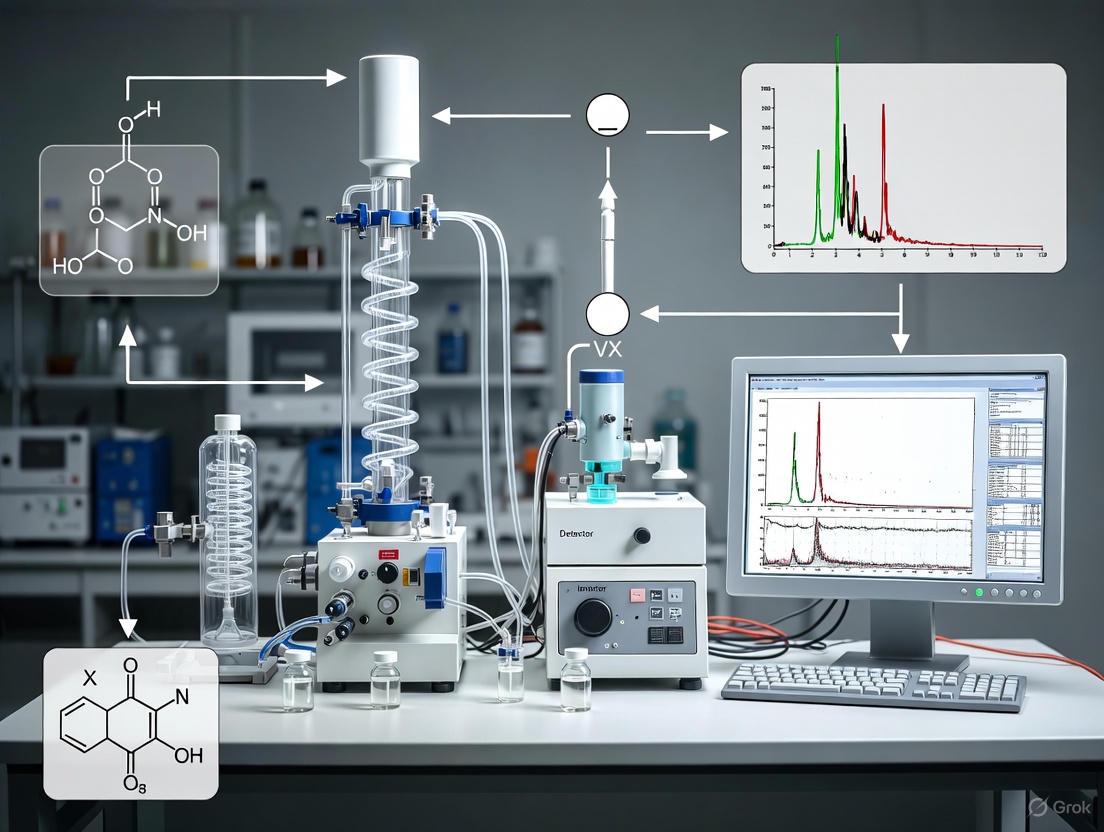
Chemical Warfare Agents (CWAs) are toxic substances intended to cause intentional death or harm through their toxic properties, with munitions and devices designed for their weaponization also falling under the definition of chemical weapons [1]. The analysis and identification of these agents are critical for international security, forensic investigations, and environmental monitoring, supporting the mandates of the Chemical Weapons Convention (CWC) [2] [3]. Gas Chromatography-Mass Spectrometry (GC-MS) has emerged as a preeminent analytical technique in this field, combining high separation efficiency with definitive identification capabilities [2] [4]. These application notes provide a structured overview of major CWA classifications, detailed experimental protocols for GC-MS analysis, and current data presentation formats essential for researchers and analytical chemists engaged in CWA identification.
CWA Classifications, Toxic Mechanisms, and Analytical Challenges
Chemical warfare agents are categorized primarily by their physiological effects on humans. The four principal classes—nerve, blister, choking, and blood agents—each present unique mechanisms of toxicity and associated analytical challenges [1] [5].
Table 1: Classification of Major Chemical Warfare Agents
| Agent Class | Representative Agents (Common Code) | Primary Toxic Mechanism | Key Physical Properties |
|---|---|---|---|
| Nerve Agents | Sarin (GB), Soman (GD), Tabun (GA), VX [1] [5] | Inhibit acetylcholinesterase (AChE), causing nervous system hyperstimulation [1] [6] | Varying volatility; G-agents are more volatile than V-agents [5] |
| Blister Agents | Sulfur Mustard (HD), Lewisite (L), Nitrogen Mustard (HN) [1] [5] | Alkylating agents causing severe skin, eye, and respiratory tract damage [5] | Persistent liquids with low to moderate volatility [5] |
| Choking Agents | Phosgene (CG), Chlorine (Cl) [1] [5] | Damage lung-blood barrier, causing pulmonary edema and asphyxia [5] | Typically gaseous or highly volatile [5] |
| Blood Agents | Hydrogen Cyanide (AC), Cyanogen Chloride (CK) [1] [5] | Inhibit cellular cytochrome c oxidase, disrupting oxygen use [1] [5] | Generally volatile with rapid effects [5] |
Advanced and Fourth-Generation Agents
A notable category beyond the traditional classes includes Fourth-Generation Agents (FGAs), also known as Novichoks. Developed to be highly toxic, untraceable, and undetectable, these low-volatility nerve agents evaporate even less readily than VX and are at least as potent [1]. The Organisation for the Prohibition of Chemical Weapons (OPCW) has added Novichok-related chemical families to Schedule 1 of the CWC's Annex on Chemicals, underscoring the need for continuous analytical method development [1].
Analytical Techniques for CWA Detection
A range of analytical techniques is employed for CWA detection, each with distinct advantages and limitations concerning sensitivity, selectivity, portability, and applicability to different sample matrices [2].
Table 2: Comparison of CWA Detection Techniques
| Analytical Technique | Key Principles | Advantages | Disadvantages/Limitations |
|---|---|---|---|
| GC-MS & Portable GC-TMS | Chrom. separation followed by mass spectral identification [2] [4] | High sensitivity & selectivity; capable of identifying unknowns; portable versions available [2] [4] | Complex sample prep; can be costly; requires skilled operators [2] |
| Ion Mobility Spectrometry (IMS) | Separation of gas-phase ions based on mobility in a drift tube [2] [3] | Rapid response; low LOD; portable and easy to operate [3] | Prone to false alarms; susceptible to contamination; poor selectivity [3] |
| Flame Photometry (FPD) | Detection of P or S species via flame excitation and optical emission [2] [7] | Highly sensitive for P- and S-containing agents (e.g., nerve & blister agents) [7] | Limited to P/S compounds; prone to false positives from other P/S sources [3] |
| Fluorescent Probes | Selective chemical reaction induces fluorescent signal change [3] | High sensitivity & selectivity; potential for real-time imaging in biological systems [3] | Requires design of specific probe molecules for each agent class [3] |
| Raman Spectroscopy | Inelastic scattering of light providing vibrational fingerprint [3] | Can detect through glass containers; non-destructive [3] | Requires a window for light; struggles with low CWA concentration in mixtures [3] |
Recent comparative studies highlight the performance of advanced GC methods. For instance, GC-ICP-MS (Inductively Coupled Plasma Mass Spectrometry) has demonstrated superior sensitivity for organophosphorus nerve agents like sarin and soman, achieving limits of detection (LODs) of ≈0.12-0.14 ng/mL, significantly lower than the ≈0.36-0.43 ng/mL LODs of GC-FPD (Flame Photometric Detection) [7]. Furthermore, comprehensive two-dimensional GC coupled with time-of-flight MS (GC×GC-TOFMS) has proven highly effective for complex tasks like impurity profiling of CWA precursors, enabling the identification of dozens of unique compounds for forensic tracking [8].
Detailed GC-MS Experimental Protocol for CWA Analysis
The following protocol details a standard methodology for the identification of trace-level CWAs in environmental samples using Gas Chromatography-Mass Spectrometry.
Safety Precautions and Sample Handling
- Personal Protective Equipment (PPE): Perform all work in a certified fume hood or biological safety cabinet. Wear appropriate PPE: lab coat, gloves, and safety glasses.
- Decontamination: Have neutralizing solutions available for immediate decontamination of spills. All waste must be disposed of as hazardous chemical waste according to institutional regulations.
Materials and Equipment
- Gas Chromatograph-Mass Spectrometer: Equipped with a split/splitless injector and an autosampler.
- Analytical Column: MXT-5 or equivalent low-bleed capillary column (5 m × 0.1 mm, 0.4 µm film thickness) [4].
- Carrier Gas: High-purity Helium (He).
- SPME Assembly: Solid-Phase Microextraction syringe with a 65-µm polydimethylsiloxane/divinylbenzene (PDMS/DVB) fiber [4].
- Vials: 2-mL glass autosampler vials with crimp-top caps and PTFE/silicone septa.
- Standards and Reagents: CWA analytical standards or simulants in suitable solvents (e.g., isopropanol, dichloromethane). Warning: Work with authentic CWAs must only be conducted in specialized, high-containment facilities.
Sample Preparation (SPME)
- Liquid Samples: For aqueous samples, transfer 1 mL to a 2-mL GC vial. For samples in organic solvent, use 0.5-1 mL.
- Headspace Sampling: For solid or soil samples, place a representative amount (e.g., 0.5 g) into a 10-mL headspace vial and seal.
- SPME Extraction:
- Pierce the vial septum with the SPME needle.
- Extend the fiber and immerse it directly into the liquid sample (for liquid samples) or expose it to the headspace (for solid samples or volatile analytes).
- Extract for a defined time (5-30 seconds) while agitating the sample if possible to enhance extraction efficiency [4].
- Retract the fiber and withdraw the assembly from the vial.
Instrumental Analysis (GC-MS)
GC Conditions [4]:
- Injector: Split injection (split ratio 1:20) at 250°C.
- Oven Program: Initial temp 50°C, ramp at 2°C/s to 270°C, hold for 1-2 minutes.
- Carrier Gas Flow: Constant helium flow, e.g., 1.0 mL/min.
- Total Run Time: Approximately 3-5 minutes, including cool-down time.
MS Conditions [4]:
- Ionization Mode: Electron Ionization (EI) at 70 eV.
- Ion Source Temperature: 230°C.
- Mass Range: 50 - 500 m/z.
- Scan Rate: 10-15 scans per second.
Sample Injection:
- Insert the SPME needle into the GC injector and rapidly expose the fiber for thermal desorption (typically 1-2 minutes).
- Retract the fiber and withdraw the needle.
Data Analysis and Compound Identification
- Peak Deconvolution: Use embedded software (e.g., Ion Signature) to deconvolute co-eluting peaks based on their unique mass spectral signatures [4].
- Library Search: Compare the mass spectrum of each chromatographic peak against commercial (e.g., NIST) and user-developed CWA spectral libraries.
- Confirmation: Confirm the identity of a detected agent by matching both its retention time and mass spectrum against an authenticated standard analyzed under identical conditions. A minimum library match factor of 80% is often used as a preliminary identification criterion, though definitive confirmation requires standard comparison.
Workflow Visualization
The following diagram illustrates the logical workflow for the GC-MS analysis of CWAs, from sample collection to final reporting.
The Scientist's Toolkit: Key Research Reagents and Materials
The following table lists essential materials and reagents for conducting GC-MS-based analysis of chemical warfare agents.
Table 3: Essential Research Reagents and Materials for CWA GC-MS Analysis
| Item | Function/Application |
|---|---|
| SPME Fibers (65µm PDMS/DVB) | Concentrates trace analytes from liquid, headspace, or solid samples for sensitive GC-MS analysis [4]. |
| MXT-5 or DB-5MS Capillary Column | Standard low-polarity stationary phase for high-resolution separation of a wide range of CWAs and related compounds [4]. |
| CWA Analytical Standards & Simulants | Essential for method development, calibration, quality control, and definitive identification of unknown agents. |
| Deuterated Internal Standards (e.g., D₅-EDPA) | Improves quantitative accuracy by correcting for variability in sample preparation and instrument response. |
| Sulfinert-Treated Vials & Liners | Provides an inert surface to prevent analyte adsorption and decomposition, crucial for labile compounds [4]. |
| CWA Mass Spectral Library | Custom or commercial library containing electron ionization (EI) spectra of CWAs and degradation products for reliable identification. |
The accurate identification and classification of chemical warfare agents remain a critical component of international security and public health protection. GC-MS, with its high separation power and definitive mass spectral identification, serves as a cornerstone technique in both laboratory and field-deployable formats. The protocols and data outlined in these application notes provide a framework for researchers to implement robust analytical methods. The field continues to advance with the development of more sensitive, selective, and portable technologies like GC-ICP-MS and comprehensive two-dimensional GC-MS to meet the evolving challenges posed by both traditional and novel threat agents [7] [8].
Organophosphorus compounds (OPs), which include many pesticides and potent chemical warfare agents (CWAs), represent a class of chemicals of significant concern for public health and military safety [9] [10]. Their primary mechanism of toxicity, common to all OPs, is the inhibition of the enzyme acetylcholinesterase (AChE) [10]. This inhibition initiates a cascade of biochemical events leading to a cholinergic crisis, which can be fatal [11] [12]. The extreme toxicity of nerve agents such as sarin, soman, and VX, with lethal doses in the ppm range, necessitates robust detection and identification methods [13]. Within the context of research focused on gas chromatography-mass spectrometry (GC-MS) identification of CWAs, a deep understanding of these toxicity mechanisms is paramount. It informs the selection of biomarkers, the development of sample preparation protocols, and the interpretation of analytical data for both forensic and diagnostic purposes. This application note details the molecular mechanisms of OP toxicity and provides standardized experimental protocols for studying these processes in a research setting.
Cholinergic Toxicity: The Primary Mechanism of Action
Acetylcholinesterase Inhibition
The fundamental toxic event in OP poisoning is the covalent inhibition of acetylcholinesterase (AChE), the enzyme responsible for terminating the signal of the neurotransmitter acetylcholine (ACh) in cholinergic synapses of the central and peripheral nervous systems, as well as neuromuscular junctions [14] [10].
- Catalytic Function of AChE: AChE exhibits an extraordinarily high catalytic activity, hydrolyzing approximately 25,000 molecules of ACh per second into acetate and choline [14]. The active site contains a catalytic triad of serine, histidine, and glutamate residues. The hydrolysis proceeds through a transesterification reaction where the serine hydroxyl group attacks the substrate, forming an acyl-enzyme intermediate [14].
- Mechanism of OP Inhibition: Organophosphorus compounds act as pseudosubstrates for AChE. They phosphorylate the critical serine hydroxyl group within the enzyme's active site, forming a stable, covalently bound organophosphate-enzyme complex (Figure 1) [14] [10]. This phosphorylation event blocks the enzyme's ability to bind and hydrolyze ACh.
Figure 1. Mechanism of AChE inhibition by organophosphorus compounds. The OP phosphorylates a serine residue in the AChE active site, forming a stable complex that prevents ACh hydrolysis, leading to neurotransmitter accumulation.
Consequences of Acetylcholine Accumulation
The inhibition of AChE results in a rapid accumulation of ACh in the synaptic cleft, causing hyperstimulation of both muscarinic and nicotinic cholinergic receptors. This overstimulation manifests as a complex clinical picture known as cholinergic toxidrome [12].
Table 1: Clinical Manifestations of Cholinergic Crisis from OP Poisoning
| Receptor Type | Location | Effects of Overstimulation |
|---|---|---|
| Muscarinic (Parasympathetic) | Glands, Smooth Muscle, Heart | SLUDGE Syndrome: Salivation, Lacrimation, Urination, Defecation, Gastrointestinal upset, Emesis. Also: Miosis, Bradycardia, Bronchospasm, Bronchorrhea [12] [15]. |
| Nicotinic | Neuromuscular Junctions | Muscle fasciculations, myoclonic jerking, flaccid paralysis, tachycardia, hypertension [12] [10]. |
| Central Nervous System | Brain | Anxiety, confusion, drowsiness, emotional lability, seizures, status epilepticus (SE), coma, and respiratory depression [12] [10] [15]. |
The primary cause of death in acute OP poisoning is respiratory failure, which results from a combination of central apnea (depression of the brainstem respiratory center), bronchospasm, bronchorrhea, and paralysis of the respiratory muscles [10].
Neurotoxicity and Secondary Mechanisms
Seizures and Status Epilepticus (SE)
A severe consequence of acute, high-dose OP exposure is the rapid induction of seizures, which can progress to status epilepticus (SE) [10]. The amygdala, particularly the basolateral nucleus, is a key brain region in the initiation of OP-induced seizures [10]. The excessive ACh leads to an imbalance in excitatory (glutamate) and inhibitory (GABA) neurotransmission, triggering self-sustaining seizure activity.
Excitotoxicity and Neuroinflammation
Prolonged SE activates secondary neurotoxic pathways that contribute to long-term brain damage (Figure 2):
- Excitotoxicity: Seizure activity causes a massive release of glutamate. The overactivation of glutamate receptors (e.g., NMDA receptors) leads to excessive calcium (Ca++) influx into neurons, triggering enzymatic processes that cause oxidative stress and cell death [10].
- Neuroinflammation: Neuronal damage activates microglia and astrocytes, leading to the release of pro-inflammatory cytokines, which can further exacerbate brain injury [10].
Figure 2. Secondary neurotoxic pathways in acute OP poisoning. Initial cholinergic hyperstimulation triggers seizures and status epilepticus (SE), which subsequently activate excitotoxicity and neuroinflammation, leading to irreversible brain damage.
Experimental Protocols for Toxicity Mechanism Studies
Protocol 1: In Vitro AChE Inhibition Assay
Objective: To quantify the inhibitory potency of an organophosphorus CWA or pesticide on acetylcholinesterase.
Principle: The rate of ACh hydrolysis by AChE is measured spectrophotometrically. Inhibition by an OP reduces the reaction rate proportionally to its concentration and potency [14].
Materials:
- Purified electric eel or human erythrocyte AChE.
- Substrate: Acetylthiocholine iodide (ATC).
- Chromogenic reagent: 5,5'-Dithio-bis-(2-nitrobenzoic acid) (DTNB, Ellman's reagent).
- Organophosphorus compound (e.g., parathion, paraoxon, or a CWA simulant).
- Phosphate buffer (0.1 M, pH 8.0).
- Microplate reader or spectrophotometer.
Procedure:
- Enzyme Incubation: Prepare a series of tubes containing a fixed activity of AChE in phosphate buffer. Add varying concentrations of the OP inhibitor. Incaculate at 25°C for a fixed time (e.g., 10-30 minutes).
- Reaction Initiation: Add DTNB and ATC to the mixture to start the enzymatic reaction.
- Kinetic Measurement: Immediately monitor the increase in absorbance at 412 nm for 2-5 minutes. The yellow anion 2-nitro-5-thiobenzoate is produced from the reaction of DTNB with thiocholine, the hydrolysis product of ATC.
- Data Analysis: Calculate the reaction velocity (ΔA/min) for each inhibitor concentration. Plot the residual enzyme activity (%) versus the log of the inhibitor concentration to determine the IC₅₀ value.
Protocol 2: Analysis of OPs in Biological Samples by GC-NPD
Objective: To extract and quantify organophosphorus compounds from serum for toxicokinetic studies.
Principle: Organophosphorus insecticides are extracted from acidified serum using an organic solvent mixture. The extract is concentrated and analyzed using Gas Chromatography with a Nitrogen-Phosphorus Detector (GC-NPD), which provides high sensitivity and selectivity for these compounds [16].
Materials:
- Serum samples.
- Organic solvents: Acetone, Diethyl Ether, n-Hexane (HPLC grade).
- 5N Hydrochloric Acid (HCl).
- Anhydrous Sodium Sulphate.
- Nitrogen evaporator.
- GC System equipped with an NPD. Column: 10% SG-30 on CHW-PW support (1.2 m x 4 mm i.d.) [16].
Procedure:
- Extraction:
- To 1 mL of serum in a glass tube, add 4 mL of acetone:diethyl ether (1:1 v/v).
- Shake vigorously for 5 minutes.
- Acidify with 0.2 mL of 5N HCl and shake again briefly.
- Separate the organic layer and repeat the extraction twice with 4 mL diethyl ether.
- Combine all organic supernatants and pass through 2 g of anhydrous sodium sulphate.
- Concentration:
- Evaporate the organic filtrate to dryness under a gentle stream of nitrogen gas.
- Reconstitute the residue in 0.2 mL of n-hexane.
- GC-NPD Analysis:
- Injector Temp.: 260°C
- Detector Temp.: 280°C
- Oven Program: 180°C for 1 min, then ramp at 6°C/min to 250°C, hold for 2 min.
- Carrier Gas (N₂) Flow: 60 mL/min
- Injection volume: 1-2 µL.
- Identification & Quantification: Identify compounds by their retention times relative to an internal standard (e.g., Diazinon). Quantify using a calibration curve prepared in the range of 0.25 - 4.0 µg/mL [16].
Advanced Detection and the Role of GC-MS in CWA Research
The extreme toxicity of CWAs demands detection technologies with exceptional sensitivity and specificity. While GC-NPD is effective for targeted analysis [16], confirmatory identification, especially in complex matrices, relies on mass spectrometry.
Table 2: Comparison of Analytical Techniques for Nerve Agent Detection
| Analytical Technique | Key Principle | Advantages | Estimated LOD for G-Agents | Primary Application |
|---|---|---|---|---|
| GC-FPD [13] | Element-specific emission from P/S in a H₂-air flame. | Selective, relatively simple, portable systems available. | ~0.36–0.43 ng/mL | Rapid field screening and environmental monitoring. |
| GC-ICP-MS [13] | Chromatographic separation with elemental (³¹P) mass spectrometric detection. | Ultra-trace sensitivity, high elemental selectivity, minimal interference. | ~0.12–0.14 ng/mL | Confirmatory analysis, forensic evidence, OPCW compliance testing. |
| GC-MS(/MS) [13] [2] | Chromatographic separation with molecular mass detection/fragmentation. | High confidence in identification, structural information, library matching. | Sub-ng/mL levels | Gold standard for confirmatory identification and forensic analysis. |
For GC-MS analysis of polar degradation products (e.g., alkyl methylphosphonic acids), a derivatization step (e.g., silylation) is required to make them volatile and amenable to GC separation [13]. The high selectivity and sensitivity of GC-ICP-MS and GC-MS/MS make them indispensable tools for verifying CWA exposure and studying their environmental fate and toxicokinetics at toxicologically relevant concentrations.
The Scientist's Toolkit: Key Research Reagents and Materials
Table 3: Essential Reagents for CWA Toxicity and Detection Research
| Reagent / Material | Function and Application in Research |
|---|---|
| Acetylcholinesterase (AChE) | Target enzyme for OPs. Used for in vitro inhibition assays to determine inhibitory potency (IC₅₀) of new compounds [14]. |
| Acetylthiocholine / DTNB | Substrate and chromogen for Ellman's assay. Allows spectrophotometric quantification of AChE activity [14]. |
| Atropine Sulfate | Muscarinic receptor antagonist. Used as an emergency antidote in in vivo studies and to probe muscarinic mechanisms [12] [15]. |
| Pralidoxime (2-PAM) Chloride | AChE reactivator. Used in experimental treatments to reverse OP-induced enzyme phosphorylation and study aging processes [12] [17]. |
| Internal Standards (e.g., D₅-DFP, Diazinon) | Isotopically labeled or structurally similar analogs. Added to samples for GC-MS or GC-NPD analysis to correct for variability in extraction and analysis [16]. |
| Silylation Derivatization Reagents | Chemicals like MTBSTFA. Used to derivative polar OP degradation products (phosphonic acids) for volatilization and analysis by GC-MS [13]. |
| Enzyme Reactivation Buffers | Specific pH buffers. Used in sample preparation for blood cholinesterase measurements to partially reverse inhibition and estimate total enzyme activity [11]. |
Gas Chromatography-Mass Spectrometry (GC-MS) stands as the undisputed reference technique for the definitive identification and quantification of chemical warfare agents (CWAs). Its unparalleled separation power, sensitivity, and specificity provide the confirmatory analysis essential for verification under the Chemical Weapons Convention (CWC), threat response, and forensic investigations. This application note details the instrumental configurations, methodologies, and performance data that solidify the status of GC-MS as an indispensable tool in the defense against chemical weapons. Framed within ongoing research on CWA identification, the protocols herein are designed for researchers, scientists, and professionals tasked with protecting public and military safety.
Chemical warfare agents (CWAs), particularly organophosphorus nerve agents such as sarin (GB), soman (GD), and VX, rank among the most toxic synthetic compounds known [2] [13]. Their high toxicity, which can be lethal at concentrations in the parts-per-million (ppm) to parts-per-billion (ppb) range, necessitates analytical methods capable of trace-level detection and unambiguous identification [13]. The analysis is further complicated by the need to detect not only the parent agents but also their degradation products and impurities in complex environmental and biological matrices [18] [19].
The Chemical Weapons Convention (CWC), overseen by the Organisation for the Prohibition of Chemical Weapons (OPCW), mandates rigorous verification of compliance, for which definitive analytical data is required [2] [20]. While a range of detection technologies exists—including ion mobility spectrometry (IMS), flame photometry, and electrochemical sensors—for rapid, on-site screening, these techniques can lack the specificity and sensitivity for conclusive identification and are prone to false positives [2] [21]. In contrast, GC-MS combines the superior separation efficiency of gas chromatography with the powerful identification capability of mass spectrometry, making it the preferred method for laboratory confirmation and forensic analysis [18] [20] [19]. Its ability to provide a unique "molecular fingerprint" for each analyte is the cornerstone of its status as the gold standard.
Comparative Sensitivity of GC-MS Techniques
The sensitivity of GC-MS systems is paramount for detecting trace-level CWAs. The following table summarizes the performance of different GC-MS configurations as demonstrated in recent research.
Table 1: Comparison of GC-MS Technique Sensitivity for CWA Analysis
| Analytical Technique | Target Analytes | Limit of Detection (LOD) | Key Advantages | Application Context |
|---|---|---|---|---|
| GC-ICP-MS [13] | Sarin, Soman, Cyclosarin | 0.12 – 0.14 ng/mL | Ultra-trace sensitivity, elemental selectivity for phosphorus | Confirmatory analysis, environmental monitoring |
| GC-FPD [13] | Sarin, Soman, Cyclosarin | 0.36 – 0.43 ng/mL | Rapid screening, cost-effective, selective for P/S | Field-portable preliminary monitoring |
| GC-MS/MS (Triple Quad) [19] | Tabun, Sarin, Soman, VX, and breakdown products | Picogram level (on-column) | High selectivity in complex matrices, robust for parent agents and derivatized products | High-confidence screening in biological samples (e.g., plasma) |
| TD-GC-MS (Full Scan) [20] | GB, VX, HD, Lewisites | Low ppt level in air | Solvent-free analysis, high enrichment factor for air samples | Field analysis of vapor hazards, on-site verification |
As evidenced by the data, GC-ICP-MS exhibits superior sensitivity for G-agent analysis, with detection limits approximately three times lower than those of GC-FPD [13]. Meanwhile, GC-MS/MS provides the exceptional selectivity required to analyze CWAs and their polar breakdown products in challenging biological matrices like plasma, achieving detection at picogram levels [19].
Detailed Experimental Protocols
Protocol A: Air Sampling and Thermal Desorption GC-MS for Vapor Analysis
This robust field method for determining traces of CWAs in air samples uses thermal desorption (TD) for high sensitivity [20].
- Principle: Air is drawn through a sorbent tube to trap and pre-concentrate analytes. The tube is then thermally desorbed in a GC inlet, transferring the entire sample to the column for separation and mass spectrometric detection.
- Workflow:
- Materials and Reagents:
- Sorbent Tubes: Glass thermal desorption liner packed with Tenax TA [20].
- GC-MS System: Agilent 7890/5975 GC-MSD or equivalent.
- GC Column: Mid-polarity capillary column (e.g., 5% Phenyl Methyl Silox, 30 m × 0.25 mm × 0.25 µm).
- Calibration Standards: Certified reference materials of target CWAs (e.g., GB, HD, VX) in appropriate solvents [20].
- Step-by-Step Procedure:
- Sample Collection: Draw a known volume of air (e.g., 1-10 L) through the Tenax TA-packed sorbent tube using a calibrated pump [20].
- Tube Installation: Place the sorbent tube into the programmable temperature vaporization (PTV) inlet of the GC.
- Thermal Desorption: Desorb the analytes by rapidly heating the PTV inlet to 270°C in splitless mode. Hold for 5-10 minutes to transfer all volatilized analytes to the GC column [20].
- Chromatographic Separation: Use a temperature ramp (e.g., 50°C for 2 min, then 20°C/min to 270°C) to achieve optimal separation.
- Mass Spectrometric Detection: Operate the mass spectrometer in full scan mode (e.g., m/z 50-500) for untargeted screening and library identification.
- Identification: Compare acquired mass spectra against certified CWA libraries (e.g., OPCW, NIST) [20].
Protocol B: Comprehensive Analysis of Nerve Agents and Breakdown Products in Plasma
This protocol uses a single GC-MS/MS method to detect both volatile nerve agents and their polar, non-volatile breakdown products in a single run, which is crucial for confirming exposure [19].
- Principle: Parent nerve agents are extracted directly from plasma. Polar breakdown products (alkyl methylphosphonic acids) are first chemically derivatized to increase their volatility before GC-MS/MS analysis.
- Workflow:
- Materials and Reagents:
- Internal Standards: Deuterated analogs of target analytes.
- Derivatization Reagent: N,O-Bis(trimethylsilyl)trifluoroacetamide (BSTFA) with 1% TMCS or similar silylation agent [13] [19].
- GC-MS/MS System: Agilent 7890A GC coupled to a 7000 series triple quadrupole mass spectrometer.
- GC Column: Mid-polarity column (e.g., DB-35ms or equivalent).
- Solid-Phase Extraction (SPE) Cartridges: For sample clean-up if necessary.
- Step-by-Step Procedure:
- Sample Preparation: Spike plasma samples with internal standards. For parent agents, proceed with liquid-liquid extraction using ethyl acetate. For breakdown products, acidify the plasma and extract the phosphonic acids [19].
- Derivatization: Evaporate the extract containing breakdown products to dryness. Add a derivatization reagent like BSTFA and heat (e.g., 70°C for 30 min) to form trimethylsilyl (TMS) derivatives [19].
- GC-MS/MS Analysis:
- Injection: 1-2 µL in pulsed splitless mode.
- GC Oven: Employ a fast temperature ramp to achieve separation within ~12.5 minutes.
- MS Detection: Operate the triple quadrupole in Multiple Reaction Monitoring (MRM) mode. Monitor specific precursor ion → product ion transitions for each parent agent and derivatized breakdown product [19].
- Identification and Quantification: Identify analytes by their specific retention times and MRM transitions. Quantify using the internal standard method with calibration curves.
The Scientist's Toolkit: Essential Research Reagents and Materials
Table 2: Key Reagents and Materials for CWA Analysis by GC-MS
| Item | Function/Application | Specific Examples |
|---|---|---|
| Mid-Polarity GC Columns | Optimal separation of diverse CWA mixtures and derivatives. | 5% Phenyl Methyl Silox, DB-35ms [22] [19] |
| Sorbent Tubes | Trapping and pre-concentration of CWAs from air/vapor samples. | Tenax TA [23] [20] |
| Derivatization Reagents | Volatilization of polar degradation products for GC analysis. | BSTFA, Pentafluoropropionic Anhydride [24] [19] |
| Solid-Phase Microextraction (SPME) | Solvent-less extraction and concentration for rapid sampling. | PDMS/DVB fiber [23] [21] |
| Certified Reference Materials | Method calibration, validation, and quality control. | Sarin, Soman, VX, Mustard Gas [23] [20] |
| Specialized Inlet Liners | For thermal desorption applications and minimizing active sites. | Tenax TA packed PTV liner [20] |
GC-MS remains the cornerstone of modern CWA analysis, a status earned through its unmatched versatility, sensitivity, and specificity. As demonstrated, its configurations range from highly sensitive confirmatory methods like GC-ICP-MS and GC-MS/MS to robust field-deployable TD-GC-MS systems. The continuous development of faster, more sensitive, and greener methods ensures that GC-MS will continue to be an indispensable tool for researchers and analysts committed to global security and the verification of the Chemical Weapons Convention. The protocols and data presented herein provide a framework for laboratories to implement this gold-standard technique for the definitive identification of chemical threats.
Chemical Warfare Agents (CWAs) represent a category of toxic chemicals used to cause intentional death or harm, posing a significant and persistent threat to global security [25]. These agents are systematically classified into several categories based on their physiological effects, including nerve agents, blister agents, choking agents, blood agents, and riot control agents [25]. The international community has established a regulatory framework centered on the Chemical Weapons Convention (CWC), which prohibits the development, production, stockpiling, and use of chemical weapons and mandates the destruction of existing stockpiles [25]. This article examines the evolution of CWA detection technologies, with a specific focus on the critical role of gas chromatography-mass spectrometry (GC-MS) and other analytical techniques in enabling precise identification and supporting international control measures. The continuous advancement of these technologies is paramount for global security, non-proliferation, and effective emergency response.
The Changing Threat Landscape and Global Response
The threat from chemical weapons has evolved significantly, with modern challenges including the potential for non-state actors to exploit technological advancements. The Global Congress on Chemical Security and Emerging Threats has highlighted concerns about the use of artificial intelligence (AI) to plan attacks or facilitate chemical synthesis, and the use of uncrewed systems like drones for dispersal, which increases their range and threat potential [26]. Fragmented regulatory controls continue to exacerbate the illegitimate diversion of chemical precursors [26].
In response, the international community has strengthened its collaborative efforts. Key institutions like the Organisation for the Prohibition of Chemical Weapons (OPCW) and INTERPOL work to cultivate a global, multi-sectoral culture of chemical security through information sharing, developing innovative strategies, and promoting cooperation [26] [25]. The global CWA detectors market, projected to reach approximately $279 million by 2025, reflects the ongoing investment in countermeasures, driven by geopolitical tensions and the need to safeguard civilian and military personnel [27].
Table 1: Categories of Chemical Warfare Agents and Their Primary Effects
| Agent Category | Primary Physiological Effects | Examples |
|---|---|---|
| Nerve Agents | Inhibit nervous system enzymes; cause seizures, paralysis, death [25] | Sarin (GB), VX, Soman (GD) [28] |
| Blister Agents | Damage eyes, respiratory tract, skin; cause severe blisters [25] | Sulfur Mustard (HD), Lewisite (L1) [29] |
| Choking Agents | Irritate lungs; cause fluid secretion (pulmonary edema) [25] | Phosgene, Chlorine [25] |
| Blood Agents | Inhibit cellular oxygen use, causing suffocation [25] | Hydrogen Cyanide [25] |
| Riot Control Agents | Irritate eyes and skin; cause temporary incapacitation [25] | Tear Gas, Pepper Spray [25] |
Evolution of CWA Detection Technologies
The methodologies for detecting CWAs have progressed from simple colorimetric tests to sophisticated instrumental analyses, each with distinct advantages and applications.
Early Detection Methods
Initial field detection relied on simple, rapid tools like M8 paper and M9 tape. M8 paper is a three-color detector that identifies liquid nerve and blister agents by changing color (yellow for G-series nerve agents, green for V-series, red for blister agents) [28]. M9 tape, worn on uniforms or equipment, provides constant monitoring by turning reddish in the presence of liquid or aerosolized nerve or blister agents, though it does not differentiate between them [28]. While critical for immediate, on-the-ground threat assessment, these methods lack the specificity and sensitivity required for definitive identification and are susceptible to false positives [21].
Advanced Instrumental Detection
Modern protocols employ advanced analytical techniques to achieve unambiguous identification. Ion Mobility Spectrometry (IMS) and various spectroscopic methods are used for on-site screening [29]. However, for definitive confirmation, gas chromatography-mass spectrometry (GC-MS) remains the gold standard due to its superior ability to separate complex mixtures and provide unique spectral fingerprints for each compound [21].
Recent technological strides have been focused on miniaturization and portability without sacrificing analytical power. Truly portable GC-MS systems, such as those utilizing toroidal ion trap mass spectrometers (TMS), are now available. These self-contained units, weighing under 28 lbs and capable of battery operation, bring laboratory-grade confidence to the field. They can detect CWAs at low concentrations with analysis cycle times of approximately 5 minutes, providing rapid, reliable data for time-sensitive decision-making [21].
Furthermore, research into other analytical techniques continues to advance. Near-Infrared Spectroscopy (NIRS) has recently been demonstrated as a viable method for CWA characterization. A 2025 study detailed a 3D-printed glass liquid cell that allows for the safe sampling and analysis of highly toxic CWAs like sarin, soman, VX, and sulfur mustard. NIRS offers practical advantages for on-site analysis, including rapid measurement (seconds), minimal sample heating, and extensive miniaturization potential, making it a promising complementary technology [29].
Table 2: Comparison of Modern CWA Detection Instrumentation
| Technology | Key Features | Analysis Time | Example Applications |
|---|---|---|---|
| Hand-Portable GC-MS [21] | High specificity/sensitivity; library-based auto-identification; ~28 lbs weight. | ~5 minutes per cycle | Field identification of CWAs and TICs in air, headspace, and liquids. |
| Benchtop LC-Orbitrap MS [30] | Ultra-high resolution (<1 ppm mass accuracy); high-throughput screening. | Varies by workflow | Forensic toxicology, food & environmental safety testing, non-targeted screening. |
| Near-Infrared (NIR) Spectroscopy [29] | Low-cost portable devices; minimal sample heating; safe for reactive materials. | Seconds | Safe, rapid characterization of liquid nerve and blister agents using a specialized cell. |
Detailed Experimental Protocols for CWA Identification
This section provides detailed methodologies for two advanced techniques relevant to modern CWA analysis.
Protocol: CWA Analysis Using Hand-Portable GC-TMS
This protocol outlines the procedure for rapid, automated detection of CWAs and Toxic Industrial Chemicals (TICs) using a hand-portable gas chromatograph coupled to a toroidal ion trap mass spectrometer (GC-TMS) [21].
- Principle: Sample components are separated by gas chromatography and then definitively identified by their unique mass spectra using a miniaturized mass spectrometer, providing a field-deployable, two-dimensional analysis.
- Key Equipment: Hand-portable GC-TMS system (e.g., GUARDION-7); SPME syringe with 65-μm polydimethylsiloxane-divinylbenzene (PDMS-DVB) fiber; LTM capillary GC column (e.g., MXT-5, 5 m × 0.1 mm, 0.4 μm df) [21].
- Procedure:
- Sample Collection: Using the SPME syringe, perform sampling by direct immersion of the fiber into a liquid sample or exposure to a vapor headspace for 5-30 seconds [21].
- Sample Injection: Insert the SPME fiber into the heated, Sulfinert-treated injection port of the GC-TMS for thermal desorption. Use a split injection method (e.g., split ratio 1:20) [21].
- Chromatographic Separation: Employ a fast temperature program (e.g., from 50 °C to 270 °C at a rate of 2 °C/s) using the Low Thermal Mass (LTM) capillary GC column. Helium is used as the carrier gas [21].
- Mass Spectrometric Detection: Detect eluting analytes with the TMS system. Set the mass scan range from 50 to 500 m/z, with a scan rate of 10-15 Hz. Typical mass resolution is 0.55 at m/z 91 [21].
- Automated Compound Identification: Use embedded deconvolution software (e.g., CHROMION-1) to automatically identify target analytes by matching both retention time and mass spectral data against a user-defined CWA/TIC library. Results are displayed in a tabular format [21].
The following workflow diagram summarizes the GC-TMS analytical process:
Protocol: Safe Liquid CWA Characterization Using NIR Spectroscopy
This protocol describes a safe method for acquiring Near-Infrared (NIR) spectra of highly toxic liquid CWAs using a custom 3D-printed glass liquid cell, as demonstrated in a 2025 study [29].
- Principle: NIR radiation interrogates a sealed liquid sample. The resulting absorption spectrum provides compound-specific information, allowing differentiation between CWA classes and individual agents within the same class.
- Key Equipment: Portable or benchtop NIR spectrometer; 3D-printed quartz glass liquid cell with PTFE spacer and insert; PTFE-coated screw caps [29].
- Safety Note: Experiments with live CWAs must be conducted in a designated High-Tox facility by specially trained personnel, in compliance with the Chemical Weapons Convention [29].
- Procedure:
- Cell Preparation: Verify that the clean, empty glass liquid cell is airtight. Remove the PTFE insert [29].
- Sample Loading: Carefully pipette approximately 100 μL of the CWA sample into the cell [29].
- Cell Sealing: Slowly reposition the PTFE insert back into the cell, ensuring the spacer creates a consistent path length (e.g., 0.5 mm). Tighten the lid to form a secure seal [29].
- Spectral Acquisition: Place the sealed cell into the NIR spectrometer. Record the NIR spectrum in diffuse reflectance mode. The analysis typically takes seconds [29].
- Data Validation: Compare the recorded spectrum against theoretical predictions (e.g., from Density Functional Theory calculations) or reference spectra to confirm agent identity [29].
The Scientist's Toolkit: Essential Research Reagents and Materials
Successful CWA analysis, particularly in a field or research setting, relies on a suite of specialized materials and reagents.
Table 3: Key Research Reagent Solutions for CWA Analysis
| Item | Function / Application |
|---|---|
| SPME Fiber (PDMS-DVB) [21] | Sample preparation; concentrates volatile/semi-volatile analytes from air, water, or solids for injection into GC-MS. |
| M8 Chemical Detection Paper [28] | Rapid field screening; colorimetrically detects and tentatively identifies liquid G-series/V-series nerve agents and blister agents. |
| M9 Chemical Detection Tape [28] | Continuous perimeter or personnel monitoring; changes color in presence of liquid or aerosolized nerve/blister agents. |
| 3D-Printed Glass Liquid Cell [29] | Safe NIR analysis; provides a sealed, single-use container for hazardous liquids, enabling safe spectral acquisition. |
| Polytetrafluoroethylene (PTFE) [29] | Reflective, chemically inert material used in NIR cells; provides a safe surface for contact with aggressive chemicals. |
| Sulfinert-Treated Injection Port [21] | GC system component; passivated surface reduces analyte adsorption and decomposition for more accurate results. |
| CWA/TIC Mass Spectral Library [21] | Data analysis; embedded user-defined library for automated compound identification based on retention time and mass spectrum. |
The evolution of CWA detection, from simple colorimetric papers to sophisticated, portable GC-MS and spectroscopic systems, mirrors the ongoing adaptation to a complex and changing threat landscape. The integration of high-specificity analytical techniques like GC-MS and LC-MS with international regulatory frameworks and global cooperation forms the cornerstone of modern chemical security. For researchers and scientists, the future lies in the continued refinement of these technologies—pushing the boundaries of sensitivity, speed, and miniaturization—while developing robust, standardized protocols. This relentless pursuit of analytical excellence is not merely a technical endeavor but a critical component of global efforts to uphold the禁令 of chemical weapons and protect human life from their devastating effects.
Within research aimed at advancing the identification of chemical warfare agents (CWAs) via gas chromatography–mass spectrometry (GC–MS), the use of live agents is precluded by extreme toxicity, stringent regulations, and the requirement for specialized containment facilities [31] [32]. Consequently, simulants—non-lethal chemicals that mimic key properties of CWAs—are indispensable tools for developing and validating analytical methods, testing decontamination procedures, and training personnel [31] [33]. This application note details essential safety protocols and analytical considerations for the rigorous and safe use of CWA simulants in a laboratory setting, framing them within the context of a GC-MS identification research workflow.
Simulant Selection and Rationale
The Imperative for Simulants
Chemical warfare agents, such as the nerve agents sarin (GB) and VX, are highly toxic synthetic chemicals whose use in research is tightly controlled by the Chemical Weapons Convention [31] [32]. Working with these live agents is dangerous and restricted to a small number of high-containment laboratories. Simulants provide a safe and ethically permissible alternative for the vast majority of research and development activities, minimizing risk to personnel and preventing contamination of equipment [31].
Common Simulants and Their Corresponding CWAs
The selection of a simulant must be driven by the specific research objective. No single simulant replicates all properties of a live agent; therefore, the choice depends on which characteristics (e.g., molecular structure, volatility, adsorption behavior) are most critical for the study. The table below summarizes well-characterized simulants for key CWAs.
Table 1: Common Chemical Warfare Agent Simulants and Their Properties
| Target CWA | Simulant | Chemical Name | Key Applications & Rationale | Safety Considerations |
|---|---|---|---|---|
| Sarin (GB), Soman (GD) | Diisopropyl fluorophosphonate (DFP) | Diisopropyl fluorophosphate | Used to study degradation pathways; contains the reactive P-F bond present in G-series agents [33]. | Toxic upon ingestion or inhalation; requires use of fume hood and appropriate PPE. |
| G-series Nerve Agents | Dimethyl methylphosphonate (DMMP) | Dimethyl methylphosphonate | Common simulant for material adsorption and degradation studies; lacks P-F bond [33]. | Low acute toxicity, but requires careful handling as a chemical hazard. |
| Soman (GD) | Diethyl malonate | Propanedioic acid, diethyl ester | Identified as suitable for human volunteer trials (HVTs) of decontamination [32]. | Low toxicity, non-corrosive, non-carcinogenic. |
| Sulfur Mustard (HD) | Methyl salicylate | 2-Hydroxybenzoic acid, methyl ester | Used in HVTs for decontamination; mimics physicochemical properties of vesicants [32]. | Oil of wintergreen; low toxicity at doses used in HVTs. |
| VX / TICs | Malathion | Diethyl 2-[(dimethoxyphosphorothioyl)sulfanyl]butanedioate | Organophosphorus pesticide; simulates structure and behavior of VX and toxic industrial chemicals [32]. | Toxic pesticide; must be handled with the same precautions as a potent chemical hazard. |
Safety and Handling Protocols
General Safety Principles
Although simulants are far less toxic than their live-agent counterparts, they are not without hazard. A rigorous safety mindset is paramount.
- Risk Assessment: Before beginning work, a comprehensive risk assessment must be conducted for each simulant, considering its toxicity, volatility, and potential routes of exposure (inhalation, dermal absorption, ingestion) [32].
- Personal Protective Equipment (PPE): Minimum PPE should include lab coats, safety glasses, and appropriate chemical-resistant gloves. Respiratory protection may be necessary when working with volatile simulants outside of a fume hood.
- Engineering Controls: All work with liquid or volatile simulants must be performed in a properly functioning chemical fume hood to prevent inhalation exposure [32].
- Hygiene: Avoid hand-to-mouth/eye contact and wash hands thoroughly after handling simulants, even when gloves are worn.
Suitability for Use
A systematic approach to simulant selection ensures both safety and experimental relevance. The following decision diagram outlines a workflow for evaluating and selecting a simulant for a given research application.
Experimental Protocols for GC-MS Analysis
Direct Analysis of Simulants Using Portable GC-TMS
For rapid, on-site analysis of simulants and toxic industrial chemicals, hand-portable gas chromatography-toroidal ion trap mass spectrometry (GC-TMS) systems offer a robust solution.
Table 2: Protocol for SPME/GC-TMS Analysis of CWA Simulants [4]
| Step | Parameter | Specification |
|---|---|---|
| Sample Introduction | Method | Solid-Phase Microextraction (SPME) |
| SPME Fiber | 65-μm polydimethylsiloxane/divinylbenzene (PDMS/DVB) | |
| Sampling | Direct immersion in liquid sample or headspace for 5–30 seconds | |
| Injection | Thermal desorption in heated GC injection port (Sulfinert-treated) | |
| Gas Chromatography | Column | MXT-5, 5 m × 0.1 mm, 0.4 μm df |
| Temperature Program | 50 °C to 270 °C at 2 °C/s | |
| Carrier Gas | Helium (onboard cartridge) | |
| Injection Split Ratio | 1:20 | |
| Mass Spectrometry | Mass Analyzer | Toroidal Ion Trap (TMS) |
| Mass Range | 50 – 500 m/z | |
| Scan Rate | 10 – 15 Hz | |
| Data Analysis | Software | CHROMION with embedded peak deconvolution |
| Identification | Automated via user-defined library (retention time & mass spectrum) |
Procedure:
- Sample Collection: Using the SPME syringe, expose the fiber to the sample's headspace or immerse it directly into a liquid sample for a predetermined time (e.g., 30 seconds) to adsorb the target analytes [4].
- Sample Injection: Insert the SPME fiber into the GC injection port for thermal desorption. The split flow ensures a narrow analyte band enters the column.
- Chromatographic Separation: The low thermal mass (LTM) GC column rapidly heats, separating the simulants over approximately 150 seconds.
- Detection & Identification: The TMS system acquires mass spectra continuously. The software automatically deconvolutes co-eluting peaks and identifies compounds by comparing acquired data against a pre-defined library of target simulants, reporting results in a tabular format [4].
Analysis of Degradation Products via Derivatization GC-MS
The analysis of acidic degradation products, such as alkylphosphonic acids (APAs) from hydrolyzed nerve agents, is critical for forensic verification of CWA use. These polar, non-volatile compounds require derivatization for GC-MS analysis.
Table 3: Protocol for Derivatization of Acidic Degradation Products with TMSDAM [34]
| Step | Parameter | Specification |
|---|---|---|
| Sample Prep | Matrix | Aqueous samples or extracts |
| Pre-treatment | Cation exchange may be required for samples with high inorganic content. | |
| Derivatization | Reagent | Trimethylsilyldiazomethane (TMSDAM), 2.0 M in hexane |
| Solvent System | Methanol added to 10-20% (v/v) to facilitate reaction | |
| Reaction | 30 minutes at 60 °C | |
| Quenching | Add acidic solvent (e.g., 0.1% formic acid in methanol) to stop reaction. | |
| GC-MS Analysis | Instrument | Standard Bench-top GC-MS System |
| Column | Standard non-polar or mid-polar capillary GC column | |
| Detection | Electron Impact (EI) Mass Spectrometry |
Procedure:
- Sample Preparation: If the aqueous sample or extract has a high salt content, pass it through a cation-exchange cartridge to reduce background interference [34].
- Derivatization: Transfer an aliquot of the sample to a derivatization vial. Add methanol to a final concentration of 10-20% (v/v). Add a molar excess of TMSDAM reagent. Cap the vial and heat at 60 °C for 30 minutes.
- Reaction Quenching: After cooling, add a small volume of 0.1% formic acid in methanol to consume any excess TMSDAM and stabilize the methylated derivatives [34].
- GC-MS Analysis: Inject the derivatized sample directly into the GC-MS. The methyl ester derivatives of the APAs (e.g., isopropyl methylphosphonic acid from sarin hydrolysis) are now volatile and chromatographically separable, enabling identification based on retention time and mass spectral fragmentation.
The Scientist's Toolkit: Research Reagent Solutions
Table 4: Essential Materials for CWA Simulant Research by GC-MS
| Item | Function/Description |
|---|---|
| Hand-Portable GC-TMS | A self-contained, battery-operated instrument for rapid, on-site separation and identification of simulants in various matrices [4]. |
| SPME Fibers (PDMS/DVB) | For solvent-free sampling and concentration of volatile and semi-volatile simulants from air, headspace, and liquids [4]. |
| Trimethylsilyldiazomethane (TMSDAM) | A safe, non-explosive alternative to diazomethane for methylating polar acidic degradation products (e.g., APAs) for GC-MS analysis [34]. |
| Low Thermal Mass (LTM) GC Columns | Enable very fast temperature programming and rapid chromatographic separations (under 3 minutes), crucial for high-throughput and field analysis [4]. |
| CWA Simulant Library | A user-defined, embedded library of target simulants containing characteristic retention times and mass spectra for automated compound identification [4]. |
| Cation Exchange Cartridges | Used for sample clean-up prior to derivatization to remove interfering inorganic ions from environmental samples [34]. |
Practical GC-MS Methodologies for CWA Detection and Analysis
Within the framework of research dedicated to the identification of chemical warfare agents (CWAs) by gas chromatography-mass spectrometry (GC-MS), sample preparation is a critical determinant of success [35]. CWAs and their degradation products are frequently present in complex environmental and industrial matrices at trace levels, necessitating efficient and robust preparation methods to isolate, concentrate, and convert analytes into a form amenable to GC-MS analysis [36] [37]. The precision of the subsequent chromatographic separation and mass spectrometric detection is fundamentally contingent upon the efficacy of these preliminary steps. This document provides detailed application notes and protocols for three pivotal sample preparation strategies—derivatization, extraction, and purge-and-trap—tailored specifically for CWA research.
Derivatization Techniques
Derivatization is employed to chemically modify polar, non-volatile, or thermally labile CWA degradation products to enhance their volatility, thermal stability, and chromatographic behavior [38] [37]. This is essential for the GC-MS analysis of many phosphorus acids, arsenicals, and other polar markers that result from the hydrolysis or oxidation of CWAs.
Key Derivatization Reagents and Protocols
Table 1: Common Derivatization Reagents for CWA Degradation Products
| Analyte Class | Example Compounds | Derivatization Reagent | Derivatization Product | Key Reference |
|---|---|---|---|---|
| Alkylphosphonic Acids | Methylphosphonic Acid | Pentyldimethylchlorosilane / N-Methylimidazole (Mediator) | Pentyldimethylsilyl esters [37] | |
| Alkylphosphonic Acids | Pinacolyl Alcohol | Phenyldimethylchlorosilane | Phenyldimethylsilyl ether [37] | |
| β-Amino Alcohols | Degradation products of VX | Silylation reagents mediated by N-Methylimidazole | Silylated derivatives [37] | |
| Lewisite Metabolite | 2-Chlorovinylarsonous Acid | 1,2- Ethanedithiol | Stable cyclic derivative [37] | |
| Weak Acids (e.g., CWAs degradation products) | Various | p-Tolyl Isocyanate | Urea-type derivatives [37] |
Detailed Protocol: Silylation of Alkylphosphonic Acids
This protocol is adapted from methods developed for the analysis of nerve agent degradation products [37].
- Reagents and Materials: Target alkylphosphonic acid, Pentyldimethylchlorosilane, N-Methylimidazole, Anhydrous Pyridine, GC-MS vial.
- Procedure:
- Transfer a dried residue of the alkylphosphonic acid standard or sample extract to a GC-MS vial.
- Add 50 µL of anhydrous pyridine to the vial.
- Add 10 µL of N-methylimidazole, followed by 10 µL of pentyldimethylchlorosilane.
- Securely cap the vial and vortex the mixture for 30 seconds.
- Heat the vial at 80°C for 30 minutes.
- Allow the vial to cool to room temperature. The derivatized sample is now ready for GC-MS analysis.
- Notes: N-Methylimidazole acts as a catalyst, enabling rapid and mild derivatization at room temperature. All reagents and solvents must be anhydrous to prevent hydrolysis of the derivatizing agent.
Detailed Protocol: Derivatization of Lewisite Metabolite
This protocol describes the derivatization of 2-chlorovinylarsonous acid (CVAA), a key metabolite of lewisite, using 1,2-ethanedithiol (EDT) [37].
- Reagents and Materials: 2-Chlorovinylarsonous acid standard, 1,2- Ethanedithiol (EDT), Sodium acetate buffer (pH 6.5), GC-MS vial.
- Procedure:
- Add 1 mL of a urine or water sample containing CVAA to a glass vial.
- Add 500 µL of sodium acetate buffer (pH 6.5).
- Add 50 µL of EDT.
- Cap the vial and mix vigorously for 2 minutes.
- Incubate the mixture at 60°C for 15 minutes.
- After cooling, extract the derivatized product with a suitable organic solvent (e.g., hexane) for GC-MS analysis.
- Notes: The reaction with EDT forms a stable, volatile cyclic dithioarsinite ideal for GC-MS, significantly improving the detectability of this lewisite biomarker.
Extraction Techniques
Extraction is fundamental for isolating target CWAs and their degradation products from complex matrices, concentrating the analytes, and reducing matrix interference [35] [39].
Solid-Phase Extraction (SPE)
SPE utilizes a solid sorbent to selectively retain analytes from a liquid sample, which are subsequently eluted with a strong solvent [40] [35].
Table 2: SPE Sorbents for CWA-Related Chemical Extraction
| Sorbent Chemistry | Mechanism | Typical Analytes | Example Product |
|---|---|---|---|
| Strong Cation Exchanger (SCX) | Cation exchange | Charged basic compounds, organic bases, catecholamines [40] | HyperSep SCX [40] |
| Strong Anion Exchanger (SAX) | Anion exchange | Weak acids, phenolic compounds, surfactants [40] | HyperSep SAX [40] |
| C18 | Reversed-phase hydrophobic | Non-polar to moderately polar compounds, trace organics in water [40] | HyperSep C18 [40] |
| Mixed-Mode (Polymeric) | Hydrophobic & ion exchange | Acidic or basic drugs of abuse from biological matrices [40] | HyperSep Retain-AX (acidic), Retain-CX (basic) [40] |
| Porous Graphitic Carbon (PGC) | Polar interactions | Highly polar and challenging species [40] | HyperSep Hypercarb [40] |
Detailed Protocol: Mixed-Mode SPE for Basic Compounds
This generic protocol is suitable for extracting basic CWA-related compounds from aqueous samples using a mixed-mode sorbent [40].
- Reagents and Materials: Aqueous sample, Mixed-mode Cation Exchange sorbent (e.g., HyperSep Retain-CX), Methanol, Acetonitrile, Deionized water, Ammonium acetate buffer, Elution solvent (e.g., dichloromethane:isopropanol:ammonium hydroxide), Vacuum manifold.
- Procedure:
- Conditioning: Condition the SPE cartridge with 3-5 mL of methanol, followed by 3-5 mL of deionized water or a weak buffer. Do not let the sorbent dry out.
- Loading: Pass the sample (pH adjusted to ensure analytes are charged) through the cartridge at a controlled flow rate of 2-5 mL/min.
- Washing: Wash the cartridge with 3-5 mL of a weak buffer (e.g., ammonium acetate) to remove salts and interfering compounds. Optionally, wash with 1-2 mL of methanol-water to remove further interferences.
- Drying: Dry the cartridge under vacuum for 5-10 minutes to remove residual water.
- Elution: Elute the target analytes with 3-5 mL of an organic elution solvent containing a small percentage of base (e.g., Dichloromethane:Isopropanol:Ammonium Hydroxide, 80:20:2 v/v/v). Collect the eluate.
- Concentration: Evaporate the eluate to dryness under a gentle stream of nitrogen and reconstitute in a small volume (e.g., 100 µL) of a solvent compatible with GC-MS injection.
Magnetic Dispersive Solid Phase Extraction (MDSPE)
MDSPE is a modern technique that utilizes magnetic microspheres as the sorbent, simplifying the extraction process by eliminating the need for centrifugation or filtration [36].
- Reagents and Materials: Iron oxide@Poly(methacrylic acid-co-ethylene glycol dimethacrylate) (Fe₂O₃@Poly(MAA-co-EGDMA)) sorbent [36], Organic liquid sample, External magnet, GC vial.
- Procedure (as applied to organic liquids like n-hexane or dodecane):
- Weigh 20 mg of the magnetic sorbent into a sample vial.
- Add 1 mL of the organic liquid sample spiked with the target CWA.
- Vortex the mixture for 2 minutes to ensure thorough contact between the sorbent and the sample.
- Separate the sorbent by applying an external magnet to the side of the vial, holding the particles while the supernatant is decanted.
- Wash the sorbent with 1 mL of a suitable solvent to remove weakly adsorbed matrix components.
- Elution: Add 1 mL of a stronger solvent (e.g., toluene) to the sorbent and vortex for 1 minute to desorb the analytes.
- Separate the eluent using the magnet and transfer it to a GC-MS vial for analysis.
Table 3: Performance Data of MDSPE for Organophosphorous Esters
| Parameter | Value/Range | Details |
|---|---|---|
| Linear Range | 0.1 - 3.0 µg mL⁻¹ | Correlation coefficient (r²) = 0.9966 - 0.9987 [36] |
| Repeatability (RSD %) | 4.5 - 7.6% | For organophosphorous esters in dodecane [36] |
| LOD (S/N=3) | 0.05 - 0.1 µg mL⁻¹ | In Selected Ion Monitoring (SIM) mode [36] |
| Recovery (%) | 53.8 - 97.3% | At spiking levels of 1 and 3 µg mL⁻¹ [36] |
Purge-and-Trap Techniques
Purge-and-trap (also known as dynamic headspace) is a solvent-free technique designed for the highly sensitive analysis of volatile organic compounds (VOCs) from liquid or solid samples [38] [39]. It is directly applicable to volatile CWAs.
Principle and Workflow
The sample is placed in a sealed vessel, and an inert gas (e.g., helium) is bubbled through it, purging the volatile analytes into the gas phase. The vapors are carried onto a trap containing an adsorbent material, which concentrates the analytes. After the purging cycle, the trap is rapidly heated and the analytes are desorbed directly onto the GC column.
Detailed Protocol: Analysis of Volatile CWAs in Water
This protocol is based on established environmental methods such as EPA Method 524.3 [41].
- Reagents and Materials: 5 mL aqueous sample, Purge-and-Trap system (e.g., Tekmar 3000), VOCarb 3000 trap or equivalent, Helium purge gas (high purity), GC-MS system.
- Instrumental Parameters:
- Purge Gas: Helium
- Purge Flow: 40 mL/min
- Purge Time: 11 minutes
- Purge Temperature: Ambient (or 35-40°C)
- Desorb Temperature: 225-250°C
- Desorb Time: 1-2 minutes
- Trap Bake: After desorption, bake trap at 260-270°C for 5-10 minutes to remove residual compounds.
- Procedure:
- Transfer a 5 mL water sample into a purge vessel.
- Connect the vessel to the purge-and-trap system.
- Initiate the method. The sample is purged with helium at the specified flow and time, transferring volatiles to the trap.
- Upon completion of the purge, the system automatically switches the trap into the desorb path, heats it rapidly, and backflushes the analytes onto the GC column with carrier gas.
- Start the GC-MS data acquisition simultaneously with the desorb process.
- Troubleshooting: Loss of highly volatile compounds like methylene chloride can indicate breakthrough [41]. Mitigation strategies include:
- Reducing purge time or volume.
- Ensuring the trap is cooling sufficiently between runs.
- Verifying trap integrity and checking for active sites.
- Confirming the trap backpressure is correctly set (e.g., 4-8 psi) to focus analytes effectively [41].
The Scientist's Toolkit
Table 4: Essential Research Reagent Solutions for CWA Analysis by GC-MS
| Reagent/Material | Function/Application | Key Considerations |
|---|---|---|
| N-Methylimidazole | Catalyst for rapid, mild silylation derivatization [37] | Enables room-temperature derivatization of amino alcohols and phosphonic acids. |
| Pentyldimethylchlorosilane | Silylation derivatizing agent [37] | Produces pentyldimethylsilyl esters for improved volatility and MS detection. |
| 1,2-Ethanedithiol (EDT) | Derivatization of Lewisite metabolites [37] | Forms a stable, cyclic complex with CVAA for sensitive GC-MS analysis. |
| Mixed-Mode SPE Sorbents | Extraction of ionic compounds from complex matrices [40] | Combines hydrophobic and ion-exchange mechanisms for high selectivity. |
| Magnetic Nanoparticle Sorbents | Dispersive solid-phase extraction [36] | Simplifies extraction workflow; no centrifugation or columns needed. |
| High-Purity Helium | Purge-and-trap carrier gas and GC carrier gas [41] | Essential for maintaining system inertness and high sensitivity. |
| Vocarb 3000 Trap | Adsorbent for volatile CWA collection in purge-and-trap [41] | A multi-bed trap designed for a broad range of VOCs. |
| Anhydrous Pyridine | Solvent for derivatization reactions [37] | Must be kept anhydrous to prevent reagent decomposition. |
Within the framework of advanced research into gas chromatography-mass spectrometry (GC-MS) for chemical warfare agent (CWA) identification, the selection of an appropriate chromatographic column is a critical determinant of analytical success. CWAs encompass diverse classes, including nerve agents, blistering agents, and incapacitating agents, each possessing distinct chemical properties that challenge separation science [18] [42]. The volatility of parent nerve agents contrasts sharply with the polar, non-volatile nature of their phosphonic acid degradation products, often necessitating derivatization prior to GC analysis [19] [42]. This application note provides a detailed protocol for selecting and optimizing GC stationary phases to achieve the rapid, sensitive, and unambiguous separation of CWAs and their markers in complex matrices, supporting forensic verification and toxicokinetic studies.
Stationary Phase Fundamentals and CWA Relevance
The efficacy of a GC separation is governed by the interactions between target analytes and the stationary phase coated onto the inner wall of a capillary column. For CWA analysis, selectivity—the ability to distinguish between co-eluting compounds of different chemical classes—is paramount [43].
- Polarity and Selectivity: A stationary phase's polarity is determined by its functional groups. The general principle of "like-dissolves-like" applies; polar phases retain polar analytes more strongly, while non-polar phases are better suited for non-polar compounds [44] [43]. However, selectivity, which arises from specific intermolecular forces (e.g., hydrogen bonding, dipole-dipole, dispersion), is often more critical than general polarity. For instance, a trifluoropropylmethyl polysiloxane phase exhibits high selectivity for analytes containing lone pair electrons, such as the phosphorus or nitrogen atoms prevalent in nerve agents [43].
- Mid-Polarity Phases as a Compromise: Research has demonstrated that mid-polarity stationary phases can serve as an effective universal choice for CWA analysis. A method utilizing a single mid-polarity column successfully achieved the rapid (12.5 min) and sensitive (picogram level) separation and detection of six nerve agents (tabun, sarin, soman, cyclosarin, VX, Russian VX) and their six corresponding, derivatized breakdown products (e.g., IMPA, PMPA, EMPA) in spiked human plasma [19]. This approach simplifies analytical procedures that would otherwise require multiple columns with different stationary phases.
Table 1: Common GC Stationary Phases and Their Applicability to CWA Analysis
| Stationary Phase Composition (USP Nomenclature) | Relative Polarity | Max Temp (°C) | Selectivity Features | Relevance to CWA Analysis |
|---|---|---|---|---|
| 100% Dimethyl polysiloxane (G1) | Non-polar | 350-400 | Separates by boiling point [43] | Good for volatile parent agents (e.g., sarin, soman) [43] |
| 5% Diphenyl/95% dimethyl polysiloxane (G27) | Low-intermediate polarity | 350-400 | Slightly increased polarity vs. 100% dimethyl | Common general-purpose phase; suitable for a wide range of CWAs [43] |
| 35% Diphenyl/65% dimethyl polysiloxane (G42) | Intermediate polarity | 320 | Enhanced selectivity for aromatic and unsaturated compounds | Useful for mustard gas and related aromatics [43] |
| 50% Diphenyl/50% dimethyl polysiloxane (e.g., Rxi-17) | Mid-polarity | 320 | Balanced selectivity | Effective single-column analysis of nerve agents and silylated breakdown products [19] |
| 14% Cyanopropylphenyl/86% dimethyl polysiloxane (G46) | Intermediate polarity | 280 | Selective for polarizable compounds (e.g., pesticides, pharmaceuticals) | Suitable for polar CWA metabolites and derivatized phosphonic acids [43] |
| Trifluoropropylmethyl polysiloxane (G6) | Medium polarity | 340-360 | High selectivity for lone-pair electron-containing compounds (halogens, N, P) | Excellent for nerve agents (P-containing) and nitrogen mustards [43] |
| Polyethylene Glycol (WAX) | Highly polar | ~250 | Strong hydrogen bond acceptor | Ideal for very polar degradation products after derivatization [44] |
Optimized Experimental Protocols
Protocol 1: Single-Column Analysis of Nerve Agents and Their Breakdown Products in Plasma
This protocol, adapted from a published single-column GC-MS/MS method, allows for the simultaneous detection of parent nerve agents and their phosphonic acid biomarkers [19].
1. Sample Preparation (Derivatization of Breakdown Products):
- Principle: Hydrolytic breakdown products (alkyl methylphosphonic acids) are polar and non-volatile. They must be chemically derivatized to volatile trimethylsilyl (TMS) esters for GC analysis [19] [42].
- Procedure: a. Extract analytes from 1 mL of plasma (e.g., via solid-phase extraction). b. Evaporate the extract to complete dryness under a gentle stream of nitrogen. c. Add 50 µL of a silylation reagent, such as N,O-Bis(trimethylsilyl)trifluoroacetamide (BSTFA) containing 1% trimethylchlorosilane (TMCS). d. Vortex vigorously and heat at 60-80°C for 15-30 minutes. e. Cool and directly inject 1-2 µL into the GC system.
2. Instrumental Parameters [19]:
- GC System: Agilent 7890A Gas Chromatograph coupled to a 7000 Triple Quadrupole MS system.
- Column: Mid-polarity fused-silica capillary column (e.g., 30 m x 0.25 mm ID, 0.25 µm film thickness). A phase equivalent to 50% diphenyl/50% dimethyl polysiloxane is recommended.
- Carrier Gas: Helium, constant flow mode (e.g., 1.0 mL/min).
- Injection: Pulsed splitless mode, injector temperature at 250°C.
- Oven Temperature Program:
- Initial: 60°C (hold 1 min)
- Ramp 1: 20°C/min to 120°C
- Ramp 2: 10°C/min to 300°C (hold 3 min)
- Total Run Time: 12.5 minutes
- Detection: MS/MS in Multiple Reaction Monitoring (MRM) mode for high selectivity and sensitivity.
Protocol 2: Thermal Desorption-GC-MS for Air Sampling of Volatile CWAs
This protocol is designed for trapping and analyzing trace-level CWA vapors in air, crucial for environmental monitoring and hazard verification [20].
1. Air Sampling:
- Principle: Air is drawn through a sorbent tube to concentrate volatile and semi-volatile CWAs.
- Procedure: a. Use a sampling tube packed with ~150 mg of Tenax TA. b. Draw a known volume of air (e.g., 1-5 liters) through the tube using a calibrated air sampling pump. c. Seal the tube with PTFE-faced caps and store refrigerated if not analyzed immediately.
2. Instrumental Analysis via Inlet Thermal Desorption [20]:
- GC-MS System: Equipped with a Programmable Temperature Vaporization (PTV) inlet.
- Column: A low-polarity column such as 5% diphenyl/95% dimethyl polysiloxane is suitable for volatile agents like sarin (GB) and soman (GD).
- Thermal Desorption Parameters:
- Desorb the Tenax tube in the PTV inlet by rapidly heating to 270°C in splitless mode.
- Hold for 5-10 minutes to transfer analytes to the column head, which is cryofocused at a low initial temperature (e.g., 40°C).
- Oven Temperature Program:
- Initial: 40°C (hold 2 min)
- Ramp: 15°C/min to 280°C (hold 5 min)
- Detection: Full-scan MS (e.g., m/z 50-450) for untargeted screening and identification.
Table 2: Key Research Reagent Solutions for CWA Analysis
| Reagent / Material | Function / Application | Key Notes |
|---|---|---|
| BSTFA / TMCS | Silylation derivatization agent | Converts polar phosphonic acid metabolites into volatile TMS derivatives for GC analysis [19] [42]. |
| Tenax TA Sorbent | Air sampling and trapping | Efficiently adsorbs a wide range of volatile CWAs from air; thermally stable for desorption [20]. |
| Diazomethane (or TMS-diazomethane) | Alkylation derivatization agent | Methylates phosphonic acids to form methyl esters; requires careful handling due to toxicity and explosiveness [42]. |
| Amberlite XAD-4 Resin | Solid-phase extraction (SPE) | Used for pre-concentrating CWAs from large-volume water samples prior to analysis [18]. |
| ChiraSil-Val-L Column | Chiral separation | Capillary column used for resolving toxic stereoisomers of nerve agents like soman in 2D-GC [18]. |
Workflow Visualization
Discussion and Concluding Remarks
The strategic selection of the GC stationary phase is a foundational element in developing robust methods for CWA identification. While a mid-polarity column (e.g., 50% diphenyl/50% dimethyl polysiloxane) offers a powerful compromise for laboratories requiring a single-method approach to analyze both parent agents and their metabolites [19], specialized phases remain essential for specific challenges. These include trifluoropropylmethyl columns for enhanced selectivity toward organophosphorus compounds and chiral columns for resolving stereoisomers of nerve agents whose toxicities can differ dramatically [18] [43]. The integration of these column selection strategies with advanced sample preparation (e.g., derivatization, thermal desorption) and detection techniques (MS/MS) enables scientists to meet the rigorous demands of modern CWA analysis, from forensic verification to toxicological research. Future directions in this field will continue to leverage comprehensive chromatographic techniques like GC×GC-TOF-MS to untangle CWA signatures in increasingly complex sample matrices without extensive clean-up [45].
Gas Chromatography-Mass Spectrometry (GC-MS) is a cornerstone analytical technique for the identification of volatile and semi-volatile organic compounds, playing a critical role in security and defense applications for the detection and confirmation of chemical warfare agents (CWAs). The ionization method employed within the GC-MS system profoundly influences the type and quality of mass spectral data obtained, directly impacting the confidence of agent identification. Within the context of CWA research, where analytical certainty is paramount, selecting the appropriate ionization technique becomes a strategic decision. This application note provides a detailed comparative analysis of three principal ionization methods—Electron Ionization (EI), Chemical Ionization (CI), and Cold EI—evaluating their performance characteristics for the analysis of different classes of hazardous agents. We present structured experimental protocols, performance data, and decision frameworks to guide researchers in method selection and implementation for this highly specialized field.
Ionization Technique Fundamentals and Mechanisms
Electron Ionization (EI)
Principle of Operation: EI is a hard ionization technique where gas-phase analyte molecules are bombarded with high-energy electrons (typically 70 eV) emitted from a heated filament [46] [47]. This collision ejects an electron from the analyte molecule (M), producing a positively charged molecular ion (M⁺•) with an odd number of electrons: M + e⁻ → M⁺• + 2e⁻ [47]. The 70 eV standard is used because it corresponds to the de Broglie wavelength of typical organic bond lengths, maximizing energy transfer and ensuring reproducible fragmentation patterns across instruments [47] [48]. The excess energy internalized during this process typically causes the molecular ion to undergo extensive and characteristic fragmentation, generating a spectrum of fragment ions [46].
Key Features: The primary strength of EI lies in its highly reproducible, compound-specific fragmentation patterns, which serve as a molecular "fingerprint" [49]. This reproducibility has enabled the creation of extensive commercial mass spectral libraries (e.g., NIST, Wiley), allowing for automated spectral matching and rapid unknown identification [50] [49]. EI is a non-selective ionization technique, applicable to virtually any compound that can be vaporized.
Chemical Ionization (CI)
Principle of Operation: CI is a softer ionization technique that relies on gas-phase ion-molecule reactions [51]. A reagent gas (e.g., methane, ammonia, or isobutane) is introduced into the ion source at a relatively high pressure (~0.1-1 Torr) and is first ionized by an electron beam. The resulting reagent gas ions (e.g., CH₅⁺ from methane, NH₄⁺ from ammonia) subsequently collide and react with neutral analyte molecules (M). The most common reaction is proton transfer, producing a protonated molecular ion ([M+H]⁺): M + CH₅⁺ → [M+H]⁺ + CH₄ [51]. The exothermicity of this reaction is controlled by the choice of reagent gas, which allows for some control over the degree of fragmentation.
Key Features: The principal advantage of CI is the reduction of fragmentation, which often preserves the molecular ion in the form of [M+H]⁺ or other adduct ions, thereby providing clear information about the molecular weight of the analyte [51] [52]. This is particularly valuable for compounds that exhibit minimal or no molecular ion under standard EI conditions. A limitation of CI is that the spectra are more dependent on source conditions and reagent gas, preventing the creation of universal, transferable spectral libraries comparable to those for EI [51].
Cold Electron Ionization (Cold EI)
Principle of Operation: Cold EI is based on interfacing the GC and MS with a supersonic molecular beam (SMB) [50] [53]. The GC effluent, mixed with helium make-up gas, expands through a small nozzle into a vacuum chamber, forming a supersonic beam. During this adiabatic expansion, the sample molecules are vibrationally and rotationally cooled via collisions with the helium atoms [50]. These vibrationally cold molecules then fly through a dual-cage, contact-free ion source where they are ionized by 70 eV electrons. The reduced internal energy of the cold molecules prior to ionization is the key to the technique's performance.
Key Features: Cold EI provides a unique combination of the benefits of both EI and CI. It offers enhanced molecular ions—often by orders of magnitude—similar to soft ionization techniques, while simultaneously retaining the rich, library-searchable fragmentation patterns characteristic of standard EI [50] [54]. Furthermore, the SMB interface allows for the use of high column flow rates, which significantly lowers elution temperatures and extends the range of analyzable compounds to include many thermally labile and low-volatility substances that would otherwise degrade in a standard GC-MS system [50] [53].
Comparative Performance Analysis
The selection of an ionization method for CWA analysis involves trade-offs between molecular ion information, spectral libraries, fragmentation detail, and the range of analyzable compounds. The following table provides a structured comparison of these key performance aspects.
Table 1: Comparative Performance of EI, CI, and Cold EI for Agent Analysis
| Performance Characteristic | Electron Ionization (EI) | Chemical Ionization (CI) | Cold EI |
|---|---|---|---|
| Ionization Mechanism | High-energy electron bombardment [47] | Ion-molecule reaction with reagent gas [51] | EI of vibrationally cold molecules in a supersonic beam [50] |
| Molecular Ion Signal | Often absent or weak due to extensive fragmentation [50] [49] | Strong; appears as [M+H]⁺ or other adducts [51] | Significantly enhanced; often by a factor of 10-100 [50] [54] |
| Fragmentation | Extensive, provides structural information [46] [49] | Reduced, limited structural information [51] [52] | Similar to EI but with amplified high-mass fragments [50] |
| Spectral Libraries | Comprehensive libraries available (NIST, Wiley) [50] [49] | Not available due to non-uniform conditions [51] | Fully compatible with EI libraries, often with improved identification probability [50] [54] |
| Analyte Range | Limited to volatile, thermally stable compounds [50] | Similar to EI, but can be more sensitive for certain classes [51] | Extended range to low-volatility and thermally labile compounds [50] [53] |
| Best Suited For | Routine screening, library-based identification of stable agents | Molecular weight determination for labile agents or when EI lacks molecular ion | High-confidence identification of novel, labile, or complex agents requiring both molecular ion and library search |
Experimental Protocols
Protocol: Standard Electron Ionization (EI) Operation
Scope: This protocol defines the standard procedure for operating a GC-MS system in EI mode for the screening of unknown chemical agents.
Reagents and Materials:
- Calibration Standard: Perfluorotributylamine (PFTBA) or similar, for mass calibration and instrument tuning [50].
- GC Column: Standard 30m x 0.25mm ID, 0.25µm film thickness DB-5MS or equivalent.
- Carrier Gas: Ultra-high-purity Helium (He), set to a constant flow of 1.0 - 1.5 mL/min [54].
Procedure:
- System Setup: Ensure the MS ion source is clean. Set the ionization energy to 70 eV and the ion source temperature to 250 - 300 °C [47] [48].
- GC Conditions: Use a standard temperature ramp (e.g., 40°C for 2 min, ramp at 20°C/min to 320°C). Use a split/splitless injector at 250°C.
- Tuning: Introduce the PFTBA tuning standard and perform automatic mass calibration and instrument tuning to ensure optimal sensitivity and mass accuracy.
- Data Acquisition: Acquire data in full-scan mode (e.g., m/z 40-550) for unknown screening. For quantitative/trace analysis, selected ion monitoring (SIM) can be used.
- Data Analysis: Compare the acquired mass spectra against the NIST or other commercial EI mass spectral library. A match factor above 800 is generally considered a good fit, though the presence of characteristic high-mass ions increases confidence [54].
Protocol: Chemical Ionization (CI) Method Development
Scope: This protocol outlines the steps for developing a CI method when molecular weight information is required for an agent that fails to show a molecular ion in EI.
Reagents and Materials:
- Reagent Gases: Methane (for strong protonation), Ammonia (softer, selective protonation), or Isobutane [51].
- GC Column: As in Protocol 4.1.
Procedure:
- Mode Selection: Switch the ion source from EI to CI mode. This may involve software commands and, on some systems, a physical valve to introduce the reagent gas.
- Reagent Gas Selection:
- Pressure Optimization: Adjust the reagent gas flow to maintain an ion source pressure of approximately 0.5 - 1.0 Torr [51]. Monitor the total ion current (TIC) to optimize for stable signal.
- Data Acquisition and Interpretation: Acquire full-scan data. The primary ion of interest will typically be the [M+H]⁺ ion. For methane CI, also look for [M+C₂H₅]⁺ and [M+C₃H₅]⁺ adducts. In Negative Chemical Ionization (NCI), look for [M-H]⁻ or other anions, which provides exceptional sensitivity for electrophilic compounds like halogenated agents [51].
Protocol: Cold EI Method for Demanding Applications
Scope: This protocol describes the use of Cold EI for the analysis of challenging compounds, such as thermally labile agents or those requiring unambiguous molecular ion confirmation.
Reagents and Materials:
- Supersonic Molecular Beam (SMB) Interface: Equipped with a supersonic nozzle and vacuum pumps [50] [53].
- GC Column: A shorter column (e.g., 15m) can be used to leverage faster analysis speeds [54].
- Carrier and Make-up Gas: Ultra-high-purity Helium. The total flow rate (column + make-up) is typically set to 50 - 60 mL/min for optimal Cold EI performance [50] [54].
Procedure:
- System Configuration: Configure the GC-MS with the SMB interface. Set the transfer line and nozzle temperature to 250 - 300 °C [50].
- Gas Flow Setup: Set a high column flow rate (e.g., 8 mL/min) and add helium make-up gas to achieve a total flow of ~60 mL/min into the supersonic nozzle [54].
- Ionization: Set the electron energy to 70 eV within the fly-through ion source. The vibrational cooling of the analytes in the SMB will lead to enhanced molecular ions.
- Fast GC Analysis: Employ a fast temperature ramp (e.g., 40°C/min) due to the high carrier gas flow and the efficiency of the SMB interface, reducing analysis time significantly [54].
- Data Analysis: Process the acquired mass spectra using the standard NIST library. The spectrum will be searchable but will display a characteristically enhanced molecular ion, leading to high identification probability [50] [54].
Ionization Method Selection Workflow
The following diagram illustrates the decision-making process for selecting the optimal ionization method based on the research objective and analyte properties.
The Scientist's Toolkit: Essential Research Reagents and Materials
Table 2: Key Reagents and Materials for GC-MS Ionization Methods
| Item | Function/Application |
|---|---|
| Perfluorotributylamine (PFTBA) | Standard calibrant for mass axis calibration and instrument performance tuning in EI and Cold EI [50]. |
| Methane (CH₄) Gas | Common reagent gas for Chemical Ionization, provides strong protonation and relatively soft ionization [51]. |
| Ammonia (NH₃) Gas | Reagent gas for softer Chemical Ionization, selective for compounds with higher proton affinity than ammonia [51]. |
| Isobutane (C₄H₁₀) Gas | Reagent gas for CI, offers an intermediate softness between methane and ammonia [51]. |
| Supersonic Molecular Beam (SMB) Interface | Key hardware component for Cold EI, enables vibrational cooling of analytes and use of high GC flow rates [50] [53]. |
| Low-Bleed GC Columns (e.g., DB-5MS) | High-quality capillary columns essential for minimizing background interference, especially critical in trace CWA analysis. |
Comprehensive two-dimensional gas chromatography coupled with time-of-flight mass spectrometry (GC×GC-TOF-MS) represents a pinnacle in separation science, delivering unparalleled resolution for analyzing complex chemical mixtures. This advanced instrumental configuration provides exceptional capability for the detailed characterization of samples where comprehensive molecular information is required, such as in the identification of chemical warfare agents (CWAs) and their attribution signatures [55] [56]. The integration of two orthogonal separation dimensions with rapid mass spectral acquisition enables the resolution of thousands of compounds in a single analysis, revealing chemical profiles essential for forensic attribution, food authenticity, environmental monitoring, and metabolomics studies [57] [58] [59].
In the specific context of CWA research, the technique's superior separation power and sensitivity are critical for addressing significant analytical challenges. These include the need to trace the origin of toxic compounds, differentiate between structurally similar analogues, and identify characteristic impurity patterns that serve as chemical fingerprints of specific synthetic routes [55] [60]. The following sections detail the fundamental principles, present a specific protocol for nerve agent analysis, and demonstrate how the data generated supports forensic attribution in chemical weapons convention compliance monitoring.
Technical Foundations of GC×GC-TOF-MS
System Configuration and Operating Principles
GC×GC-TOF-MS combines two discrete gas chromatography separations with rapid full-scan mass spectrometry detection. The system fundamentally comprises a primary GC column, a modulator, a secondary GC column, and a TOF-MS detector [58] [56]. The modulator serves as the heart of the system, trapping, focusing, and reinjecting effluent from the first dimension (1D) onto the second dimension (2D) column. This process occurs in rapid cycles (typically 2-8 seconds), preserving the separation achieved in the first dimension while adding a complementary separation in the second [57] [61].
The separation mechanism utilizes two columns with different stationary phase chemistries. A common configuration employs a non-polar primary column (e.g., 100% polyethylene glycol or DB-XLB) that separates compounds primarily by volatility, coupled with a more polar secondary column (e.g., 50% phenyl polysilphenylene-siloxane or OV1701) that separates by polarity [57] [61]. This orthogonality spreads chemical components across a two-dimensional chromatographic plane, dramatically increasing peak capacity compared to one-dimensional GC [58].
The time-of-flight mass spectrometer is ideally suited for detection in GC×GC due to its ability to acquire complete mass spectra at very high speeds (50-200 Hz), which is necessary to accurately define the narrow peaks (100-200 ms at base) produced by the fast secondary separation [56] [62]. Unlike scanning instruments like quadrupoles, TOF-MS simultaneously transmits all ions, ensuring no loss of sensitivity in full-spectrum mode and enabling reliable deconvolution of co-eluting peaks [62] [63].
Visualizing the GC×GC-TOF-MS Workflow
The following diagram illustrates the complete analytical workflow from sample introduction to data interpretation, particularly in the context of CWA analysis:
Figure 1: Complete GC×GC-TOF-MS workflow for CWA analysis. The process encompasses sample introduction, two-dimensional separation, high-speed detection, and advanced chemometric data processing to extract chemical attribution signatures.
Application Note: Forensic Attribution of Organophosphorus Nerve Agents
Experimental Protocol: Signature Profiling of Ethyltabun (EGA) and VM
The following detailed protocol is adapted from published forensic methodology for chemical attribution signature (CAS) analysis of organophosphorus nerve agents (OPNAs) [55].
Materials and Reagents
Table 1: Essential research reagents and materials for OPNA analysis
| Reagent/Material | Specification | Function/Purpose |
|---|---|---|
| Sorbent Tubes | Chemically inert, Tenax TA | Trapping volatile analytes during thermal desorption |
| Internal Standard | Deuterated or fluorinated OPNAs | Quantification and quality control |
| n-Alkane Series | C9-C25 in cyclohexane (100 mg/L) | Retention index calibration |
| Derivatization Reagents | MSTFA with 1% TMCS | Silylation of polar degradation products |
| Solvents | HPLC-grade cyclohexane, dichloromethane | Sample preparation and dilution |
| Calibration Standards | Certified reference materials of target OPNAs | Quantitative method calibration |
- Thermal Desorption Setup: Employ a chemically inert thermal desorption system with twin reciprocally operated focusing traps for continuous monitoring or batch analysis [60].
- Sample Collection: Collect airborne analytes using sorbent tubes at flow rates of 10-200 mL/min, with total sample volumes typically ranging from 0.1 to 10 liters depending on expected concentration levels.
- Internal Standard Addition: Spike samples with appropriate internal standards (e.g., deuterated analogues) prior to desorption to correct for analytical variability.
- Thermal Desorption Parameters:
- Primary desorption: 250-300°C for 5-10 minutes with helium flow 30-60 mL/min
- Cold trap focusing: -30°C to -10°C (electrically cooled)
- Secondary desorption: Rapid heating to 250-300°C for 1-5 minutes
- Split ratio: Adjustable from 1:1 to 1:100 depending on concentration
GC×GC-TOF-MS Instrumental Parameters
Table 2: Instrumental parameters for OPNA signature profiling
| Parameter | Setting | Rationale |
|---|---|---|
| GC×GC System | ||
| 1D Column | SolGel-Wax (30 m × 0.25 mm × 0.25 µm) | Separation by polarity |
| 2D Column | OV1701 (2 m × 0.1 mm × 0.10 µm) | Orthogonal separation mechanism |
| Modulation Period | 3-4 seconds | Optimal coverage of 1D peaks |
| Carrier Gas | Helium, constant flow 1.3 mL/min | Optimal separation efficiency |
| Oven Program | 40°C (2 min) to 240°C at 3.5°C/min (hold 10 min) | Balanced resolution and analysis time |
| TOF-MS System | ||
| Ionization Mode | Electron ionization (70 eV) | Standardized library-compatible spectra |
| Mass Range | 40-300 m/z | Optimized for OPNAs and related compounds |
| Acquisition Rate | 50-100 Hz | Sufficient data points for narrow 2D peaks |
| Ion Source Temperature | 250°C | Optimal ionization efficiency |
| Transfer Line Temperature | 270°C | Prevent analyte condensation |
Data Processing and Chemometric Analysis
- Raw Data Preprocessing: Use instrument software for baseline correction, peak finding, and spectral deconvolution. Tile-based approaches divide chromatograms into rectangular sections to mitigate retention time misalignment and enhance signal-to-noise ratio [58].
- Chemical Attribution Signature Extraction:
- Pattern Recognition: Employ multivariate statistical methods including principal component analysis (PCA) and hierarchical clustering to classify samples based on their impurity profiles [55] [56].
Results and Data Interpretation
In a recent study applying this methodology, researchers systematically cataloged chemical attribution signatures for two structurally homologous OPNAs—ethyltabun (EGA) and VM—synthesized via three distinct routes each [55]. The analysis revealed 160 route-specific markers for EGA and 138 for VM, providing a robust foundation for forensic tracing of these compounds.
The following table summarizes quantitative data from this investigation:
Table 3: Chemical attribution signature profiling results for nerve agents [55]
| Analyte | Synthetic Routes | Total CAS Identified | Key Route-Specific Markers | Common Molecules (Between Agents) |
|---|---|---|---|---|
| EGA (Ethyltabun) | 3 distinct routes | 160 | Phosphorus-containing compounds, N,N-diethylamine derivatives | 11 common molecules identified |
| VM | 3 distinct routes | 138 | Diethylaminoethoxy structural analogs | Including 2 ethoxyphosphates and 6 ethoxyphosphonates |
| Forensic Utility | Enables discrimination between synthetic pathways with high confidence | Critical for chemical weapons convention verification |
The tile-based F-ratio analysis approach proved particularly effective in discovering these class-distinguishing analytes, successfully handling the high dimensionality of the GC×GC-TOF-MS data while maintaining sensitivity for low-abundance impurities that serve as crucial synthetic route markers [58].
Advanced Data Handling and Chemometric Tools
The complex data generated by GC×GC-TOF-MS requires sophisticated chemometric tools for proper interpretation. Several advanced approaches have been developed specifically for this technique:
Tile-Based F-Ratio Analysis
This supervised method discovers class-distinguishing analytes by calculating Fisher ratios on the summed signal within rectangular sections ("tiles") of the chromatograms. This approach mitigates retention time misalignment issues common in pixel-based methods and provides signal-to-noise enhancement compared to peak table-based approaches [58]. The method has been successfully applied to various fields including metabolomics, fuel quality assessment, and CWA forensic profiling [55] [58].
Untargeted/Targeted (UT) Fingerprinting
Based on template matching principles, UT fingerprinting extends investigation potential to both unknown and targeted analytes simultaneously. This approach is particularly valuable for identifying novel chemical attribution signatures not previously documented in databases [57].
Data Processing Workflow
The following diagram illustrates the advanced chemometric processing workflow for extracting meaningful information from complex GC×GC-TOF-MS data:
Figure 2: Advanced chemometric workflow for GC×GC-TOF-MS data. The process encompasses both untargeted screening for novel compounds and targeted quantification of known analytes, with validation ensuring analytical robustness.
GC×GC-TOF-MS has established itself as an indispensable analytical platform for the characterization of complex mixtures, with particular demonstrated utility in the challenging field of chemical warfare agent forensic profiling. The technique's exceptional separation power, combined with advanced chemometric data processing tools, enables the detection and identification of chemical attribution signatures that are critical for synthetic route determination and source apportionment of toxic compounds. The continuous development of more sophisticated pattern recognition algorithms, including emerging machine learning approaches, promises to further enhance the capabilities of this powerful analytical technique in safeguarding global security and supporting chemical weapons convention verification.
Gas Chromatography-Mass Spectrometry (GC-MS) is a cornerstone analytical technique for the separation, identification, and quantification of volatile and semi-volatile organic compounds. Its application is critical in high-stakes fields requiring definitive chemical analysis. This article details specialized GC-MS protocols and applications within forensic science, environmental monitoring, and chemical weapons verification, providing researchers with detailed methodologies for trace-level analysis in complex matrices.
Experimental Protocols
Protocol 1: Detection of Organophosphorus Chemical Warfare Agents (CWAs) in Environmental Samples Using GC-ICP-MS and GC-FPD
This protocol describes a comparative method for the ultra-trace detection of G-series nerve agents (sarin, soman, cyclosarin) using two detection techniques [13].
Materials and Reagents
- Analytical Standards: Certified reference materials of sarin (GB), soman (GD), and cyclosarin (GF).
- Solvents: HPLC-grade or higher n-hexane, acetone, and methanol.
- Calibration Standards: Prepare serial dilutions in n-hexane to create a calibration curve ranging from 0.1 ng/mL to 10 µg/mL.
- Gas Chromatograph: Equipped with a split/splitless injector.
- Detectors:
- Inductively Coupled Plasma Mass Spectrometer (ICP-MS).
- Flame Photometric Detector (FPD) with a phosphorus-specific optical filter (526 nm).
- GC Column: Fused-silica capillary column (e.g., 30 m x 0.25 mm ID, 0.25 µm film thickness) with a low-bleed stationary phase.
Detailed Procedure
Sample Preparation (Water/Soil):
- Water Samples: Extract 100 mL of water with 3 x 10 mL of n-hexane. Combine the organic layers and concentrate to 1 mL under a gentle stream of nitrogen gas.
- Soil Samples: Extract 10 g of soil with 20 mL of acetone:n-hexane (1:1, v/v) via ultrasonication for 15 minutes. Centrifuge, evaporate the supernatant to near dryness, and reconstitute in 1 mL of n-hexane.
Instrumental Analysis (GC-ICP-MS):
- GC Conditions: Injector temperature: 250°C; Carrier gas: Helium, constant flow of 1.0 mL/min; Oven program: Initial 40°C (hold 2 min), ramp to 280°C at 15°C/min (hold 5 min).
- ICP-MS Conditions: Monitor phosphorus at m/z 31; RF power: 1550 W; Plasma gas flow: 15 L/min; Auxiliary gas flow: 0.9 L/min.
- Injection: 1 µL in splitless mode.
- Quantification: Use external calibration curves. The expected Limit of Detection (LOD) is 0.12-0.14 ng/mL [13].
Instrumental Analysis (GC-FPD):
- GC Conditions: Identical to those used for GC-ICP-MS to ensure comparability.
- FPD Conditions: Hydrogen flow: 75 mL/min; Air flow: 100 mL/min.
- Injection: 1 µL in splitless mode.
- Quantification: Use external calibration curves. The expected LOD is 0.36-0.43 ng/mL [13].
Data Interpretation
GC-ICP-MS provides superior sensitivity and elemental selectivity for phosphorus, minimizing matrix interferences and making it the preferred confirmatory method. GC-FPD serves as a robust and cost-effective technique for preliminary screening [13].
Protocol 2: Forensic Analysis of Complex Matrices Using Comprehensive Two-Dimensional GC-MS (GC×GC–MS)
This protocol is adapted for the analysis of complex evidence such as lubricants and paints, where superior separation is required to resolve co-eluting compounds [64] [65].
Materials and Reagents
- Samples: Forensic evidence (e.g., lubricant residue, paint chips).
- Solvents: HPLC-grade n-hexane, dichloromethane.
- Extraction Supplies: Glass vials, micropipettes, solvent-resistant filters.
- GC×GC–MS System: Gas chromatograph with a dual-stage thermal modulator or flow modulator, coupled to a quadrupole or time-of-flight mass spectrometer.
- GC Columns:
- 1st Dimension: Standard non-polar column (e.g., 100% polydimethylsiloxane, 30 m x 0.25 mm ID, 0.25 µm df).
- 2nd Dimension: Polar column (e.g., 50% phenyl polysilphenylene-siloxane, 1-2 m x 0.15 mm ID, 0.14 µm df).
Detailed Procedure
Sample Preparation:
- Lubricants: Extract residue with 1 mL of n-hexane via vortexing for 60 seconds. Filter the solution before analysis [64] [65].
- Paint/Pyrolysis: For paint chips or tire rubber, use a pyrolysis probe. Place ~50 µg of sample in the probe, which is then heated from 50°C to 750°C at 50°C/s and held for 2 seconds, directly introducing the pyrolysate into the GC injector [64] [65].
Instrumental Analysis (GC×GC–TOF-MS):
- GC Conditions: Injector temperature: 280°C; Carrier gas: Helium.
- Oven Program: Initial 50°C (hold 2 min), ramp to 280°C at 5°C/min (hold 10 min).
- Modulator: Thermal modulator offset +15°C relative to the oven, with a modulation period of 4-6 s.
- MS Conditions: TOF-MS acquisition rate: 100-200 spectra/second; Mass range: m/z 40-600; Ion source temperature: 230°C.
Data Analysis:
- Process the data using GC×GC software to generate 2D contour plots.
- Identify compounds based on their unique first- and second-dimension retention times and mass spectral library matching.
- Compare the "fingerprint" of the sample against reference standards.
Protocol 3: Monitoring of Environmental Pollutants in Water
This standard operational protocol targets volatile and semi-volatile organic pollutants in water samples [66].
Materials and Reagents
- Water Samples: 1 L of water collected in amber glass bottles with zero headspace.
- Internal Standards: Deuterated analogs of target analytes (e.g., d10-phenanthrene, d8-naphthalene).
- Extraction: Solid Phase Extraction (SPE) cartridges (C18, 1 g/6 mL) or Solid Phase Microextraction (SPME) fibers.
- GC-MS System: Single quadrupole GC-MS.
- GC Column: Mid-polarity capillary column (e.g., 35% phenyl polysilphenylene-siloxane, 30 m x 0.25 mm ID, 0.25 µm film thickness).
Detailed Procedure
Sample Preparation (SPE):
- Spike 1 L of water with internal standards.
- Condition the SPE cartridge with 5 mL of methanol followed by 5 mL of reagent water.
- Load the sample at a flow rate of 5-10 mL/min.
- Dry the cartridge under vacuum for 20 minutes.
- Elute analytes with 2 x 5 mL of dichloromethane.
- Concentrate the eluent to 1 mL under a nitrogen stream.
Instrumental Analysis (GC-MS):
- GC Conditions: Injector temperature: 270°C; Carrier gas: Helium, constant flow of 1.2 mL/min.
- Oven Program: Initial 40°C (hold 2 min), ramp to 320°C at 10°C/min (hold 5 min).
- MS Conditions: Ionization mode: Electron Impact (EI, 70 eV); Ion source temperature: 230°C; Quadrupole temperature: 150°C.
- Data Acquisition: Use Selected Ion Monitoring (SIM) mode for high sensitivity in quantitative targeted analysis.
Data Presentation
Table 1: Comparative Sensitivity of GC-ICP-MS and GC-FPD for Nerve Agent Analysis [13]
| Compound (CWA) | Parameter | GC-ICP-MS | GC-FPD |
|---|---|---|---|
| Sarin (GB) | LOD (ng/mL) | 0.12 | 0.36 |
| LOQ (ng/mL) | 0.40 | 1.20 | |
| Soman (GD) | LOD (ng/mL) | 0.14 | 0.43 |
| LOQ (ng/mL) | 0.46 | 1.43 | |
| Cyclosarin (GF) | LOD (ng/mL) | 0.13 | 0.40 |
| LOQ (ng/mL) | 0.43 | 1.33 |
Table 2: Key Research Reagent Solutions for GC-MS Analysis of CWAs and Environmental Pollutants
| Reagent / Material | Function / Application |
|---|---|
| Deuterated Internal Standards | Corrects for analyte loss during sample preparation and matrix effects during instrumental analysis, ensuring quantification accuracy [13]. |
| Certified CWA Reference Materials | Essential for instrument calibration, method validation, and achieving definitive identification in forensic and OPCW-related analyses [13]. |
| SPE Cartridges (C18) | Extracts and concentrates a wide range of semi-volatile organic pollutants from water samples, improving method sensitivity [66]. |
| SPME Fibers | Provides a solvent-free alternative for extracting volatile organic compounds (VOCs) from water and air samples via headspace sampling [66]. |
| Derivatization Reagents (e.g., MTBSTFA) | Chemically modifies polar degradation products of CWAs (e.g., alkyl methylphosphonic acids) to volatile, chromatographically stable species for GC analysis [13]. |
Workflow Visualization
Optimizing GC-MS Performance: Critical Parameters and Troubleshooting Guide
GC Column and Flow Rate Optimization for Enhanced CWA Separation
The analysis of chemical warfare agents (CWAs) represents one of the most challenging applications in analytical chemistry due to the extreme toxicity, environmental persistence, and complex chemical behavior of these compounds. Within the broader thesis research on gas chromatography-mass spectrometry (GC-MS) for CWA identification, method robustness hinges on the optimal selection of chromatographic conditions [18]. This application note provides detailed protocols for maximizing CWA separation efficiency through systematic column selection and carrier gas flow rate optimization, enabling researchers to achieve the high-resolution separations necessary for definitive identification and quantification of these hazardous compounds.
Column Selection for CWA Separation
Stationary Phase Considerations
The choice of stationary phase fundamentally governs the separation factor (α), which has the greatest impact on resolution [67]. For CWA analysis, particularly organophosphorus nerve agents, selectivity toward specific functional groups is paramount.
Key Stationary Phase Properties:
- Polarity and Selectivity: The stationary phase polarity should complement analyte polarity. Stronger attractive forces between similar polarities increase retention and often improve resolution [67]. For nerve agents containing phosphorus and unique organic structures, phases with specific selectivities are beneficial.
- Temperature Stability: Highly polar stationary phases typically have lower maximum operating temperatures [67]. Since many CWAs require elevated temperatures for elution, selecting a column with an appropriate temperature ceiling is critical.
- Application-Specific Phases: Where available, application-specific columns provide the best resolution in the shortest time [67]. For trace CWA analysis and MS work, high-performance columns exhibiting outstanding inertness, low bleed, and high reproducibility are essential [67].
Table 1: Stationary Phase Selection Guide for CWA Analysis
| Stationary Phase Composition | Polarity Level | Key Selectivity Features | Max Temp (°C) | Suitability for CWAs |
|---|---|---|---|---|
| 100% Dimethyl polysiloxane | Non-polar | Boiling point separation | 350-400 | General screening |
| 5% Diphenyl/95% dimethyl polysiloxane | Non-polar | Improved polarity handling | 350-400 | Broad-range CWA analysis |
| 35% Diphenyl/65% dimethyl polysiloxane | Mid-polar | Enhanced for aromatics | 320 | Vesicants, metabolites |
| 50% Cyanopropylphenyl/50% dimethyl polysiloxane | Polar | High polarity selectivity | 240 | Polar degradation products |
| Trifluoropropylmethyl polysiloxane | Polar | Selective for lone pair electrons | 340-360 | Nerve agents (P, F atoms) |
| Polyethylene glycol (WAX) | Highly polar | H-bonding, polar compounds | 250 | Polar metabolites |
Column Dimensions
The physical dimensions of the GC column directly impact efficiency (N) and analysis time, requiring careful balance for optimal CWA separation.
- Length: Longer columns (30-60 m) provide higher theoretical plates and resolution for complex mixtures but increase analysis time. For rapid screening of known CWAs, shorter columns (10-15 m) may be preferable [67].
- Internal Diameter (ID): Narrow-bore columns (0.18-0.25 mm ID) offer higher efficiency but require higher operating pressures. Standard 0.32 mm ID columns provide a good balance between efficiency and capacity [67].
- Film Thickness: Thicker films (1.0-3.0 µm) retain analytes longer, improving separation of early eluting compounds and increasing capacity. Thin films (0.1-0.25 µm) reduce retention times for high-boiling point CWAs [67].
Carrier Gas Flow Rate Optimization
Fundamental Flow Relationships
Carrier gas flow critically impacts retention times, peak shape, and overall separation efficiency in CWA analysis. The relationship between holdup time (tₘ) and average linear velocity (ū) is governed by the equation [68]:
[ \bar{u} = \frac{L}{t_M} ]
where L is the column length and tₘ is the holdup time. The retention factor (k) is calculated as:
[ k = \frac{tR - tM}{t_M} ]
where tᵣ is the retention time. Optimal separations typically occur when k is between 2 and 10 [68]. At lower k values, the influence of tₘ (and thus flow rate) becomes more significant, necessitating precise flow control.
Measuring and Setting Flow Parameters
Holdup Time Measurement Protocol:
- Disconnect the autosampler if present and set up a manual method.
- Inject 1-5 µL of butane vapor from a lighter using a gas-tight syringe.
- Record the retention time of the symmetrical butane peak as tₘ.
- For thick-film columns where butane may be retained, use methane instead.
- Calculate average linear velocity using the equation above [68].
Electronic Flow Meter Method:
- Connect an electronic flow meter to the column outlet.
- For capillary columns, ensure the system is at operating temperature.
- Record the volumetric flow rate (typically 1-2 mL/min for capillary columns).
- Verify that the measured flow matches the data system calculations [68].
Table 2: Optimal Flow Conditions for CWA Analysis
| Column Dimension | Recommended Average Linear Velocity (cm/s) | Recommended Volumetric Flow (mL/min) | Holdup Time Measurement Method |
|---|---|---|---|
| 0.18-0.25 mm ID | 25-35 | 0.8-1.5 | Methane injection |
| 0.32 mm ID | 30-40 | 1.5-2.5 | Butane injection |
| 0.53 mm ID | 20-30 | 4-8 | Butane injection |
Constant Pressure vs. Constant Flow Mode
Modern GC systems offer two operational modes for carrier gas control:
- Constant Pressure Mode: Maintains consistent inlet pressure throughout the analysis. As oven temperature increases, gas viscosity increases, causing flow rate to decrease [68].
- Constant Flow Mode: Electronically adjusts inlet pressure to maintain consistent volumetric flow rate despite temperature changes. This provides more consistent retention times and improved reproducibility for CWA analysis [68].
For temperature-programmed analysis of CWAs, constant flow mode is generally recommended as it provides more predictable retention times and better peak shape consistency across the chromatogram.
Experimental Protocols for CWA Method Development
Column Selection and Conditioning Protocol
Materials:
- GC system with mass spectrometric detection
- Candidate GC columns (varying stationary phases and dimensions)
- CWA standard mixtures (including tabun, sarin, soman, cyclosarin, VX)
- Deuterated internal standards ([²H₈]-bis-(2-chloroethyl)sulfide for sulfur mustard) [69]
- Methane or butane for tₘ determination
Procedure:
- Install and condition new columns according to manufacturer specifications, typically 2-4 hours at maximum temperature with carrier gas flow.
- Inject 1 µL of CWA standard mixture (10-100 ng/µL each component) using split or splitless injection based on expected concentrations.
- Perform initial temperature programming: 40°C (hold 2 min), 10°C/min to 280°C (hold 5 min).
- Evaluate separation quality based on resolution of critical pairs, peak symmetry, and overall analysis time.
- For chiral separations of nerve agent stereoisomers (critical due to differing toxicities), employ a chiral stationary phase such as ChiraSil-Val as the second dimension in comprehensive GC×GC [18].
- Select the column providing baseline resolution of all target CWAs with the shortest analysis time.
Flow Rate Optimization Protocol
Materials:
- Optimized GC column from previous protocol
- Electronic flow meter
- Butane lighter or methane standard
- CWA standard mixture
Procedure:
- Set initial flow rate to manufacturer recommendations for the column dimensions.
- Measure actual tₘ using butane or methane as described in section 3.2.
- Calculate current average linear velocity.
- Analyze CWA standard at 5 different flow rates/linear velocities (±25% of initial value).
- Construct a van Deemter plot (HETP vs. linear velocity) for each critical CWA component.
- Identify the optimal linear velocity providing minimum HETP (maximum efficiency).
- Set the method to constant flow mode at the optimized flow rate.
- Verify separation quality with CWA standard mixture.
Comprehensive 2D-GC for Complex CWA Mixtures
For samples containing CWAs along with degradation products and matrix interferents, comprehensive 2D-GC provides enhanced separation power [18].
Materials:
- GC×GC system with thermal modulator or flow modulator
- Primary column: mid-polarity (e.g., 35% diphenyl/65% dimethyl polysiloxane, 30 m × 0.25 mm × 0.25 µm)
- Secondary column: polar (e.g., polyethylene glycol, 2 m × 0.18 mm × 0.18 µm) or chiral for stereoisomer separation
- CWA mixture in complex matrix (soil, water extract)
Procedure:
- Connect columns using a press-fit Y-union or modulator.
- Optimize primary column temperature program to spread CWAs across the separation space.
- Set secondary column temperature offset +5-10°C relative to primary oven.
- Adjust modulation period (3-8 s) based on primary column peak widths.
- Inject 1 µL of CWA matrix sample using splitless injection.
- Use cryofocusing if necessary for volatile CWAs (sarin, soman) [18].
- Detect using time-of-flight mass spectrometry for rapid spectral acquisition.
- Process data using 2D contour plots to visualize separation and identify co-elutions.
Detector Selection for CWA Analysis
The extreme toxicity of CWAs demands exceptional detection sensitivity and selectivity. While MS detection provides definitive identification, element-specific detectors offer complementary benefits.
Table 3: Detector Comparison for CWA Analysis
| Detector Type | Detection Limit | Selectivity | Advantages | Limitations |
|---|---|---|---|---|
| GC-ICP-MS [13] | 0.12-0.14 ng/mL | Element-specific (³¹P) | Ultra-trace detection, minimal interference | High cost, specialized operation |
| GC-FPD [13] | 0.36-0.43 ng/mL | P/S heteroatoms | Rugged, cost-effective | Susceptible to quenching |
| GC-MS (SIM) | 0.1-1.0 ng/mL | Mass-selective | Definitive identification, library searchable | Matrix interference possible |
| GC-NPD | ~10 pg | N/P compounds | Sensitive for nerve agents | Limited linear dynamic range |
The Scientist's Toolkit: Essential Research Reagents and Materials
Table 4: Essential Research Reagents and Materials for CWA Analysis
| Item | Function/Application | Examples/Specifications |
|---|---|---|
| Internal Standards [69] | Quantitative accuracy by correcting for sample preparation variations | Deuterated [²H₈]-bis-(2-chloroethyl)sulfide for mustard gas; Dipinacolyl methyl phosphonate for nerve agents |
| CWA Reference Standards [69] | Method development, calibration, and identification | O-ethyl-N,N-dimethylphosphoramidocyanidate (tabun, GA); O-isopropylmethylphosphonofluoridate (sarin, GB); O-(3,3-dimethyl-2-butyl)methylphosphonofluoridate (soman, GD) |
| Derivatization Reagents [13] | Volatilization of polar degradation products for GC analysis | Silylation reagents (e.g., TBDMS) for phosphonic acids |
| Solid-Phase Extraction Sorbents [18] | Sample cleanup and concentration of CWAs from environmental matrices | Amberlite XAD-4, Tenax-TA for air sampling |
| Deactivation Reagents | Column and liner inertness maintenance | Silanizing reagents for reducing active sites |
| Carrier Gases [68] | Mobile phase for chromatographic separation | Ultra-high purity helium (standard), hydrogen (for faster analysis), nitrogen (for packed columns) |
Workflow and Decision Pathway Diagrams
Diagram 1: CWA Method Development Workflow
Diagram 2: Detector Selection Decision Pathway
Optimal GC column selection and flow rate optimization are foundational to successful CWA analysis by GC-MS. Through systematic application of the protocols outlined in this document, researchers can develop robust methods capable of resolving complex CWA mixtures, including challenging stereoisomers, at trace levels required for protection of public and military safety. The implementation of orthogonal detection approaches, particularly the combination of GC-FPD for screening with GC-ICP-MS for confirmatory analysis, provides both practical monitoring capability and the highest level of analytical confidence [13].
In the critical field of chemical warfare agent (CWA) identification, the reliability of gas chromatography-mass spectrometry (GC-MS) data is paramount. Proper instrument tuning ensures that detected peaks can be accurately identified and quantified, which is especially crucial when analyzing trace levels of toxic compounds in complex environmental samples. The autotune procedure serves as a fundamental performance verification step, optimizing mass spectrometer parameters to maintain sensitivity, resolution, and mass accuracy across the analytical range. For CWA research, where results may have significant consequences, a properly tuned instrument provides the analytical integrity required for confident identification of target compounds such as sulfur mustard, VX, and other toxic chemicals. This application note details comprehensive autotune procedures and performance verification protocols specifically framed within CWA research requirements.
Theoretical Background of GC-MS Tuning
The Role of the Tuning Compound
GC-MS tuning is performed using a reference compound with well-characterized mass spectral fragmentation patterns. The most commonly used compound is perfluorotributylamine (PFTBA), which introduces characteristic ions at m/z 69, 219, and 502 into the ion source when vaporized [70]. These ions are distributed across the mass range, allowing the instrument to optimize parameters for low, medium, and high masses simultaneously. The autotune algorithm adjusts voltages on ion lenses, the electron multiplier, and other components to achieve optimal ion transmission, peak shape, and mass assignment.
Key Tuning Parameters and Their Significance
The tuning process optimizes several critical parameters that directly impact analytical performance in CWA detection. Mass axis calibration ensures that detected ions are assigned the correct mass-to-charge ratios, which is fundamental for accurate compound identification through library searching. Peak width resolution (typically measured at 50% peak height, or PW50) affects the instrument's ability to distinguish between closely spaced masses, with optimal values around 0.55 ± 0.1 atomic mass units [70]. The electron multiplier voltage is optimized to provide sufficient detection sensitivity without unnecessarily shortening detector lifetime. Additionally, the relative abundance ratios of the key PFTBA ions provide insight into the linearity of response across the mass range, which is crucial for both qualitative and quantitative analysis of CWAs and their degradation products.
Autotune Procedure Protocol
Pre-Tuning Requirements
Before initiating the autotune procedure, ensure the following conditions are met:
- The instrument has achieved adequate vacuum levels (typically ≤ 2 × 10⁻⁵ Torr for the analyzer)
- The ion source temperature is stabilized at the normal operating temperature (often 230°C for Agilent systems)
- The GC system is cooled and idle, with no gas flow through the column when using the built-in PFTBA reservoir
- The PFTBA reservoir is properly installed and contains sufficient tuning compound
- The instrument has been warmed up for at least 1-2 hours after maintenance or extended shutdown periods
Step-by-Step Autotune Protocol
Open the Tune Interface: In the MSD Chemstation software, select "Tune & Vacuum Control" under the "View" menu [71].
Initiate Autotune: Select "Autotune" from the "Tune" menu to begin the comprehensive tuning process [71]. This initiates an automated sequence that adjusts ratio, peak width, and mass alignment parameters throughout the mass range.
Monitor Progress: Observe the tuning process as the instrument collects data and optimizes parameters. The autotune typically requires 5-10 minutes to complete.
Save Tune Parameters: Upon completion, save the tune parameters—this critical step ensures the optimized settings are applied to subsequent analyses [71]. Failure to save will result in the method using previous parameters, potentially compromising sensitivity.
Evaluate Tune Results: Select "Tune Evaluation" under the "Tune" menu to assess performance [71].
Document the Tune: Save the complete tune report and associate it with the analytical sequence or project documentation. For CWA research, maintaining a tune report archive provides traceability and supports data defensibility.
Table 1: Comparison of Tune Types for Agilent GC-MS Systems
| Tune Type | Scope of Adjustment | Typical Duration | Recommended Use Case |
|---|---|---|---|
| Autotune | Comprehensive: adjusts EM voltage, peak width, mass alignment, and lens corrections | 5-10 minutes | After maintenance; before critical analyses; performance verification |
| Standard Tune | Sets standard response values across mass range | 3-5 minutes | Routine system checks; when instrument performance is known to be stable |
| Quicktune | Limited: adjusts only EM voltage and peak width | 1-2 minutes | Rapid verification between analytical runs |
Performance Verification and Tune Report Interpretation
Critical Tune Parameters and Acceptance Criteria
Systematic evaluation of the autotune report is essential for verifying instrument performance suitable for CWA analysis. The following parameters should be examined against established acceptance criteria [70]:
Mass Assignment Accuracy: Verify that the tune has correctly assigned the masses of key PFTBA peaks with the following tolerances for unit mass resolution: m/z 69 (68.8–69.2), m/z 219 (218.8–219.2), and m/z 502 (501.8–502.2).
Peak Width Resolution: The mass peak widths (PW50) should be 0.55 ± 0.1 atomic mass units at half height. Broader peaks may indicate ion source contamination or misalignment, while overly narrow peaks can suggest improper mass calibration.
Spectral Peak Shape: Peaks should be smooth and approximately Gaussian. Minor shoulders are acceptable but should reflect natural isotopic abundance patterns (particularly C13). Significant distortions may indicate instrumental issues.
Electron Multiplier Voltage: A clean source with a relatively new electron multiplier typically requires 1400–1600 V. As the detector ages or the source becomes contaminated, this value may increase to 2800–3000 V, indicating need for maintenance [70].
Relative Abundance Ratios: The relative ratios of the characteristic PFTBA ions should fall within specified ranges: 219/69 (20–35%) and 502/69 (0.5–1.0%). Significant deviations may indicate mass-dependent sensitivity issues.
Absolute Abundance: The absolute abundance of mass 69 should typically be ≥200,000 counts but ≤400,000 counts, reflecting appropriate instrument sensitivity without signal saturation.
System Suitability Verification for CWA Analysis
Beyond the standard tune parameters, CWA analysis requires additional verification to ensure detection capability at trace levels:
Diagram 1: Performance verification workflow for CWA analysis
Background Contamination Assessment: The tune report should show low background signals across the mass range. Elevated backgrounds can arise from column bleed, septum degradation, pump oil contamination, or previous high-concentration samples, all of which can interfere with trace CWA detection [70].
Air and Water Leak Verification: Check for significant peaks at m/z 18 (water), 28 (nitrogen), and 32 (oxygen), which indicate system leaks that can compromise sensitivity and increase chemical noise. Most data systems include specific air and water checks for this purpose [70].
Detection Limit Verification: For CWA analysis specifically, analyze a surrogate standard at concentrations near the required detection limits (e.g., 50 ng for many CWAs in full scan mode [72]) to verify adequate sensitivity after tuning.
Table 2: Troubleshooting Common Tune Report Anomalies in CWA Analysis
| Anomaly | Potential Causes | Impact on CWA Analysis | Corrective Actions |
|---|---|---|---|
| High EM Voltage (>2500 V) | Aging electron multiplier; contaminated ion source | Reduced dynamic range; potentially missed low-level agents | Clean ion source; replace electron multiplier if voltage exceeds 3000 V [70] |
| Elevated m/z 18, 28, 32 | Air/water leak in GC-MS interface or column | Increased chemical noise; reduced sensitivity for early-eluting compounds | Check column connections; replace septa; verify carrier gas seals |
| Incorrect Abundance Ratios | Mass calibration drift; source contamination | Inaccurate library matching; potential misidentification | Perform manual mass calibration; clean ion source and lenses |
| Broad Peak Widths (>0.65 amu) | Source contamination; misaligned components | Reduced mass resolution; interference from co-eluting compounds | Clean or realign ion source; verify tune parameters |
| High Background | Column bleed; contaminated inlet; previous samples | Elevated baseline; interference with target ion detection | Condition/change column; clean inlet; run solvent blanks |
Application in Chemical Warfare Agent Identification Research
Implications for CWA Detection Methods
Proper instrument tuning is particularly critical for CWA research applications where analytes may be present at trace levels in complex matrices. Thermal desorption GC-MS methods, commonly used for air sampling of CWAs, require optimized instrument performance to achieve detection limits sufficient for protective monitoring (e.g., 50 ng for most CWAs in full scan mode) [72]. The tuning parameters established during autotune directly impact the ability to detect and correctly identify CWAs and their degradation products, especially when these compounds appear as small peaks in the presence of significant hydrocarbon backgrounds from field sampling [73].
Research has demonstrated that optimal thermal desorption temperatures (e.g., 270°C for GB and HD) significantly enhance peak area recovery when combined with a properly tuned MS system [73]. Furthermore, the storage conditions of sampling tubes affect analyte recovery, with VX showing particular sensitivity to storage time and environmental conditions [73]. These matrix and sample handling effects make consistent instrument performance through proper tuning essential for obtaining reproducible, reliable data in CWA research.
Quality Assurance Through Tune Monitoring
In longitudinal CWA research studies, tracking tune parameters over time provides valuable insights into instrument performance degradation and helps establish preventive maintenance schedules. Consistent drift in parameters such as EM voltage or abundance ratios often indicates the need for source cleaning or component replacement before analytical performance is compromised [70]. Documenting tune reports as part of the quality assurance protocol establishes a performance baseline and supports the defensibility of CWA identification in research publications or verification activities.
For retrospective identification of CWAs, where samples may be reanalyzed months or years after collection, having archived tune reports from the original analysis provides critical context for interpreting historical data [72]. This practice is particularly important in treaty verification scenarios where results may have significant political or legal implications.
The Scientist's Toolkit: Essential Research Reagents and Materials
Table 3: Key Research Reagent Solutions for GC-MS Tuning and CWA Analysis
| Reagent/Material | Function/Application | Usage Notes |
|---|---|---|
| PFTBA (Perfluorotributylamine) | GC-MS tuning compound | Provides characteristic ions at m/z 69, 219, 502 for mass calibration and sensitivity optimization [70] |
| Tenax TA Sorbent Tubes | Air sampling for CWA collection | Used in thermal desorption methods; optimal desorption temperature ~270°C for many CWAs [73] |
| CWA Analytical Standards | Method calibration and verification | Certified reference materials for target CWAs; required for retention time determination and sensitivity verification |
| Deuterated Internal Standards | Quantitation and sample integrity monitoring | Correct for matrix effects and sample preparation variations; essential for accurate quantitation |
| Silylation Derivatization Reagents | Analysis of degradation products | Improve chromatographic behavior of polar degradation products; enable simultaneous detection of multiple analyte classes |
Robust autotune procedures and comprehensive performance verification form the foundation of reliable GC-MS analysis in chemical warfare agent identification research. By systematically implementing the protocols outlined in this application note—including regular autotune execution, critical assessment of tune report parameters against established criteria, and documentation for quality assurance—researchers can maintain instrument performance suitable for detecting trace levels of CWAs in challenging sample matrices. The integration of these tuning practices with matrix-specific validation protocols ensures the analytical integrity required for confident CWA identification and supports the critical role of GC-MS in chemical weapons verification and defense research.
EI and CI Source Variable Optimization for Maximum Sensitivity
Within the critical field of chemical warfare agent (CWA) identification research, the detection and definitive identification of trace-level analytes is paramount for military, forensic, and public safety applications [2]. Gas Chromatography-Mass Spectrometry (GC-MS) stands as a cornerstone technique in this domain, prized for its high separation power and reliable identification capabilities [2] [13]. The sensitivity of a GC-MS method, which directly influences the ability to detect ultra-trace levels of hazardous substances such as organophosphorus nerve agents, is profoundly affected by the configuration and operation of its ion source [74]. The ion source is the heart of the mass spectrometer, where neutral sample molecules are converted into ions, making them amenable to mass analysis [74]. Electron Ionization (EI) and Chemical Ionization (CI) are two principal ionization techniques employed in GC-MS. EI, characterized by its high-energy (typically 70 eV) electron bombardment, provides rich, reproducible fragmentation spectra ideal for library matching [74]. In contrast, CI is a softer ionization technique that often produces molecular ion species with less fragmentation, which is crucial for confirming the molecular weight of an unknown compound [75]. The performance of both EI and CI sources is not static; it is highly dependent on the optimization of several key operational variables. This document provides detailed application notes and protocols for the optimization of EI and CI source parameters, with a specific focus on achieving maximum sensitivity for the detection of CWAs within the framework of a comprehensive thesis on GC-MS based identification.
The fundamental differences between EI and CI sources necessitate distinct optimization strategies. The choice between them often involves a trade-off between the desire for structural information (favored by EI) and the need for molecular ion confirmation (favored by CI). Table 1 summarizes the core variables and their optimal configuration for each ionization mode in the context of sensitive CWA detection.
Table 1: Optimization Guidelines for EI and CI Source Variables
| Variable | Electron Ionization (EI) | Chemical Ionization (CI) |
|---|---|---|
| Ionization Energy | 70 eV (standard for library matching) [74]. | Adjustable; lower energies (e.g., 50-200 eV) common for softer ionization [75]. |
| Filament Current | ~150 μA collector current typical; set via feedback circuit to maintain stable electron flux [74]. | Higher emission currents (e.g., 200-500 μA) may be needed to sustain ionization at higher source pressures [74]. |
| Source Temperature | 180-220 °C to prevent sample condensation and maintain source cleanliness [74]. | Often similar to EI (180-250 °C), but must be high enough to prevent reagent gas condensation. |
| Emission Voltage | Filament held at -70 V relative to source block to accelerate electrons [74]. | Similar configuration to EI for electron generation. |
| Reagent Gas | Not applicable. | Methane: Strong proton donor, can cause some fragmentation.Isobutane: Softer than methane, yields clearer [M+H]+ ions.Ammonia: Very soft, highly selective for basic compounds. |
| Source Pressure | Low (~10⁻⁶ Torr), similar to instrument base pressure [74]. | High (~0.1-1.0 Torr) to facilitate ion-molecule reactions [74]. |
Experimental Protocols for Source Optimization
Protocol for EI Source Tuning and Sensitivity Maximization
This protocol is designed to systematically optimize an EI source for the detection of trace-level organophosphorus CWAs, such as sarin (GB) or soman (GD).
1. Initial Setup and Calibration:
- Instrument Preparation: Ensure the GC-MS system is thoroughly calibrated and leak-free. Perform an initial autotune procedure using a standard calibrant gas (e.g., perfluorotributylamine, PFTBA) to establish a baseline instrument response [75].
- Standard Preparation: Prepare a calibration standard containing a CWA simulant (e.g., dimethyl methylphosphonate, DMMP) or a target nerve agent at a low concentration (e.g., 1-10 ng/µL) in a suitable solvent.
- Chromatography: Employ a GC method capable of achieving baseline separation of the target analytes. A common configuration is a 30 m × 0.25 mm ID, 0.25 µm film thickness, 5% phenyl methyl polysiloxane column [22].
2. Iterative Optimization of Key Parameters:
- Filament Current and Emission: The goal is a stable electron beam. Monitor the ion source's collector current and adjust the filament emission current to achieve the manufacturer's recommended value, typically around 150 μA [74]. A feedback circuit usually maintains this automatically, but verify stability.
- Source Temperature: Inject the standard and monitor the signal-to-noise (S/N) ratio for the primary quantitation ion of the analyte. Systematically increase the source temperature from 150 °C to 250 °C in 10-20 °C increments. An optimum is typically found between 180 °C and 220 °C, which minimizes analyte adsorption and maintains source cleanliness without catalyzing thermal decomposition [74].
- Electron Energy: While 70 eV is standard for generating reproducible, library-searchable spectra, marginally increasing or decreasing the electron energy (e.g., from 50 to 80 eV) can, in some cases, enhance the abundance of a specific high-mass ion. For maximum sensitivity across a range of compounds, 70 eV is recommended.
3. Final Method Validation:
- After establishing optimal parameters, run a series of calibration standards to determine the method's limit of detection (LOD), limit of quantification (LOQ), linearity, and precision. For CWA analysis, LODs at or below the ng/mL level are often targeted, which techniques like GC-ICP-MS can achieve [13].
Protocol for CI Source Optimization and Reagent Gas Selection
CI optimization focuses on selecting an appropriate reagent gas and tuning the source for efficient ion-molecule reactions.
1. Reagent Gas System Setup:
- Select and install a source designed for CI operation, capable of handling higher pressures.
- Choose a reagent gas based on the analytical goal. For proton-affinity determination of unknown CWAs, methane is a good starting point. For maximum sensitivity and a dominant [M+H]+ ion, isobutane or ammonia are often superior choices.
2. Pressure and Temperature Optimization:
- Introduce the reagent gas to achieve a source pressure of approximately 0.5-1.0 Torr [74]. The optimal pressure is a balance: too low, and EI-like fragmentation will occur; too high, and the ion beam may be unstable, or sensitivity may be lost due to excessive collisions.
- Optimize the source temperature as described in the EI protocol, ensuring it is sufficient to prevent condensation of the reagent gas and analyte.
3. Tuning for Molecular Ion Response:
- Inject the CWA standard and tune the ion source parameters (lens voltages, electron energy) while monitoring the abundance of the [M+H]+ ion or other adduct ions (e.g., [M+NH4]+ with ammonia). The goal is to maximize the signal for the quasi-molecular ion while minimizing fragmentation.
- The quadrupole mass analyzer may also require tuning for the specific mass range of interest to maximize transmission and detection efficiency for the ions produced [75].
The Scientist's Toolkit: Essential Research Reagents and Materials
Successful GC-MS analysis of CWAs relies on a suite of specialized materials and reagents. The following table details key items for this field of research.
Table 2: Essential Research Reagents and Materials for CWA Analysis by GC-MS
| Item | Function / Application |
|---|---|
| CWA Simulants (e.g., DMMP) | Less toxic, structurally similar compounds used for method development and training in lieu of live agents [2]. |
| Silylation Derivatization Reagents (e.g., MTBSTFA) | Chemical agents used to convert polar, non-volatile CWA degradation products (e.g., alkyl methylphosphonic acids) into volatile, chromatographable derivatives for trace analysis [13]. |
| High-Purity Reagent Gases (Methane, Isobutane, Ammonia) | Used in CI mode to generate reagent ions for soft ionization and the production of quasi-molecular ions [74]. |
| GC Capillary Columns (e.g., 5% Phenyl Methyl Siloxane) | The stationary phase for chromatographic separation; a mid-polarity column is versatile for separating a wide range of CWA-related compounds [22]. |
| Tuning Standard (PFTBA) | A perfluorinated compound used for mass calibration, instrument tuning, and performance verification of the mass spectrometer [75]. |
| Inert Carrier Gas (Helium) | The mobile phase for GC, transporting vaporized samples through the column to the mass spectrometer [22]. |
Workflow and Interrelationships in CWA Identification
The process from sample to identification is a multi-stage workflow where the performance of each stage is critical to the next. The following diagram logically maps the pathway and key decision points, highlighting the central role of ion source selection and optimization.
Within the framework of thesis research dedicated to the identification of chemical warfare agents (CWAs) by gas chromatography-mass spectrometry (GC-MS), the optimization of data acquisition parameters is a critical determinant of success. This balance is not merely a technical consideration but an operational necessity. The need for rapid analysis is paramount for first responders and in security applications where timely information dictates life-saving measures [23] [21]. Conversely, the unequivocal identification of lethal substances like VX or sarin demands high spectral quality and confidence, which can be compromised by overly aggressive speed-oriented methods [50] [8].
This application note details protocols for achieving an optimal equilibrium between these competing demands. We present a systematic approach to parameter selection, enabling fast, reliable identification of CWAs, which is crucial for defense applications and emergency response [23] [21].
Critical Data Acquisition Parameters and Optimization Strategies
The following parameters most directly influence the trade-off between analysis speed and spectral quality. Their optimal settings are highly dependent on the specific instrument platform and the physicochemical properties of the target analytes.
Table 1: Key GC-MS Data Acquisition Parameters and Their Impact on Speed and Quality
| Parameter | Impact on Spectral Quality | Impact on Analysis Speed | Optimization Strategy for CWA Analysis |
|---|---|---|---|
| GC Temperature Program Rate | Higher rates may reduce chromatographic resolution, leading to co-elution and impure spectra. | Directly increases speed. Shorter run times. | Use fast programming rates (e.g., 2°C/s [21]) with short, narrow-bore columns (e.g., 5-10m) to maintain resolution. |
| Carrier Gas Flow Rate | Excessive flow can degrade chromatographic separation. | Higher flow reduces analysis time. | Employ constant linear velocity mode. Consider high flow rates with low thermal mass (LTM) GC for rapid heating/cooling [21]. |
| MS Scan Rate (Hz) | Higher scan rates increase the number of data points across a peak, improving spectral fidelity and deconvolution. | Faster scanning allows for narrower chromatographic peaks, enabling faster separations. | Use high scan rates (e.g., 10-15 Hz [21]) to adequately define fast-eluting peaks from rapid GC methods. |
Mass Range (m/z) |
A wider range captures more ions, providing more complete spectral information for library matching. | A narrower range allows for a higher scan rate or improved sensitivity for a fixed scan rate. | Limit mass range to a relevant window (e.g., 50-500 m/z [21]) to maximize scan speed and sensitivity for CWA-relevant masses. |
| Electron Energy (eV) | Standard 70 eV ensures compatibility with large commercial libraries (e.g., NIST). | Not a direct factor. | Use 70 eV for library-based identification. For enhanced molecular ions, consider cold EI [50]. |
Experimental Protocol: Rapid Screening of CWA Simulants via Fast GC-MS
This protocol is adapted from methodologies used for the field detection of CWAs and toxic industrial chemicals, utilizing a hand-portable GC-TMS (Toroidal Ion Trap Mass Spectrometer) system [21].
Research Reagent Solutions and Essential Materials
Table 2: Essential Research Reagents and Materials for CWA Analysis
| Item | Function / Explanation | Example / Specification |
|---|---|---|
| SPME Fiber | Solvent-free extraction and pre-concentration of analytes from vapor, liquid, or solid samples. Protects the GC-MS from overload. | 65 µm PDMS/DVB (Polydimethylsiloxane/Divinylbenzene) [23] [21] |
| GC Capillary Column | Rapid separation of analyte mixtures. Short, narrow-bore columns are key for fast GC. | MXT-5, 5 m × 0.1 mm i.d. × 0.4 µm film thickness [21] |
| High-Purity Helium | Carrier gas for GC. Onboard cartridges are used for field-portable systems. | Purity: >99.999% [21] |
| CWA Standards/Simulants | For method development, calibration, and quality control. Handle only in OPCW-designated facilities. | Sarin (GB), Soman (GD), Sulfur Mustard (HD), and their simulants [23] |
| Sulfinert-Treated Liner | GC injection port liner; deactivated surface minimizes thermal degradation of sensitive analytes. | Critical for preserving the integrity of labile CWAs during thermal desorption [21] |
| Tuning Standard | For mass calibration and instrument performance verification (e.g., PFTBA). | Perfluorotributylamine (PFTBA) [50] |
Step-by-Step Procedure
Sample Collection and Pre-concentration:
- For air/vapor sampling, expose the SPME fiber (65 µm PDMS/DVB) to the headspace for a defined period (e.g., 5-30 seconds) [21].
- For liquid samples, use direct immersion of the SPME fiber.
- This step concentrates trace-level analytes, enhancing detection limits.
Instrument Setup and Injection:
- Install and condition the fast GC column (e.g., 5m, 0.1mm i.d.).
- Set the GC injection port to an appropriate temperature (e.g., 250-300°C) for rapid thermal desorption.
- Inject the sample by inserting the SPME fiber into the injection port using a split ratio of 1:20 for 5-10 seconds to desorb analytes onto the column [21].
Fast Gas Chromatographic Separation:
- Use a rapid temperature program to achieve speedy separation. Example parameters:
- Initial Temp: 50°C
- Ramp Rate: 2°C/sec
- Final Temp: 270°C
- Hold Time: 0 seconds [21]
- Maintain a constant helium carrier gas flow rate of ~2 mL/min.
- Use a rapid temperature program to achieve speedy separation. Example parameters:
Mass Spectrometric Detection:
- Set the mass spectrometer to scan a range of m/z 50 to 500.
- Ensure a high scan rate (e.g., 10-15 Hz) to adequately capture the narrow peaks produced by the fast GC method [21].
- Use standard 70 eV electron ionization (EI) for library search compatibility.
Data Analysis and Compound Identification:
- Process the acquired data using instrument software.
- Perform automated compound identification by searching against a user-defined CWA/TIC library.
- Utilize embedded peak deconvolution software to resolve co-eluted peaks based on their unique mass spectra [21].
Advanced Techniques for Enhanced Performance
Accelerating Spectral Library Searching
As library sizes grow, search times can become a bottleneck. Implementing a two-stage pre-search algorithm significantly accelerates compound identification without sacrificing accuracy [76] [77].
- Step 1 (m/z Filtering): The m/z values of the three most intense peaks from the query spectrum are compared to those in the library. Spectra without matches are discarded.
- Step 2 (Intensity/Mass Filtering): The intensities and m/z values of the top N peaks (e.g., N=10) in the remaining candidate spectra are compared to the query spectrum.
- Final Scoring: Only the small subset of spectra passing both pre-search steps undergoes a full, computationally intensive similarity calculation (e.g., cosine correlation) [77]. This workflow can improve search speed by a factor of four or more [76].
Improving Spectral Quality with Cold EI
A major limitation of standard EI is the frequent absence of a molecular ion, complicating identification. Gas Chromatography-Mass Spectrometry with Cold Electron Ionization (Cold EI) addresses this by using supersonic molecular beams (SMB) to vibrationally cool analytes prior to ionization [50].
- Principle: Sample molecules are cooled in a supersonic jet of helium, reducing their internal energy. When subjected to 70 eV electrons in this cold state, they fragment less, leading to significantly enhanced molecular ions [50].
- Benefit for CWA Research: The enhanced molecular ion provides definitive molecular weight confirmation, increasing confidence in identifying unknown compounds and differentiating between closely related CWA precursors or degradation products. This is particularly valuable for impurity profiling and forensic tracking of synthesis pathways [8].
Balancing spectral quality and analysis speed in GC-MS for CWA identification is a multifaceted challenge. By implementing the protocols outlined herein—employing fast GC parameters, efficient spectral library search algorithms, and leveraging advanced techniques like cold EI—researchers can achieve the rapid, reliable results demanded by this critical field. The optimized methods ensure that speed does not come at the cost of confidence, which is non-negotiable when dealing with chemical warfare agents.
In the high-stakes field of gas chromatography-mass spectrometry (GC-MS) analysis for chemical warfare agent (CWA) identification, data integrity is paramount. Peak tailing, carryover, and sensitivity loss represent three of the most prevalent challenges that can compromise analytical results, potentially leading to false identifications or missed detections. These issues are particularly critical when analyzing CWAs and their degradation products, as the complex matrices and trace-level concentrations push analytical systems to their limits. This application note provides a systematic, symptom-based troubleshooting guide with detailed protocols to diagnose, resolve, and prevent these common analytical problems within CWA research.
Understanding and Resolving Peak Tailing
Symptom Diagnosis and Root Causes
Peak tailing occurs when chromatographic peaks exhibit asymmetry, with a gradual trailing edge toward the baseline. This phenomenon reduces resolution between closely eluting peaks and compromises quantitative accuracy, which is especially problematic when separating complex mixtures of CWAs and their degradation products [78] [79].
The pattern of tailing provides critical diagnostic information, as systematically outlined in the troubleshooting workflow below:
Figure 1: Diagnostic workflow for identifying the root causes of peak tailing in GC-MS analysis.
Experimental Protocols for Peak Tailing Resolution
Protocol 2.2.1: Correcting Universal Tailing (All Peaks)
When all peaks in the chromatogram exhibit tailing, the issue is typically physical in nature rather than chemical [78].
- Inspect and Re-cut Column Ends: Remove 10-30 cm from both the inlet and detector ends of the column. Use a certified column cutter to ensure a clean, square cut with no jagged edges or debris [78] [79].
- Verify Column Positioning:
- Check Installation Components: Verify that correct ferrules (graphite/Vespel) are used for your column dimensions and that nuts are tightened to manufacturer specifications (typically finger-tight plus ¼-½ turn) [78].
- Address Severe Contamination: If tailing persists after steps 1-3, trim an additional 20 cm from the inlet end. If performance improves but doesn't fully restore, additional trimming may be necessary as a temporary measure until column replacement [78].
Protocol 2.2.2: Correcting Selective Tailing (Specific Peaks)
When only specific peaks tail, particularly polar or acidic/basic compounds, chemical interactions are likely the cause [78]. This is common when analyzing CWA degradation products which are often more polar than their parent compounds [80].
- Employ Highly Inert System Components:
- Replace standard inlet liner with a chemically deactivated liner.
- Use a guard column or retention gap of deactivated fused silica.
- For CWA hydrolysis products, consider a highly inert liner packing material such as Tenax or Deactivated Carbon [78].
- Perform Preventive Maintenance:
- Replace inlet liner regularly, especially after aqueous injections which accelerate deactivation loss.
- Trim 10-20 cm from the inlet side of the column during routine maintenance.
- Address Thermal Decomposition:
- Reduce inlet temperature by 50°C and re-assess peak shape.
- If using splitless injection, apply a small split (5:1 or 10:1) to reduce analyte residence time in the inlet [78].
Protocol 2.2.3: Optimizing Splitless Time for Solvent Tailing
When only the solvent peak and very early eluting analytes tail, the splitless time requires optimization [78].
- Determine Minimum Effective Splitless Time:
- Inject a test mixture containing your earliest eluting analyte of interest.
- Starting with an excessively long splitless time (e.g., 3 minutes), measure the peak area of the early eluter.
- Gradually decrease the splitless time in 0.2-minute increments, measuring the peak area at each interval.
- Plot peak area versus splitless time; the minimum effective splitless time is the shortest time after which peak area remains constant [78].
- Apply Pressure Pulsing (if available): Use pressure pulsed injection to increase inlet pressure during injection and reduce the risk of solvent vapor overload [81].
Table 1: Troubleshooting Guide for Common Peak Tailing Scenarios
| Tailing Pattern | Root Cause | Corrective Actions | Preventive Measures |
|---|---|---|---|
| All peaks tail | Physical system issues: poor column cut, incorrect positioning, dead volumes [78] | Re-cut and properly reposition column; use correct ferrules; trim contaminated inlet section [78] [79] | Use quality column cutter; follow manufacturer installation specs; regular maintenance [78] |
| Specific peaks tail | Chemical interactions with active sites; thermal decomposition [78] | Use deactivated liners/columns; add active site blockers; lower inlet temperature; apply small split [78] | Regular liner replacement; column trimming; use highly inert components [78] |
| Late eluting peaks tail | Stationary phase degradation or contamination [78] | Trim column inlet; condition or replace column; use temperature programming [78] [79] | Use guard column; avoid non-volatile samples; proper column storage [79] |
| Solvent/early peaks tail | Splitless time too long; solvent vapor overload [78] | Optimize splitless/purge time; use pressure pulsed injection; reduce injection volume [78] [81] | Calculate minimum splitless time; verify liner volume adequacy [78] |
Managing and Eliminating Carryover
Understanding Carryover Mechanisms
Carryover occurs when components from a previous injection appear in subsequent blank injections, potentially leading to false positives in trace-level CWA analysis. The primary mechanisms include backflash, split line contamination, and active site adsorption [81].
Figure 2: Systematic approach to diagnosing and resolving GC-MS carryover problems.
Experimental Protocols for Carryover Elimination
Protocol 3.2.1: Addressing Backflash-Related Carryover
Backflash occurs when the expanding sample vapor volume exceeds the available liner volume, causing vapor to escape into cooler areas of the inlet where high-boiling point compounds condense [81].
- Calculate Vapor Volume: Use online vapor volume calculators to determine the expanded vapor volume for your injection parameters (solvent, volume, inlet temperature/pressure).
- Compare with Liner Volume: Ensure the liner volume exceeds the calculated vapor volume. If not, select a liner with higher internal volume.
- Implement Pressure Pulsed Injection:
- Set pressure pulse 20-30 psi above normal operating pressure.
- Match pulse time to splitless time (typically 0.5-2 minutes).
- Alternative Approaches:
- Reduce injection volume (e.g., from 2 μL to 1 μL).
- Apply a small split flow (5:1 to 10:1) during splitless injection.
Protocol 3.2.2: Cleaning Contaminated Split Lines
The unheated split line can accumulate less volatile components that gradually desorb during subsequent injections [81].
- Steam Cleaning Procedure:
- Set injector temperature to 250°C.
- Set split flow to 150-200 mL/min.
- Make 3-5 consecutive 2-5 μL injections of deionized water.
- Follow with 2-3 injections of ethyl acetate for non-polar contaminants.
- Assess Effectiveness: Run a blank injection after cleaning to verify carryover elimination.
Protocol 3.2.3: Optimizing Autosampler Wash Protocols
Syringe-related carryover can be minimized through optimized washing procedures [81].
- Wash Solvent Selection: Use at least two wash solvents of different polarity:
- Primary wash: Match sample solvent polarity.
- Secondary wash: Match polarity of problematic analytes (e.g., polar solvent for CWA degradation products).
- Increase Wash Cycles: Implement 3-5 wash cycles both pre- and post-injection for methods with severe carryover issues.
- Syringe Priming: Include 3-5 prime cycles with sample before injection to ensure syringe volume is fully equilibrated.
Table 2: Carryover Troubleshooting and Prevention Strategies
| Carryover Source | Diagnostic Indicators | Corrective Protocols | Preventive Measures |
|---|---|---|---|
| Backflash | Random appearance of peaks from several injections prior; more severe with splitless injection and higher volumes [81] | Pressure pulsed injection; reduce injection volume; use higher volume liner [81] | Calculate vapor volume pre-method; use appropriate liner; consider LVI techniques for large volumes [81] |
| Split line contamination | Consistent carryover peaks across multiple injections; reduced after high-temperature, high-flow operation [81] | Steam cleaning with water; solvent washing with ethyl acetate; replace charcoal trap [81] | Regular split line cleaning schedule; use split flow during method development |
| Active site adsorption | Carryover primarily with polar compounds; intermittent based on solvent polarity [81] | Replace with deactivated liner; remove packing materials; clean inlet body [78] [81] | Use highly deactivated liners; avoid quartz wool; regular inlet maintenance [78] |
| Autosampler syringe | Consistent low-level carryover; improves with thorough manual cleaning [81] | Optimize wash solvent selection; increase wash cycles; use fast injection mode [81] | Implement robust wash protocols; regular syringe inspection/replacement; use co-solvents for problematic compounds |
Diagnosing and Restoring Sensitivity Loss
Systematic Approach to Sensitivity Issues
Sensitivity loss in GC-MS analysis for CWAs significantly impacts detection limits, potentially causing critical misses in identification. A systematic diagnostic approach is essential.
Figure 3: Diagnostic pathway for investigating sensitivity loss in GC-MS systems.
Experimental Protocols for Sensitivity Restoration
Protocol 4.2.1: Inlet Maintenance and Liner Replacement
Inlet contamination is a leading cause of sensitivity loss, particularly for high-boiling point compounds like CWAs [79].
- Inlet Liner Replacement:
- Shut down carrier gas flow and cool the inlet.
- Replace contaminated liner with a deactivated, unpacked liner for CWA analysis.
- Ensure proper orientation and seating of the new liner.
- Reinstall column with correct positioning and re-check for leaks.
- Column Trimming:
- Remove 10-30 cm from the inlet end to eliminate contaminated stationary phase.
- Use a certified column cutter for a clean, square cut.
- Reinstall with new ferrule if necessary.
Protocol 4.2.2: MS System Maintenance
- Ion Source Cleaning:
- Follow manufacturer protocols for ion source removal.
- Clean meticulously with appropriate solvents (e.g., methanol, acetone, distilled water).
- Use non-abrasive tools to remove debris; ensure thorough drying before reinstallation.
- Detector Maintenance:
- For FID systems: clean or replace collector jet and insulator.
- For MS systems: perform routine tuning and calibration; consider electron multiplier replacement if significantly aged.
Protocol 4.2.3: Carrier Gas System Verification
- Leak Testing:
- Perform comprehensive leak check, especially after column installation or maintenance.
- Use electronic leak detectors or leak detection fluid at all connections.
- Gas Purity Verification:
- Ensure use of ultra-high purity carrier gas (99.999% minimum).
- Replace gas filters/traps regularly (oxygen/moisture/hydrocarbon traps).
- Install additional trap if contamination suspected [79].
Research Reagent Solutions for CWA Analysis
Table 3: Essential Research Reagents and Materials for GC-MS Analysis of Chemical Warfare Agents
| Reagent/Material | Function/Purpose | Application Notes for CWA Research |
|---|---|---|
| Deactivated Inlet Liners | Minimizes active sites that cause adsorption/tailing of polar compounds [78] | Critical for analyzing polar CWA degradation products; choose single-goose neck design for splitless injection |
| High-Purity Solvents (LC-MS grade) | Sample preparation and dilution; autosampler wash solutions [82] | Ensure compatibility with CWA stability; avoid chlorinated solvents for phosphorous-containing agents |
| Certified Column Cutter | Provides clean, square column cuts without debris [78] | Essential for preventing peak tailing from turbulent flow paths at column ends |
| Ultra Inert Liners/Packing | Specifically deactivated surfaces for challenging compounds [78] | Use Tenax or deactivated carbon packing for complex CWA mixtures in soil/environmental samples [78] [80] |
| Guard Columns/Retention Gaps | Pre-column segment to trap non-volatile materials [79] | Extends analytical column life when analyzing dirty samples; deactivated fused silica for best inertness |
| CWA Reference Standards | Qualitative and quantitative comparison | Include parent compounds and known degradation products for comprehensive identification [80] |
| Deactivated Ferrules | Proper column sealing without creating active sites [78] | Graphite/Vespel composite recommended for most applications; ensure correct size for column dimensions |
| Gas Purification Traps | Removes oxygen, moisture, hydrocarbons from carrier gas [79] | Essential for maintaining column performance and MS detector sensitivity; replace every 6-12 months |
Effective troubleshooting of peak tailing, carryover, and sensitivity loss requires a systematic, symptom-based approach that understands the underlying physical and chemical mechanisms. For CWA identification research, where analytical reliability is critical, implementing these detailed protocols and preventive maintenance schedules ensures data integrity and minimizes instrumental downtime. Regular documentation of system performance and proactive replacement of key consumables forms the foundation of robust, reproducible GC-MS analysis for chemical warfare agents and their degradation products.
Method Validation and Technology Comparison: Ensuring Reliable CWA Identification
Within the critical field of chemical warfare agent (CWA) identification research, the reliability of analytical data is paramount. Gas chromatography-mass spectrometry (GC-MS) serves as a cornerstone technique for the detection and verification of CWAs and their related compounds [19] [18]. The complexity of these analyses, often involving trace levels of agents in challenging matrices like air or biological samples, demands rigorously validated methods to ensure results are trustworthy and defensible [20] [18]. This application note details the essential validation protocols—specificity, linearity, accuracy, and precision—framed within the context of GC-MS analysis for CWA research, providing researchers and scientists with detailed methodologies for establishing robust analytical procedures.
Core Validation Parameters and Protocols
The following parameters form the foundation of a credible GC-MS analytical method for CWA identification.
Specificity
Definition and Importance: Specificity is the ability of an analytical method to unambiguously identify and resolve the target analyte in the presence of other components that may be expected to be present, such as impurities, degradants, or matrix interferences [83] [84]. In CWA analysis, where complex samples may contain closely related degradation products or background compounds, lack of specificity can lead to false positives or underestimation of target compounds.
Experimental Protocol:
- Sample Analysis: Separately inject the following into the GC-MS system:
- A blank sample (e.g., clean solvent or matrix).
- A standard reference material of the target CWA(s).
- A sample spiked with the target CWA(s) at the intended working concentration.
- A sample spiked with potential interferents (e.g., other CWAs, hydrolysis products, or matrix components).
- Chromatographic Examination: Compare the chromatograms to confirm that:
- The blank shows no interfering peaks at the retention time of the analyte.
- The analyte peak is baseline resolved from any other peaks.
- Spectroscopic Confirmation: For GC-MS, peak identity is confirmed not only by retention time but also by the mass spectrum. The use of a triple quadrupole mass spectrometer in Multiple Reaction Monitoring (MRM) mode can provide an exceptional degree of specificity by monitoring specific precursor-to-product ion transitions [19].
- Acceptance Criterion: The method is considered specific if there is no interference observed at the retention time of the target analyte, and the mass spectrum or MRM transition is unequivocal [85].
Linearity and Range
Definition and Importance: Linearity is the ability of the method to obtain test results that are directly proportional to the concentration of the analyte. The range is the interval between the upper and lower concentration levels over which this linearity, as well as acceptable precision and accuracy, are demonstrated [84] [85]. Establishing a wide linear range is crucial for CWA analysis, as concentrations can vary from trace levels in environmental samples to higher levels in toxicokinetic studies [18].
Experimental Protocol:
- Calibration Standard Preparation: Prepare a minimum of five standard solutions at different concentration levels across the anticipated range. For CWA analysis, this range must be carefully selected to encompass from the Limit of Quantitation (LOQ) to at least 120% of the expected sample concentrations [85].
- Analysis and Data Collection: Analyze each calibration standard in a random order.
- Calibration Curve Construction: Plot the instrument response (e.g., peak area) against the concentration of the standard.
- Statistical Analysis: Calculate a regression line using the least-squares method. The correlation coefficient (r) should be ≥ 0.999 to demonstrate acceptable linearity [85]. The residuals (difference between the actual and back-calculated concentrations) should be randomly scattered.
Table 1: Example Linear Range for CWA Analysis
| CWA | Linear Range | Matrix | Correlation Coefficient (r²) |
|---|---|---|---|
| Sarin (GB) | LOQ to 200 µg/L | Water | >0.999 |
| VX | LOQ to 50 µg/L | Plasma | >0.999 |
| Sulfur Mustard (HD) | LOQ to 100 µg/m³ | Air | >0.999 |
Accuracy
Definition and Importance: Accuracy expresses the closeness of agreement between the measured value and a value accepted as a true or reference value [83] [84]. In the context of CWA analysis, it verifies that the method can recover the true amount of an agent from a given sample matrix, which is vital for accurate risk assessment and forensic verification.
Experimental Protocol (Recovery Study):
- Sample Preparation: Prepare a minimum of nine determinations over a minimum of three concentration levels (e.g., low, mid, and high within the linear range) [84]. For each level, spike a known amount of the CWA standard into the sample matrix (e.g., plasma, water, soil extract).
- Analysis: Analyze the spiked samples using the validated GC-MS method.
- Calculation: Calculate the percent recovery for each sample using the formula:
- Recovery (%) = (Measured Concentration / Spiked Concentration) × 100
- Acceptance Criterion: The mean recovery is typically required to be within 98-102%, though this may vary slightly depending on the matrix and concentration level [85].
Table 2: Typical Accuracy Acceptance Criteria
| Concentration Level | Number of Replicates | Acceptable Mean Recovery |
|---|---|---|
| Low (near LOQ) | 3 | 85-115% |
| Mid (within range) | 3 | 98-102% |
| High (upper range) | 3 | 98-102% |
Precision
Definition and Importance: Precision is the closeness of agreement between a series of measurements obtained from multiple sampling of the same homogeneous sample under prescribed conditions [84]. It is a measure of the method's reproducibility and is typically investigated at three levels: repeatability, intermediate precision, and reproducibility [84] [86].
Experimental Protocol:
- Repeatability (Intra-assay Precision):
- Intermediate Precision:
- Demonstrate the impact of random events on the analysis by varying conditions such as day, analyst, or equipment.
- Have a second analyst prepare and analyze the same sample set on a different day and/or with a different GC-MS system.
- The RSD for the combined data from both analysts should be < 3% [85]. The results can be subjected to a Student's t-test to check for a statistically significant difference between the means.
Table 3: Precision Parameters and Testing
| Precision Level | Conditions Varied | Acceptance Criterion (RSD%) |
|---|---|---|
| Repeatability | None (within-run) | < 2.0% |
| Intermediate Precision | Day, Analyst, Instrument | < 3.0% |
| Reproducibility | Between laboratories | As per collaborative study |
Experimental Workflow for GC-MS Method Validation
The following diagram outlines the logical sequence for validating a GC-MS method for CWA analysis.
Diagram 1: GC-MS Method Validation Workflow. This workflow outlines the key stages in validating an analytical method, from initial specificity testing to final robustness evaluation.
The Scientist's Toolkit: Essential Research Reagents and Materials
The following table details key materials and reagents essential for conducting GC-MS analysis and validation for CWA research.
Table 4: Essential Research Reagents and Materials for CWA Analysis
| Item | Function/Application | Example from Literature |
|---|---|---|
| Tenax TA Sorbent Tubes | Trapping and concentrating volatile CWAs from air samples for thermal desorption-GC-MS analysis. | Used for sensitive on-site air sampling and analysis of nerve agents and sulfur mustard [20] [18]. |
| Derivatization Reagents (e.g., Silylating agents, Thiols) | Converting non-volatile or reactive CWA degradation products (e.g., alkylphosphonic acids) into volatile, stable derivatives suitable for GC-MS. | Monothiols used for in-tube derivatization of Lewisites 1 & 2 to enable their detection [20]. |
| Mid-Polarity GC Capillary Column | Providing optimal chromatographic separation for a wide range of CWAs and their products, which differ significantly in volatility and polarity. | A single mid-polarity column used for the rapid analysis of nerve agents and their silylated breakdown products [19]. |
| Certified Reference Standards | Calibrating the GC-MS system, determining accuracy (recovery), and establishing the linearity of the method. | Pure analytical standards of GB, VX, HD, etc., are essential for quantitative method development and validation [20]. |
| Programmable Temperature Vaporization (PTV) Inlet | Acting as a thermal desorption unit for sorbent tubes, enabling efficient transfer of analytes to the column and improving sensitivity for trace analysis. | A Tenax-packed GC liner was desorbed directly in a PTV inlet for robust field analysis of CWAs in air [20]. |
| Triple Quadrupole Mass Spectrometer (GC-MS/MS) | Providing highly selective and sensitive detection by monitoring specific precursor-product ion transitions, reducing matrix interference. | Applied for the rapid analysis of nerve agents and breakdown products in spiked human plasma at picogram levels [19]. |
The rigorous validation of GC-MS methods is non-negotiable in the field of chemical warfare agent research. By systematically establishing specificity, linearity, accuracy, and precision, as detailed in this application note, researchers can ensure their analytical procedures yield reliable, defensible, and high-quality data. These protocols are fundamental not only for compliance with international standards and verification procedures but also for the critical work in toxicokinetic studies, medical countermeasure development, and forensic investigation of alleged CWA use. Adherence to these validation principles forms the bedrock of scientific confidence in this high-stakes analytical domain.
The reliable identification of chemical warfare agents (CWAs) represents a critical challenge in analytical chemistry, where the extreme toxicity of these compounds demands detection systems capable of operating at trace levels. Within the framework of gas chromatography-mass spectrometry (GC-MS) research, achieving the necessary sensitivity for early warning systems requires careful selection of instrumental techniques and methodologies. The foundation of effective chemical defense lies in detecting organophosphorus nerve agents such as sarin (GB), soman (GD), and cyclosarin (GF) at concentrations significantly below their toxicity thresholds, often in complex environmental matrices where interference compounds may be present [2] [13]. This application note details the advanced GC-MS techniques and experimental protocols essential for achieving the trace-level sensitivity required for effective early warning systems in CWA identification.
Comparative Sensitivity of Analytical Techniques
The selection of detection methodology dramatically influences the achievable detection limits for CWA analysis. Multiple Reaction Monitoring (MRM) using GC-MS/MS provides superior selectivity and sensitivity compared to full scan or Selected Ion Monitoring (SIM) modes, primarily through significant reduction of chemical noise from complex matrices [87]. The evolution from conventional GC-MS to more sophisticated coupling like GC-ICP-MS (Inductively Coupled Plasma Mass Spectrometry) demonstrates a clear trajectory toward enhanced sensitivity required for trace-level detection.
Table 1: Comparison of Detection Limits for GC-Based Techniques in CWA Analysis
| Analytical Technique | Typical Detection Limits | Key Advantages | Primary Applications |
|---|---|---|---|
| GC-Full Scan MS | High ppb range (≈100 ng/L) [87] | Universal screening capability | Preliminary compound identification |
| GC-SIM | Low ppb (≈5-10 ng/L) [87] | Improved sensitivity over full scan | Targeted analysis in relatively clean matrices |
| GC-MRM (MS/MS) | Sub-ppb to ppt (0.1-1 ng/L) [87] | Superior selectivity in complex matrices | Trace-level confirmation in environmental/biological samples |
| GC-FPD | ≈0.36-0.43 ng/mL [13] [7] | Cost-effective, selective for P/S compounds | Rapid field screening |
| GC-ICP-MS | ≈0.12-0.14 ng/mL [13] [7] | Exceptional sensitivity, element-specific detection | Ultra-trace confirmatory analysis |
For organophosphorus nerve agents, recent comparative studies demonstrate that GC-ICP-MS achieves detection limits of approximately 0.12-0.14 ng/mL for sarin, soman, and cyclosarin, representing approximately a 3-fold improvement over GC-FPD (Flame Photometric Detection), which achieved detection limits of approximately 0.36-0.43 ng/mL [13] [7]. This enhanced sensitivity is particularly valuable for forensic attribution and compliance monitoring under the Chemical Weapons Convention.
Advanced Methodologies for Enhanced Sensitivity
MS/MS Operation and MRM Optimization
The implementation of tandem mass spectrometry (MS/MS) with MRM represents the gold standard for achieving maximum sensitivity and specificity in complex matrices. The fundamental principle involves two stages of mass selection: Q1 isolates the precursor ion specific to the target analyte, which undergoes controlled fragmentation in the collision cell (Q2), followed by monitoring of specific product ions in Q3 [87]. This two-dimensional mass filtration dramatically reduces background interference, thereby improving signal-to-noise ratios even when the absolute signal intensity may be lower than in SIM mode [88].
Critical parameters for MRM optimization include:
- Precursor Ion Selection: Choose high-mass, unique ions characteristic of the target CWA to reduce potential interferences.
- Collision Energy Optimization: Systematically optimize energy to maximize product ion abundance for each transition.
- Dwell Time Management: Utilize smart MRM technologies that dynamically adjust dwell times, acquiring data primarily during peak elution to maximize signal quality [88].
For early warning systems, MRM typically provides 5-10 times lower detection limits compared to SIM, often reaching parts-per-trillion (ppt) levels that are essential for detecting highly toxic CWAs like VX and soman well below their hazardous concentrations [87].
Comprehensive Two-Dimensional Gas Chromatography (GC×GC)
For chemical forensic applications requiring the highest level of characterization, comprehensive two-dimensional GC coupled to time-of-flight mass spectrometry (GC×GC-TOFMS) provides unprecedented separation power and sensitivity for impurity profiling of CWAs and their precursors. This technique enhances detection by separating co-eluting compounds that would be unresolved in conventional 1D-GC, thereby reducing mass spectral overlap and improving compound-specific detection limits [89] [8].
In the analysis of tabun (GA) precursors, GC×GC-TOFMS successfully identified 58 unique impurity compounds that served as chemical attribution signatures for synthetic pathway determination, achieving traceability at impurity levels as low as 0.5% [8]. The enhanced sensitivity of this approach stems from the cryofocusing modulation process, which creates sharp, concentrated peaks ideal for TOFMS detection.
Programmed Temperature Vaporizing (PTV) inlets enable large-volume injection without requiring offline preconcentration steps, providing a 10-100 fold sensitivity enhancement over conventional splitless injection [88]. The PTV process involves injecting the sample into a cool liner, evaporating the solvent under high gas flow, and then rapidly heating to transfer analytes to the column. This approach is particularly valuable for aqueous environmental samples where direct injection of large volumes (10-100 µL) can achieve detection limits in the low ng/L range.
For solid-phase extraction (SPE) of pesticides and chemically related compounds, the use of analyte protectants (APs) such as D-sorbitol and gluconolactone has proven essential for minimizing matrix effects and improving chromatographic performance at trace levels. These compounds reduce analyte degradation and adsorption during injection, thereby enhancing signal intensity and enabling more accurate quantification [90].
Experimental Protocols
Protocol: GC-MS/MS Method Development for Nerve Agent Detection
This protocol outlines the systematic development of a GC-MS/MS method for trace-level detection of G-series nerve agents (sarin, soman, cyclosarin) using MRM.
Research Reagent Solutions:
- Standard Solutions: Certified reference materials of target CWAs in appropriate solvents at known concentrations (e.g., 1-100 µg/mL in methanol or acetonitrile).
- Internal Standards: Stable isotope-labeled analogs of target CWAs (e.g., D₅-sarin, D₅-soman) for quantification.
- Derivatization Reagents: For degradation product analysis: silylation reagents like N,O-Bis(trimethylsilyl)trifluoroacetamide (BSTFA) with 1% TMCS.
- Matrix Modifiers: Analyte protectants including D-sorbitol and gluconolactone solutions in methanol [90].
Instrumentation and Conditions:
- GC System: Equipped with PTV inlet capable of large-volume injection (5-50 µL).
- Column: Mid-polarity stationary phase (e.g., 35%-phenyl-arylenee), 30 m × 0.25 mm i.d., 0.25 µm film thickness.
- MS System: Triple quadrupole mass spectrometer with electron ionization (EI) source.
- Temperature Program: 40°C (hold 1 min), ramp 20°C/min to 280°C (hold 5 min).
- Carrier Gas: Helium, constant flow 1.2 mL/min.
Step-by-Step Procedure:
- MRM Transition Identification: Inject individual CWA standards (100 ng/µL) in full scan mode (m/z 50-350) to identify characteristic precursor ions.
- Product Ion Optimization: For each precursor ion, perform product ion scans at multiple collision energies (5-35 eV) to identify optimal transitions.
- Method Building: Establish 2-3 MRM transitions per compound, ensuring at least one quantitative and one qualifying transition.
- Dwell Time Optimization: Adjust dwell times to achieve 10-15 data points across the peak while maintaining adequate cycle time.
- Calibration Curve Construction: Prepare calibration standards ranging from 0.1-100 ng/µL, including internal standards at constant concentration.
- Method Validation: Evaluate linearity (R² > 0.995), precision (RSD < 15%), and detection limits (S/N ≥ 3) [87] [13] [88].
Protocol: Impurity Profiling of CWA Precursors Using GC×GC-TOFMS
This protocol describes a non-targeted screening approach for establishing chemical attribution signatures of CWA precursors through impurity profiling.
Research Reagent Solutions:
- Precursor Compounds: Methylphosphonothioic dichloride or other CWA precursors from different synthetic routes.
- Sample Dilution Solvents: High-purity dichloromethane or chlorobenzene.
- Internal Standard Mix: Deuterated polyaromatic hydrocarbons or other isotopically labeled compounds covering the volatility range of expected impurities.
Instrumentation and Conditions:
- GC×GC System: Comprehensive two-dimensional GC with thermal modulator.
- Primary Column: Non-polar phase (e.g., 100% dimethylpolysiloxane), 30 m × 0.25 mm i.d., 0.25 µm df.
- Secondary Column: Mid-polar phase (e.g., 35%-phenyl-arylenee), 2 m × 0.15 mm i.d., 0.15 µm df.
- Modulator Offset: +15°C relative to primary oven.
- TOFMS Conditions: Acquisition rate 100-200 Hz, mass range m/z 45-500.
Step-by-Step Procedure:
- Sample Preparation: Dilute precursor samples to approximately 1 mg/mL in appropriate solvent, add internal standard mixture.
- GC×GC-TOFMS Analysis: Inject 1 µL in split mode (split ratio 20:1), using optimized temperature program.
- Data Deconvolution: Process raw data using specialized software to resolve co-eluting compounds and extract pure mass spectra.
- Peak Identification: Compare deconvoluted spectra against commercial and custom libraries for identification.
- Chemometric Analysis: Subject peak table data to multivariate statistical analysis (PCA, OPLS-DA) to identify route-specific impurities.
- Marker Validation: Confirm the diagnostic value of identified impurities through analysis of samples from verified synthetic routes [89] [8].
Visualization of Workflows
Achieving the trace-level sensitivity necessary for effective early warning systems against chemical warfare agents requires a multifaceted approach combining advanced instrumentation, optimized methodologies, and rigorous sample preparation. The data presented demonstrates that GC-MS/MS operated in MRM mode typically provides detection limits in the low ppt range, sufficient for most monitoring applications, while emerging techniques like GC-ICP-MS and GC×GC-TOFMS offer even greater sensitivity and specificity for the most challenging analytical scenarios. The implementation of these protocols enables researchers to establish robust detection systems capable of identifying trace levels of CWAs in complex environmental and biological matrices, thereby supporting both public safety and chemical forensics investigations in compliance with the Chemical Weapons Convention.
Gas chromatography coupled with mass spectrometry (GC-MS) is a cornerstone analytical technique for the separation, identification, and quantification of volatile and semi-volatile organic compounds. Its application is critical in fields ranging from metabolomics to forensic science, including the identification of Chemical Warfare Agents (CWAs) and their precursors [2]. However, the analysis of complex samples, such as biological extracts or synthetic reaction mixtures, often exceeds the peak capacity and resolution of traditional one-dimensional GC-MS. Comprehensive two-dimensional gas chromatography (GC×GC-MS) was developed to address this limitation by coupling two capillary columns with orthogonal separation mechanisms, thereby providing a dramatic increase in peak capacity and resolution [91] [92]. This application note provides a detailed comparative analysis of GC-MS and GC×GC-MS, focusing on their quantitative performance in peak capacity and metabolite coverage, framed within the context of CWA research. Supported by experimental data and protocols, this document serves as a guide for researchers and scientists in selecting the appropriate analytical platform for complex mixture analysis.
Theoretical Background and Technical Comparison
The fundamental difference between the two techniques lies in their chromatographic separation dimensionality. GC-MS employs a single separation column, where compounds are separated based on their partitioning between a stationary and a mobile gas phase. In contrast, GC×GC-MS utilizes two separate columns, typically with different stationary phases (e.g., a non-polar primary column and a more polar secondary column), connected via a special interface known as a thermal modulator [91] [92].
The thermal modulator traps effluent from the first dimension (1D) column in brief, sequential pulses and re-injects them into the second dimension (2D) column. Each modulation produces a fast, high-resolution secondary separation. The result is a two-dimensional chromatogram where peaks are spread across a plane, characterized by their first- and second-dimension retention times (1tR and 2tR) [92]. This orthogonal separation mechanism provides two primary advantages: a significant increase in peak capacity and enhanced sensitivity due to the focusing effect of the modulator.
Peak capacity is the maximum number of chromatographic peaks that can be resolved in a given separation time. The theoretical peak capacity of GC×GC-MS is the product of the peak capacities of the first and second dimensions, leading to a system with a peak capacity often an order of magnitude greater than that of GC-MS [92]. This directly translates to a higher probability of resolving complex mixtures into individual, pure components.
Table 1: Comparative Technical Specifications of GC-MS and GC×GC-MS
| Feature | GC-MS | GC×GC-MS |
|---|---|---|
| Separation Dimensions | One (1D) | Two (2D) |
| Typical Peak Capacity | ~ 400 [92] | ~ 4000 (10x increase) [92] |
| Modulator | Not Applicable | Thermal modulator (essential) |
| Detection Sensitivity | Standard | 5-10x higher due to peak focusing [93] |
| Data Structure | Linear chromatogram (1tR) | Two-dimensional chromatogram (1tR, 2tR) |
| Major Advantage | Simplicity, standardized protocols | Superior resolution for complex mixtures |
| Major Challenge | Peak co-elution in complex matrices | Complex data analysis, longer processing time |
The following diagram illustrates the core instrumental setup and the logical flow of analysis for both techniques, highlighting the key differentiator—the modulator in GC×GC-MS.
Quantitative Performance Comparison
A direct comparative study analyzing 109 human serum samples on both GC-MS and GC×GC-MS platforms provides clear quantitative evidence of the performance gap. The data, derived from quality control samples, demonstrates the superior capability of GC×GC-MS for metabolite biomarker discovery [91].
Table 2: Quantitative Metabolite Profiling Performance: GC-MS vs. GC×GC-MS [91]
| Performance Metric | GC-MS | GC×GC-MS | Performance Ratio (GC×GC / GC-MS) |
|---|---|---|---|
| Detected Peaks (SNR ≥ 50) | ~500-800 (estimated) | Approx. 3x more than GC-MS | 3 : 1 |
| Identified Metabolites (Rsim ≥ 600) | ~100-150 (estimated) | Approx. 3x more than GC-MS | 3 : 1 |
| Statistically Significant Biomarkers | 23 metabolites | 34 metabolites | 1.5 : 1 |
The study attributed the difference in discovered biomarkers primarily to the limited chromatographic resolution of GC-MS, which results in severe peak overlap. This co-elution makes subsequent spectrum deconvolution, a process critical for accurate metabolite identification and quantification, difficult or impossible [91]. Manual verification confirmed that several biomarkers detected by GC×GC-MS were simply obscured by larger peaks in the GC-MS data.
This advantage is not limited to metabolomics. In the context of CWA research, GC×GC-TOFMS coupled with advanced chemometrics has been successfully used for the impurity profiling of methylphosphonothioic dichloride, a key precursor to V-series nerve agents. The platform identified 58 unique compounds and achieved 100% classification accuracy for synthetic pathways, with traceability established for impurities at levels as low as 0.5% [8]. This exceeds the verification standards of the Organization for the Prohibition of Chemical Weapons (OPCW) and demonstrates the power of GC×GC-MS for forensic tracking.
Detailed Experimental Protocols
This protocol is adapted from a comparative metabolomics study and is relevant for generating samples for both GC-MS and GC×GC-MS analysis.
I. Materials and Reagents
- Ice-cold Methanol/Chloroform (3:1, v/v) extraction solvent. Function: To precipitate proteins and extract a broad range of metabolites.
- Internal Standard Solution. Function: To correct for variations in sample preparation and instrument analysis. Example: Heptadecanoic acid and norleucine (10 µg/mL each).
- Derivatization Reagents: Function: To increase metabolite volatility and thermal stability.
- Methoxyamine in pyridine (20 mg/mL)
- N-methyl-N-(trimethylsilyl)trifluoroacetamide (MSTFA) with 1% trimethylchlorosilane (TMCS)
- High-purity Nitrogen Gas. Function: For gentle sample drying.
II. Procedure
- Protein Precipitation and Extraction:
- Pipette 100 µL of serum into a microcentrifuge tube.
- Add 1 mL of ice-cold extraction solvent containing the internal standards.
- Vortex the mixture vigorously for 60 seconds.
- Centrifuge at 18,000 rcf for 15 minutes at 4°C to pellet proteins and debris.
- Transfer 1 mL of the supernatant to a fresh glass vial.
Sample Drying:
- Dry the supernatant overnight at room temperature under a gentle stream of nitrogen gas.
Chemical Derivatization:
- Methoximation: Add 50 µL of methoxyamine solution to the dried sample. Shake at 1400 rpm for 90 minutes at 30°C.
- Silylation: Add 50 µL of MSTFA (+1% TMCS) to the sample. Shake at 1400 rpm for 60 minutes at 70°C.
- Cool the derivatized sample to -20°C for approximately 1 hour before analysis.
I. Instrument Configuration
- GC System: Agilent 7890A or equivalent.
- Columns:
- 1D Column: DB-5 ms UI (60 m × 0.25 mm × 0.25 µm). Function: Primary separation based on analyte volatility.
- 2D Column: DB-17 ms (1 m × 0.25 mm × 0.25 µm). Function: Secondary separation based on polarity.
- Modulator: Dual stage quad-jet thermal modulator.
- Mass Spectrometer: LECO Pegasus time-of-flight (TOF) mass spectrometer or equivalent.
II. Method Parameters
- GC Conditions:
- Carrier Gas: Helium, constant flow of 1.0 mL/min.
- Inlet Temperature: 250°C.
- Oven Program: 60°C (hold 1 min), ramp to 300°C at +5°C/min (hold 12 min).
- GC×GC-Specific Settings:
- Modulator Period: 2.5 s (0.5 s hot pulse, 0.75 s cold pulse).
- Secondary Oven Offset: +10°C relative to primary oven.
- MS Conditions:
- Ionization: Electron Impact (EI) at -70 eV.
- Ion Source Temperature: 230°C.
- Acquisition Rate: 200 spectra/second (essential for capturing fast 2D peaks).
- Mass Range: m/z 45–1000.
The workflow for data acquisition and analysis in a comparative study is summarized below.
The Scientist's Toolkit: Essential Reagents and Materials
Table 3: Key Research Reagent Solutions for GC-MS/GC×GC-MS Metabolomics
| Item | Function / Application |
|---|---|
| DB-5 ms UI GC Column | Standard non-polar (5%-phenyl)-methylpolysoxane column used for primary separation in both GC-MS and as the 1D column in GC×GC-MS [91]. |
| DB-17 ms GC Column | Mid-polarity ((50%-phenyl)-methylpolysiloxane) column used as the 2D column in GC×GC-MS for orthogonal separation [91]. |
| Methoxyamine Hydrochloride | Derivatization reagent; protects carbonyl groups (aldehydes, ketones) by forming methoximes, reducing ring formation in sugars [94]. |
| MSTFA with 1% TMCS | Silylation reagent; replaces active hydrogens (-OH, -COOH, -NH) with trimethylsilyl groups, making metabolites volatile and stable for GC analysis [91] [94]. |
| Alkane Retention Index Standard (C10-C40) | A mixture of straight-chain alkanes; used to calculate retention indices for metabolites, enabling identification via retention index libraries [91]. |
| NIST/EPA/NIH Mass Spectral Library | Reference library containing electron ionization (EI) mass spectra of hundreds of thousands of compounds, essential for metabolite identification [94]. |
Advanced Applications in Chemical Warfare Agent Research
The enhanced resolution and sensitivity of GC×GC-MS make it particularly valuable for the trace-level detection and identification of CWAs and their precursors. While traditional GC-MS remains a reliable workhorse, GC×GC-MS offers distinct advantages for complex forensic and environmental samples.
Impurity Profiling: A primary application is the identification of synthetic route-specific impurities in CWA precursors. For example, a 2025 study on methylphosphonothioic dichloride used GC×GC-TOFMS with chemometrics (PCA, HCA, OPLS-DA) to identify 58 impurity compounds, achieving 100% classification accuracy between two different synthetic pathways [8]. This "impurity fingerprint" is crucial for attributing the origin of a seized material.
Ultra-Sensitive Detection: Comparative studies of detection techniques for organophosphorus nerve agents (sarin, soman, cyclosarin) show that advanced GC-coupled methods are essential for trace analysis. GC coupled with Inductively Coupled Plasma Mass Spectrometry (GC-ICP-MS) has demonstrated detection limits as low as 0.12-0.14 ng/mL, outperforming GC with Flame Photometric Detection (GC-FPD) [7]. While GC×GC-MS was not directly tested in this particular study, its superior chromatographic separation reduces matrix effects, which can further enhance the effective sensitivity and reliability of these detectors in complex samples.
Limitations of Traditional GC-MS: The disadvantages of standard GC-MS, such as limited resolution leading to peak overlap and difficulty distinguishing closely related compounds or isomers, can be a critical bottleneck in CWA analysis [95]. GC×GC-MS directly addresses these limitations by providing the necessary peak capacity to resolve complex mixtures of degradation products, precursors, and matrix components.
The comparative data unequivocally demonstrates that GC×GC-MS provides a substantial advantage over GC-MS in terms of peak capacity, metabolite coverage, and ability to discover biomarkers in complex samples. The approximately three-fold increase in detected peaks and identified metabolites, coupled with a 50% increase in significant biomarkers in a serum metabolomics study, underscores its power for untargeted profiling [91]. For targeted, ultra-sensitive quantification, techniques like GC-ICP-MS and GC-MS/MS offer exceptional performance [7] [93]. The choice of platform should be guided by the specific analytical goals: GC-MS for routine, targeted analyses, and GC×GC-MS for comprehensive, untargeted discovery and the resolution of highly complex mixtures, such as those encountered in CWA precursor profiling and metabolomics.
Within the rigorous demands of chemical warfare agent (CWA) identification research, gas chromatography-mass spectrometry (GC-MS) represents a gold standard for precise analysis and confirmation [96] [2]. However, the evolving landscape of field detection and laboratory analysis necessitates a critical evaluation of complementary and alternative techniques. This application note provides a structured benchmark of three prominent methods—Ion Mobility Spectrometry (IMS), Raman Spectroscopy, and Electrochemical Methods—framed within a GC-MS-centric research context. We summarize their performance metrics against GC-MS, detail standardized experimental protocols for cross-technique validation, and visualize their operational workflows, thereby equipping researchers with the data needed to select context-appropriate methodologies for CWA identification and related forensic applications.
The following table provides a comparative summary of the key analytical techniques benchmarked against GC-MS for CWA detection.
Table 1: Performance Benchmarking of CWA Detection Techniques Against GC-MS
| Technique | Key Principles | Key Strengths | Key Limitations | Typical Analysis Time | Representative Sensitivity |
|---|---|---|---|---|---|
| GC-MS | Separation by volatility and polarity, identification by mass-to-charge ratio [96]. | High sensitivity and specificity; gold standard for confirmation [96] [2]. | Complex, non-portable equipment; requires skilled operation [2]. | Minutes to hours [96] | Low ppm range (e.g., 1-10 ppm for degradation products) [96] |
| Ion Mobility Spectrometry (IMS) | Separation of gas-phase ions by size, shape, and charge in an electric field [97] [98]. | Very fast analysis; high sensitivity; portable devices available [98] [2]. | Limited resolving power; can be affected by humidity and interferents [97] [2]. | Milliseconds to seconds [98] | High ppt to ppb range [2] |
| Raman Spectroscopy | Inelastic light scattering providing a vibrational fingerprint of molecules [99] [100]. | Minimal sample preparation; suitable for solids, liquids, and gases; can be configured for portability [101] [99]. | Weak signal; susceptible to fluorescence interference (e.g., from matrices) [102] [100]. | Seconds to minutes [102] | Varies; can achieve trace detection with SERS enhancement [2] |
| Electrochemical Methods | Measurement of electrical signals (current, potential) from chemical reactions at a modified electrode [2]. | Excellent potential for miniaturization; low cost; rapid response [2]. | Selectivity can be an issue; sensor fouling and limited lifespan [2]. | Seconds [2] | Sub-ppb to ppb range for nerve agents [2] |
Detailed Experimental Protocols
Protocol for IMS Analysis of Nerve Agent Simulants
Principle: IMS separates ionized molecules in the gas phase based on their drift time through a carrier gas under an electric field, providing a characteristic mobility spectrum [97] [98].
Materials:
- Equipment: Standalone IMS spectrometer or IMS-MS system.
- Reagents: Nerve agent simulant (e.g., methyl parathion, dimethyl methylphosphonate).
- Consumables: Glass sampling vials, inert gas tubing, certified calibration standards.
Procedure:
- Instrument Calibration: Introduce a certified standard with a known collision cross section (CCS) into the IMS. The reduced mobility value (K₀) or CCS of the standard is used to calibrate the system [97].
- Sample Introduction: For volatile simulants, use headspace sampling. Place the liquid simulant in a sealed vial and allow it to equilibrate. Using a gastight syringe, draw 0.1-0.5 mL of the headspace vapor and inject it into the IMS inlet.
- Data Acquisition: The sample is ionized, typically using a radioactive ⁶³Ni source or a corona discharge [98] [2]. Ions are pulsed into the drift tube filled with an inert buffer gas (e.g., N₂). The drift time of the resulting ions is measured at the detector.
- Data Analysis: Identify the peak corresponding to the simulant based on its calibrated drift time or CCS value. The intensity of the signal can be correlated with concentration for quantitative analysis.
Protocol for Raman Spectroscopy with Surface-Enhancement (SERS) for Nerve Agent Vapors
Principle: This protocol enhances the inherently weak Raman signal by adsorbing target molecules onto nanostructured metal surfaces, leading to dramatic signal amplification ideal for trace detection [2].
Materials:
- Equipment: Portable or benchtop Raman spectrometer equipped with a laser (e.g., 785 nm to minimize fluorescence), SERS substrate.
- Reagents: SERS substrate (e.g., Au or Ag nanoparticles on a solid support), nerve agent simulant.
- Consumables: Glass vials, sealed sample cells.
Procedure:
- Substrate Preparation: Place the SERS substrate inside a sealed sampling cell. Ensure the substrate is clean and free from contaminants.
- Vapor Exposure: Introduce a controlled volume of nerve agent simulant vapor into the sampling cell containing the SERS substrate. Allow sufficient time (e.g., 5-15 minutes) for the vapor to adsorb onto the substrate.
- Spectral Acquisition: Position the sampling cell in the Raman spectrometer. Focus the laser beam onto the SERS substrate. Acquire spectra with an integration time of 1-10 seconds to avoid sample degradation.
- Data Analysis: Identify the characteristic Raman peaks of the simulant (e.g., P–O–C and P=O stretches for organophosphorus agents) [2]. Compare the fingerprint region to a library of known spectra for identification.
Protocol for Electrochemical Detection of Nerve Agent Simulants
Principle: Organophosphorus (OP) nerve agents inhibit the enzyme acetylcholinesterase (AChE). This protocol measures the decrease in enzymatic activity electrochemically, which is proportional to the amount of OP compound present [2].
Materials:
- Equipment: Potentiostat, screen-printed carbon electrode (SPCE).
- Reagents: AChE enzyme, acetylthiocholine (ATCh) substrate, nerve agent simulant, buffer salts.
- Consumables: Electrolyte cells, micropipettes.
Procedure:
- Electrode Modification: Immobilize AChE onto the surface of the SPCE.
- Baseline Measurement: Place the modified electrode in a buffer solution containing ATCh. The enzyme hydrolyzes ATCh, producing thiocholine, which is electroactive. Apply a suitable potential and measure the steady-state amperometric current as a baseline.
- Inhibition Phase: Incubate the AChE-modified electrode in a solution containing the nerve agent simulant for a fixed time (e.g., 10 minutes). The simulant will inhibit the enzyme.
- Post-Inhibition Measurement: Rinse the electrode and place it back in the ATCh solution. Measure the amperometric current again. The percentage decrease in current is proportional to the concentration of the simulant.
- Data Analysis: Quantify the simulant concentration using a calibration curve constructed from the % inhibition values of standards with known concentrations.
Workflow and Relationship Visualization
Diagram 1: Technique selection workflow for CWA analysis. GC-MS serves as the confirmatory core, with alternative techniques acting as rapid screening tools that can trigger more detailed GC-MS analysis.
Essential Research Reagent Solutions
Table 2: Key Research Reagents for CWA Detection Research
| Reagent / Material | Function in Research | Example Application |
|---|---|---|
| Derivatization Reagents (e.g., BSTFA, MSTFA) | Chemically modifies polar degradation products (e.g., alkyl methylphosphonic acids) to increase volatility and thermal stability for GC-MS analysis [96]. | Analysis of hydrolysis products from nerve agents in environmental samples [96]. |
| Enzymes (e.g., Acetylcholinesterase - AChE) | Serves as a biological recognition element; its inhibition by organophosphorus nerve agents is the basis for highly sensitive biosensors [2]. | Electrochemical and colorimetric detection of nerve agent simulants [2]. |
| SERS Substrates (e.g., Au/Ag Nanoparticles) | Provides massive signal enhancement (10⁶–10⁸) for Raman scattering, enabling trace-level detection of target molecules adsorbed to the metal surface [2]. | Portable, sensitive detection of nerve agent vapors and surface contaminants [2]. |
| IMS Dopants (e.g., Acetone, Nicotinamide) | Introduced into the drift gas to selectively ionize target analytes, enhancing sensitivity and selectivity by promoting specific ionization pathways [98]. | Improving the detection of chemical warfare agents and explosives in complex backgrounds [98]. |
| Solid-Phase Extraction (SPE) Sorbents | Pre-concentrates target analytes and removes interfering matrix components from liquid samples prior to instrumental analysis [40]. | Clean-up and pre-concentration of CWAs or their degradation products from water samples for GC-MS or LC-MS analysis [40]. |
Within the high-stakes field of chemical warfare agent (CWA) identification using gas chromatography-mass spectrometry (GC-MS), a robust quality control (QC) framework is not merely a best practice but an analytical necessity. The definitive identification of trace-level organophosphorus nerve agents, such as sarin (GB) and soman (GD), demands that laboratories can demonstrate the utmost reliability and validity of their data [2] [7]. This document outlines detailed application notes and protocols for implementing QC samples and proficiency testing (PT), framed within a research context focused on GC-MS identification of CWAs. These procedures are designed to ensure that analytical results are accurate, reproducible, and defensible.
Theoretical Framework: Quality Control in the CWA Laboratory
A comprehensive Quality Management System (QMS) integrates various tools to assure data quality. For CWA research, this involves a multi-layered approach where internal QC checks are continuously validated through external assessment.
The Role of Proficiency Testing
Proficiency Testing (PT) is an essential external quality assessment tool. It involves the analysis of characterized materials, or "test panels," provided by an accredited PT provider. These samples are designed to represent typical sample matrices and analyte targets but are treated as "unknowns" by the participating laboratory [103]. The primary objective is to objectively evaluate the competency of the laboratory, its staff, and the performance of its analytical methods through interlaboratory comparison [103] [104]. For ISO/IEC 17025 accredited laboratories, participation in PT programs from providers accredited to ISO 17043 is a mandatory requirement [103].
QC Samples and Reference Materials
In conjunction with PT, internal quality control relies on the routine use of several types of samples:
- Certified Reference Materials (CRMs): These are obtained from providers accredited to ISO 17034 and have certified values with stated uncertainties. They are used for method validation and periodic verification of measurement accuracy [103].
- Quality Control Samples: These are typically prepared in-house from secondary standards and are run with each batch of analytical samples to monitor the stability and precision of the analytical system over time.
The relationship between these elements is foundational. A successfully validated method, verified using CRMs and monitored with routine QC samples, provides the groundwork for achieving acceptable PT results.
Experimental Protocols
Protocol 1: Preparation and Analysis of QC Samples for GC-MS
This protocol describes the procedure for implementing ongoing QC for the GC-MS analysis of CWAs and their degradation products.
1. Key Research Reagent Solutions Table 1: Essential reagents for CWA analysis via GC-MS.
| Reagent/Material | Function |
|---|---|
| Certified Reference Materials (CRMs) | Method validation and establishing measurement traceability. Must be from an ISO 17034 accredited provider [103]. |
| Organophosphorus Compound Standards | Used for daily calibration and preparation of in-house QC samples (e.g., simulants like dimethyl methylphosphonate (DMMP)). |
| Derivatization Reagents | Compounds such as trimethyloxonium tetrafluoroborate to derivative phosphonic and sulfonic acids for enhanced detection and identification [7]. |
| Internal Standard Solution | A deuterated or otherwise structurally similar analog to the target analyte, added to all samples and standards to correct for instrumental variance. |
| PT Sample Panels | Characterized samples from an accredited PT provider (e.g., SigSci PT Program) for external performance assessment [104]. |
2. Procedure
- Step 1: Calibration: Prepare a fresh calibration curve daily using the CRM. A minimum of a five-point calibration curve is recommended.
- Step 2: QC Sample Analysis: Within each analytical batch, include a minimum of one QC sample, prepared at a mid-range concentration. The acceptance criteria for the QC sample should be established during method validation (e.g., ±15% of the known value).
- Step 3: System Suitability: Prior to sample analysis, perform a system suitability test to ensure the GC-MS system meets predefined criteria for sensitivity (signal-to-noise ratio), resolution, and retention time reproducibility.
- Step 4: Data Acceptance: The analytical batch is considered acceptable only if the QC sample result falls within the established control limits.
Protocol 2: Participating in a Proficiency Testing Scheme
This protocol outlines the steps for the successful execution of a PT exercise, specifically referencing programs like the SigSci GC-MS Identity of Unknowns PT [104].
1. Key Research Reagent Solutions Table 2: Key components of a proficiency testing scheme.
| Component | Function |
|---|---|
| PT Sample Panel | Contains four unique solid or liquid samples simulating real-world scenarios (e.g., explosives precursors, CWA-related compounds) [104]. |
| Laboratory's Validated Method | The established, documented GC-MS method used for routine sample analysis. PT is not a means for method validation [103]. |
| Statistical Evaluation Software | Programmed algorithms used by the PT provider to calculate z-scores or En-values for participant grading [103]. |
2. Procedure
- Step 1: Sample Receipt and Storage: Upon receipt, inspect the PT shipment for damage and store the samples according to the provider's instructions.
- Step 2: Sample Treatment: Process the PT samples exactly as routine samples are processed, using the laboratory's standard validated GC-MS method [103].
- Step 3: Analysis and Reporting: Analyze the samples within the designated timeframe (typically 4 weeks). Report the identified unknowns and their concentrations, along with any required uncertainty estimates, to the PT provider confidentially [104].
- Step 4: Evaluation and Response: The provider will issue a report comparing your results to the assigned reference values. Review the scores and implement a root cause analysis and corrective action for any unacceptable results [103].
Data Presentation and Evaluation
Statistical Evaluation of Proficiency Testing
PT providers use standardized statistical methods to evaluate participant performance. The two primary methods per ISO 13528 are the z-score and the En-value [103].
Table 3: Statistical methods for evaluating proficiency test results.
| Statistical Method | Formula | Interpretation |
|---|---|---|
| Z-Score | ( z = \frac{Xi - \mu}{s} ) Where ( Xi ) is the lab's result, ( \mu ) is the assigned value (mean), and ( s ) is the standard deviation. | ( |z| \leq 2.0 ): Acceptable ( 2.0 < |z| < 3.0 ): Questionable ( |z| \geq 3.0 ): Unacceptable [103] |
| En-Value | ( En = \frac{Xi - X{ref}}{\sqrt{U{lab}^2 + U{ref}^2}} ) Where ( X_{ref} ) is the reference value and ( U ) is the expanded uncertainty. | ( |En| \leq 1.0 ): Acceptable ( |En| > 1.0 ): Unacceptable [103] |
Performance Benchmarks in CWA Analysis
Recent research provides context for the sensitivity required in CWA analysis. A 2025 comparative study of GC techniques demonstrated that while GC-FPD achieved limits of detection (LOD) of 0.36-0.43 ng/mL for nerve agents, the more advanced GC-ICP-MS technique achieved significantly lower LODs of 0.12-0.14 ng/mL [7]. These values represent the cutting-edge performance that QC measures must underpin.
Workflow Visualization
The following diagrams, generated using DOT language, illustrate the core workflows for routine QC and PT response.
Routine QC and PT Workflow
PT Failure Corrective Action Protocol
The Scientist's Toolkit: Research Reagent Solutions
Table 4: Essential research reagents and materials for CWA analysis and QC.
| Item | Function & Importance |
|---|---|
| ISO 17043 Accredited PT Provider | Provides independently characterized test samples for unbiased assessment of analytical competency, crucial for laboratory accreditation [103]. |
| ISO 17034 Certified Reference Materials | Ensures the highest level of traceability and accuracy for instrument calibration and method validation, forming the metrological foundation of all data [103]. |
| GC-ICP-MS Instrumentation | Offers ultra-sensitive confirmatory detection of organophosphorus CWAs at sub-ng/mL levels, as demonstrated in recent research [7]. |
| Derivatization Reagents | Critical for converting polar, hard-to-detect degradation products of CWAs into volatile, chromatographable species for comprehensive analysis [7]. |
| Quality Control Charting Software | Enables real-time tracking of QC sample data against control limits for early detection of analytical drift or systematic errors. |
Conclusion
GC-MS remains the cornerstone technology for chemical warfare agent identification, offering unparalleled specificity and sensitivity when properly optimized and validated. The integration of advanced techniques like GC×GC-TOFMS significantly enhances detection capabilities for complex samples, while rigorous method validation ensures reliability in critical security and forensic applications. Future directions will focus on developing more portable systems for field deployment, improving detection limits for early threat identification, and creating comprehensive spectral libraries for emerging threat agents. These advancements will further strengthen global security infrastructure against chemical threats while supporting biomedical research into countermeasures and treatments.
References
- 1. Chemical Warfare Agents [https://www.epa.gov/emergency-response/chemical-warfare-agents]
- 2. Current status of research on chemical warfare agent ... [https://www.sciencedirect.com/science/article/abs/pii/S0165993625003425]
- 3. Fluorescent probes for the detection of chemical warfare agents [https://pubs.rsc.org/en/content/articlehtml/2023/cs/d2cs00650b]
- 4. Rapid Field Detection of Chemical Warfare Agents and ... [https://www.spectroscopyonline.com/view/rapid-field-detection-chemical-warfare-agents-and-toxic-industrial-chemicals-using-hand-portable-g-0]
- 5. What are the most common types of chemical warfare ... [https://www.argonelectronics.com/blog/what-are-the-most-common-types-of-chemical-warfare-agent-cwa]
- 6. List of chemical warfare agents [https://en.wikipedia.org/wiki/List_of_chemical_warfare_agents]
- 7. Implications for Chemical Warfare Agent Detection [https://pubmed.ncbi.nlm.nih.gov/41157102/]
- 8. Decoding synthetic pathways of chemical warfare precursors [https://pubs.rsc.org/en/content/articlehtml/2025/ay/d5ay00870k]
- 9. History of Organophosphorus Compounds in the Context of ... [https://pmc.ncbi.nlm.nih.gov/articles/PMC11990821/]
- 10. Mechanisms of Organophosphate Toxicity and the Role ... [https://www.mdpi.com/2305-6304/11/10/866]
- 11. Cholinesterase inhibition by organophosphorus compounds ... [https://pmc.ncbi.nlm.nih.gov/articles/PMC2428032/]
- 12. Organophosphate Toxicity - StatPearls - NCBI Bookshelf - NIH [https://www.ncbi.nlm.nih.gov/books/NBK470430/]
- 13. Implications for Chemical Warfare Agent Detection - PMC [https://pmc.ncbi.nlm.nih.gov/articles/PMC12565916/]
- 14. Acetylcholinesterase Inhibitors: Pharmacology and Toxicology [https://pmc.ncbi.nlm.nih.gov/articles/PMC3648782/]
- 15. Organophosphate poisoning: Symptoms and treatment [https://www.medicalnewstoday.com/articles/320350]
- 16. Rapid method for the determination of some ... [https://pmc.ncbi.nlm.nih.gov/articles/PMC2847332/]
- 17. Comparative analysis of organophosphorus versus carbamate ... [https://pmc.ncbi.nlm.nih.gov/articles/PMC10978098/]
- 18. Gas Chromatographic Techniques for the Analysis ... [https://www.chromatographyonline.com/view/gas-chromatographic-techniques-analysis-chemical-warfare-agents]
- 19. The Application of a Single-Column GC-MS-MS Method for ... [https://pubmed.ncbi.nlm.nih.gov/30364974/]
- 20. Gas chromatography–full scan mass spectrometry ... [https://www.sciencedirect.com/science/article/abs/pii/S0021967312000404]
- 21. Rapid Field Detection of Chemical Warfare Agents and ... [https://www.chromatographyonline.com/view/rapid-field-detection-chemical-warfare-agents-and-toxic-industrial-chemicals-using-hand-portable-gc]
- 22. BAGI-assessed green GC-MS method for rapid analysis of ... [https://www.nature.com/articles/s41598-025-25388-4]
- 23. Real-time monitoring and verification of scheduled-I ... [https://www.sciencedirect.com/science/article/abs/pii/S2468170925000293]
- 24. Development, validation and applications of a GC/MS ... [https://www.sciencedirect.com/science/article/pii/S0379073825001070]
- 25. Chemical Warfare Agents (CH0903) [https://www.undrr.org/understanding-disaster-risk/terminology/hips/ch0903]
- 26. Evolving threat of chemical weapons calls for united front [https://www.interpol.int/en/News-and-Events/News/2025/Evolving-threat-of-chemical-weapons-calls-for-united-front]
- 27. Chemical Warfare Agent Detectors 2025 to Grow at 6.7 CAGR ... [https://www.archivemarketresearch.com/reports/chemical-warfare-agent-detectors-795950]
- 28. Understanding Chemical Agent Detection Paper, Tape & Kits [https://luxfermagtech.com/understanding-chemical-warfare-agent-detection-paper-and-tape/]
- 29. Rapid On-Site NIR Spectroscopic Characterization of CWA ... [https://pmc.ncbi.nlm.nih.gov/articles/PMC12489889/]
- 30. Orbitrap LC-MS Comparison Chart [https://www.thermofisher.com/us/en/home/industrial/mass-spectrometry/liquid-chromatography-mass-spectrometry-lc-ms/lc-ms-systems/orbitrap-lc-ms/orbitrap-lc-ms-comparison-chart.html]
- 31. Simulants for Chemical and Explosive Threats [https://www.jhuapl.edu/technical-digest/issues/vol-37-no-4-2025/simulants-chemical-and-explosive-threats]
- 32. Chemical warfare agent simulants for human volunteer ... [https://pmc.ncbi.nlm.nih.gov/articles/PMC5725685/]
- 33. Degradation of chemical warfare simulants over CeO 2 and ... [https://pubs.rsc.org/en/content/articlehtml/2025/lf/d4lf00390j]
- 34. Identification of acidic degradation products of chemical ... [https://jast-journal.springeropen.com/articles/10.1186/s40543-022-00338-1]
- 35. Sample-preparation techniques for the analysis of ... [https://www.sciencedirect.com/science/article/abs/pii/S0165993612001458]
- 36. Analysis of chemical warfare agents in organic liquid ... [https://pubmed.ncbi.nlm.nih.gov/27113675/]
- 37. Analysis of chemical warfare agents by gas ... [https://www.osti.gov/pages/biblio/1374096]
- 38. Sample Preparation – GC-FID [https://pccl.chem.ufl.edu/sample-preparation-gc/]
- 39. GC Sample Preparation - Techniques and Challenges [https://www.drawellanalytical.com/gc-sample-preparation-techniques-and-challenges/]
- 40. GC-MS Sample Preparation [https://www.thermofisher.com/us/en/home/industrial/mass-spectrometry/mass-spectrometry-learning-center/gas-chromatography-mass-spectrometry-gc-ms-information/gc-ms-sample-preparation.html]
- 41. Purge and trap - loss of methylene chloride [http://www.chromforum.org/viewtopic.php?t=48454]
- 42. Analysis of chemical warfare agents by gas ... [https://www.degruyterbrill.com/document/doi/10.1515/revac-2017-0007/html?lang=en&srsltid=AfmBOooSjLy9vuDv7BCW2Ytdb6QmiQ5rz9us2gv_YyKrUEtrhN-n2AIR]
- 43. Guide to GC Column Selection and Optimizing Separations [https://www.restek.com/articles/guide-to-gc-column-selection-and-optimizing-separations?srsltid=AfmBOopCurQkLiaoifQccKxMmlhrkcw59u1HPFellqF6hM6rS2_I9vpp]
- 44. 27.3: Gas Chromatographic Columns and Stationary Phases [https://chem.libretexts.org/Bookshelves/Analytical_Chemistry/Instrumental_Analysis_(LibreTexts)/27%3A_Gas_Chromatography/27.03%3A_Gas_Chromatographic_Columns_and_Stationary_Phases]
- 45. Detection and Identification of Chemical Warfare Agents ... [https://etheses.whiterose.ac.uk/id/eprint/24078/]
- 46. Electron Ionization for GC–MS [https://www.chromatographyonline.com/view/electron-ionization-gc-ms]
- 47. Electron ionization - Wikipedia [https://en.wikipedia.org/wiki/Electron_ionization]
- 48. Ionization Modes: EI [https://www.shimadzu.com/an/service-support/technical-support/gas-chromatograph-mass-spectrometry/essental_knowledge/three_modes.html]
- 49. Electron Ionization in GC-MS: The Gold Standard for ... [https://www.metwarebio.com/electron-ionization-gc-ms-volatile-compound-analysis/]
- 50. Gas Chromatography–Mass Spectrometry (GC–MS) with Cold Electron Ionization (EI): Bridging the Gap Between GC–MS and LC–MS [https://www.spectroscopyonline.com/view/gas-chromatography-mass-spectrometry-gc-ms-with-cold-electron-ionization-ei-bridging-the-gap-between-gc-ms-and-lc-ms]
- 51. Chemical ionization [https://en.wikipedia.org/wiki/Chemical_ionization]
- 52. An Overview of Different GC-MS Ionization Techniques [https://www.jeolusa.com/NEWS-EVENTS/Blog/an-overview-of-different-gc-ms-ionization-techniques]
- 53. Improving GC–MS with Cold EI [https://www.chromatographyonline.com/view/improving-gc-ms-with-cold-ei]
- 54. Cool Classical EI - A New Standard in EI and Its Many Benefits [https://pmc.ncbi.nlm.nih.gov/articles/PMC11311530/]
- 55. GC×GC-TOFMS-Based Signature Profiling of Ethyltabun ... [https://www.sciencedirect.com/science/article/abs/pii/S0039914025012846]
- 56. Advanced chemometric and data handling tools for GC×GC-TOF-MS: Application of chemometrics and related advanced data handling in chemical separations - ScienceDirect [https://www.sciencedirect.com/science/article/abs/pii/S0165993621000741]
- 57. GC×GC–TOF-MS and Comprehensive Fingerprinting of Volatiles in Food: Capturing the Signature of Quality | LCGC International [https://www.chromatographyonline.com/view/gc-gc-tof-ms-and-comprehensive-fingerprinting-volatiles-food-capturing-signature-quality]
- 58. New Perspectives on Comparative Analysis for ... [https://www.chromatographyonline.com/view/new-perspectives-on-comparative-analysis-for-comprehensive-two-dimensional-gas-chromatography]
- 59. Qualitative and Quantitative Metabolite Comparison of ... [https://pmc.ncbi.nlm.nih.gov/articles/PMC12382752/]
- 60. Chemical Warfare Agents and Use of Thermal Desorption ... [https://www.chromatographyonline.com/view/chemical-warfare-agents-and-use-thermal-desorption-gc-ms-achieve-improved-trace-level-detection]
- 61. Powerful GC-TOF-MS Techniques for Screening, Identification and Quantification of Halogenated Natural Products - PMC [https://www.ncbi.nlm.nih.gov/pmc/articles/PMC3810361/]
- 62. Gas Chromatography–Time-of-Flight Mass Spectrometry in ... [https://www.chromatographyonline.com/view/gas-chromatography-time-flight-mass-spectrometry-food-analysis-0]
- 63. Principles and Applications of Gas Chromatography ... [https://www.sciencedirect.com/science/article/abs/pii/B9780444626233000113]
- 64. GC×GC–MS for Forensic Analysis [https://www.spectroscopyonline.com/view/gc-gc-ms-forensic-analysis-1]
- 65. GC×GC–MS for Forensic Analysis [https://www.chromatographyonline.com/view/gc-gc-ms-forensic-analysis]
- 66. Buyer's Guide: GC-MS for Monitoring Environmental ... [https://www.labcompare.com/10-Featured-Articles/607292-Buyer-s-Guide-GC-MS-for-Monitoring-Environmental-Pollutants/]
- 67. Guide to GC Column Selection and Optimizing Separations [https://www.restek.com/articles/guide-to-gc-column-selection-and-optimizing-separations?srsltid=AfmBOooKpVsT5vhNshRMCsZ3rbk5fYVayj7kENVKrhKcHmq8pCAM6dZu]
- 68. Go With the Flow: Thinking About Carrier Gas Flow in GC [https://www.chromatographyonline.com/view/go-flow-thinking-about-carrier-gas-flow-gc]
- 69. Internal Standards for Quantitative Analysis of Chemical ... [https://pmc.ncbi.nlm.nih.gov/articles/PMC7439166/]
- 70. What a GC–MS Tune Report Can Tell You [https://www.chromatographyonline.com/view/what-gc-ms-tune-report-can-tell-you-0]
- 71. TECH TIP: Steps to Autotune Your Agilent 5975 GC/MSD [https://gentechscientific.com/tech-tips-steps-to-autotune-your-agilent-5975-gc-msd/]
- 72. Retrospective identification of chemical warfare agents by ... [https://pubmed.ncbi.nlm.nih.gov/11519809/]
- 73. Gas chromatography–full scan mass spectrometry ... [https://www.academia.edu/53975250/Gas_chromatography_full_scan_mass_spectrometry_determination_of_traces_of_chemical_warfare_agents_and_their_impurities_in_air_samples_by_inlet_based_thermal_desorption_of_sorbent_tubes]
- 74. Electron Ionization Sources: The Basics [https://www.spectroscopyonline.com/view/electron-ionization-sources-basics]
- 75. The Secrets of Optimizing GC-MS Methods [https://www.chromatographyonline.com/view/the-secrets-of-optimizing-gc-ms-methods]
- 76. Memory-Efficient Searching of Gas-Chromatography Mass ... [https://pmc.ncbi.nlm.nih.gov/articles/PMC9229217/]
- 77. Two-step spectral library pre-search: A novel approach for ... [https://www.sciencedirect.com/science/article/abs/pii/S1387380617300246]
- 78. The LCGC Blog: GC Diagnostic Skills I | Peak Tailing [https://www.chromatographyonline.com/view/lcgc-blog-gc-diagnostic-skills-i-peak-tailing]
- 79. Maintenance and Troubleshooting of GC Columns [https://www.phenomenex.com/knowledge-center/gc-knowledge-center/gc-column-troubleshooting-maintenance?srsltid=AfmBOoo_MkdxVCqp1CMGxuU2M-rb1QEXIhI7V5nb3lAJRFwrOKCH0_3-]
- 80. Mass spectrometric analysis of chemical warfare agents ... [https://pubmed.ncbi.nlm.nih.gov/15100471/]
- 81. GC Carryover Problems [https://www.elementlabsolutions.com/uk/chromatography-blog/post/gc-carryover-problems]
- 82. Effective LC Troubleshooting: Symptom-Based Strategies ... [https://www.restek.com/articles/effective-lc-troubleshooting-symptom-based-strategies-and-solutions?srsltid=AfmBOor5kHvSS3EatArifyUjBB95sajnBYCzJp3S_PrTLEmdV1sPd7wY]
- 83. The 6 Key Aspects of Analytical Method Validation [https://www.elementlabsolutions.com/uk/chromatography-blog/post/6-key-aspects-of-analytical-method-validation]
- 84. Analytical Method Validation: Back to Basics, Part II [https://www.chromatographyonline.com/view/analytical-method-validation-back-basics-part-ii]
- 85. Conducting GC Method Validation Using High Accuracy ... [https://www.environics.com/2024/08/23/gc-method-validation-high-accuracy-standards/]
- 86. A Practical Guide to Immunoassay Method Validation - PMC [https://pmc.ncbi.nlm.nih.gov/articles/PMC4541289/]
- 87. Detection Limits By GC/MS/MS in SIM And MRM - Blogs [https://www.alwsci.com/news/detection-limits-by-gc-ms-ms-in-sim-and-mrm-85210153.html]
- 88. Gaining Sensitivity in Environmental GC–MS [https://www.chromatographyonline.com/view/gaining-sensitivity-environmental-gc-ms]
- 89. Chemometrics-assisted analysis of chemical impurity ... [https://www.sciencedirect.com/science/article/abs/pii/S0021967322008342]
- 90. Automated SPE and GC-MS/MS for trace-level pesticide ... [https://www.sciencedirect.com/science/article/abs/pii/S0039914025010069]
- 91. Comparison of GC-MS and GC×GC-MS in the Analysis ... [https://pmc.ncbi.nlm.nih.gov/articles/PMC4849136/]
- 92. Recent advances in comparative analysis for ... [https://www.sciencedirect.com/science/article/abs/pii/B978044313261200014X]
- 93. Flying High with Sensitivity and Selectivity:- GC–MS to ... [https://www.chromatographyonline.com/view/flying-high-with-sensitivity-and-selectivity---gc-ms-to-gc-ms-ms]
- 94. Metabolomics by Gas Chromatography-Mass Spectrometry [https://pmc.ncbi.nlm.nih.gov/articles/PMC4829120/]
- 95. Limitations and disadvantages of GC-MS [https://labioscientific.com/limitations-and-disadvantages-of-gc-ms/]
- 96. Analysis of chemical warfare agents by gas ... [https://www.degruyterbrill.com/document/doi/10.1515/revac-2017-0007/html?lang=en&srsltid=AfmBOop47KdK427vCeXDbpI0q0nLJPCVAmLvARsBJcXHeM9x6eUhYz5d]
- 97. Ion Mobility Spectrometry: Fundamental Concepts ... [https://pmc.ncbi.nlm.nih.gov/articles/PMC6832852/]
- 98. Ion mobility spectrometry [https://en.wikipedia.org/wiki/Ion_mobility_spectrometry]
- 99. What is Raman Spectroscopy? [https://www.horiba.com/usa/scientific/technologies/raman-imaging-and-spectroscopy/raman-spectroscopy/]
- 100. Raman spectroscopy [https://en.wikipedia.org/wiki/Raman_spectroscopy]
- 101. A comparative study of fluorescence and Raman ... [https://www.sciencedirect.com/science/article/abs/pii/S0956713523006503]
- 102. Comparing the Performance of Raman and Near-Infrared ... [https://pmc.ncbi.nlm.nih.gov/articles/PMC10534500/]
- 103. Passing the Test: Understanding Proficiency Testing [https://www.chromatographyonline.com/view/passing-the-test-understanding-proficiency-testing]
- 104. GCMS-PT Details - Signature Science Proficiency Testing [https://sigscipt.com/pages/gcms-pt-details]